WB, W-S, IP, IHC-P, IF-IC, ChIP
H Mk
Endogenous
200
Rabbit IgG
#P51531
6595
Product Information
Product Usage Information
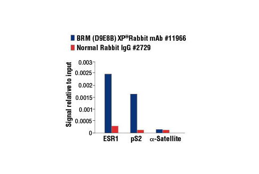
For optimal ChIP results, use 5 μl of antibody and 10 μg of chromatin (approximately 4 x 10⁶ cells) per IP. This antibody has been validated using SimpleChIP® Enzymatic Chromatin IP Kits.
| Application | Dilution |
|---|---|
| Western Blotting | 1:1000 |
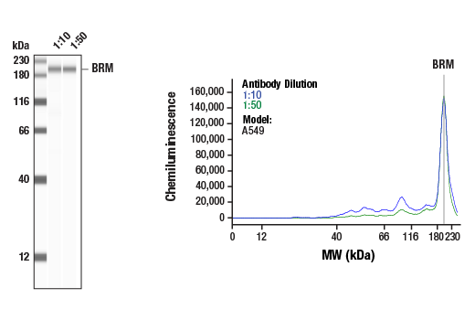
| Simple Western™ | 1:10 - 1:50 |
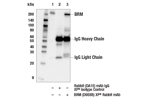
| Immunoprecipitation | 1:50 |
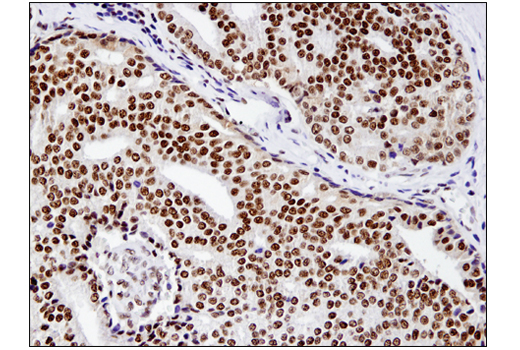
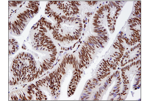
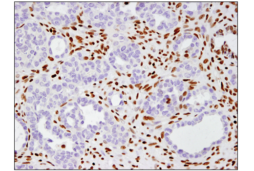
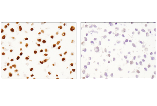
| Immunohistochemistry (Paraffin) | 1:400 - 1:1600 |
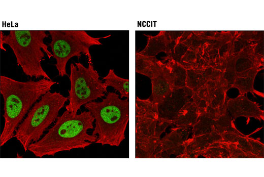
| Immunofluorescence (Immunocytochemistry) | 1:3200 - 1:6400 |
| Chromatin IP | 1:100 |
Storage
Specificity / Sensitivity
Species Reactivity:
Human, Monkey
Species predicted to react based on 100% sequence homology
The antigen sequence used to produce this antibody shares
100% sequence homology with the species listed here, but
reactivity has not been tested or confirmed to work by CST.
Use of this product with these species is not covered under
our
Product Performance Guarantee.
Dog
Source / Purification
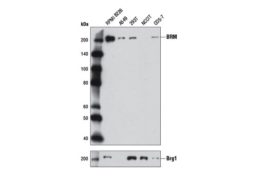
Monoclonal antibody is produced by immunizing animals with a synthetic peptide corresponding to residues surrounding Gly264 of human BRM protein.
Background
ATP-dependent chromatin remodeling complexes play an essential role in the regulation of various nuclear processes, such as gene expression, DNA replication, and repair (1,2). The SWI/SNF chromatin remodeling complex consists of more than 10 subunits with a single molecule of the ATPase catalytic subunit BRM or BRG1, but not both. The activities of these two subunits drive the disruption of histone-DNA contacts that lead to changes in accessibility of crucial regulatory elements within chromatin (2-5). The BRM/BRG1 containing SWI/SNF complexes are recruited to target promoters by transcription factors, such as nuclear receptors, p53, RB, and BRCA1 to regulate gene activation, cell growth, the cell cycle, and differentiation processes (1,6-9). BRM and BRG1 are also considered to be tumor suppressors and their expression levels are severely reduced in several cancer cell lines (10-13).
- Ho, L. and Crabtree, G.R. (2010) Nature 463, 474-84.
- Becker, P.B. and Hörz, W. (2002) Annu Rev Biochem 71, 247-73.
- Eberharter, A. and Becker, P.B. (2004) J Cell Sci 117, 3707-11.
- Bowman, G.D. (2010) Curr Opin Struct Biol 20, 73-81.
- Gangaraju, V.K. and Bartholomew, B. (2007) Mutat Res 618, 3-17.
- Lessard, J.A. and Crabtree, G.R. (2010) Annu Rev Cell Dev Biol 26, 503-32.
- Morettini, S. et al. (2008) Front Biosci 13, 5522-32.
- Wolf, I.M. et al. (2008) J Cell Biochem 104, 1580-6.
- Simone, C. (2006) J Cell Physiol 207, 309-14.
- Yamamichi, N. et al. (2005) Oncogene 24, 5471-81.
- Reisman, D.N. et al. (2002) Oncogene 21, 1196-207.
- Shen, H. et al. (2008) Cancer Res 68, 10154-62.
- Weissman, B. and Knudsen, K.E. (2009) Cancer Res 69, 8223-30.
Species Reactivity
Species reactivity is determined by testing in at least one approved application (e.g., western blot).
Western Blot Buffer
IMPORTANT: For western blots, incubate membrane with diluted primary antibody in 5% w/v nonfat dry milk, 1X TBS, 0.1% Tween® 20 at 4°C with gentle shaking, overnight.
Applications Key
WB: Western Blotting W-S: Simple Western™ IP: Immunoprecipitation IHC-P: Immunohistochemistry (Paraffin) IF-IC: Immunofluorescence (Immunocytochemistry) ChIP: Chromatin IP
Cross-Reactivity Key
H: human M: mouse R: rat Hm: hamster Mk: monkey Vir: virus Mi: mink C: chicken Dm: D. melanogaster X: Xenopus Z: zebrafish B: bovine Dg: dog Pg: pig Sc: S. cerevisiae Ce: C. elegans Hr: horse GP: Guinea Pig Rab: rabbit All: all species expected
Trademarks and Patents
Limited Uses
Except as otherwise expressly agreed in a writing signed by a legally authorized representative of CST, the following terms apply to Products provided by CST, its affiliates or its distributors. Any Customer's terms and conditions that are in addition to, or different from, those contained herein, unless separately accepted in writing by a legally authorized representative of CST, are rejected and are of no force or effect.
Products are labeled with For Research Use Only or a similar labeling statement and have not been approved, cleared, or licensed by the FDA or other regulatory foreign or domestic entity, for any purpose. Customer shall not use any Product for any diagnostic or therapeutic purpose, or otherwise in any manner that conflicts with its labeling statement. Products sold or licensed by CST are provided for Customer as the end-user and solely for research and development uses. Any use of Product for diagnostic, prophylactic or therapeutic purposes, or any purchase of Product for resale (alone or as a component) or other commercial purpose, requires a separate license from CST. Customer shall (a) not sell, license, loan, donate or otherwise transfer or make available any Product to any third party, whether alone or in combination with other materials, or use the Products to manufacture any commercial products, (b) not copy, modify, reverse engineer, decompile, disassemble or otherwise attempt to discover the underlying structure or technology of the Products, or use the Products for the purpose of developing any products or services that would compete with CST products or services, (c) not alter or remove from the Products any trademarks, trade names, logos, patent or copyright notices or markings, (d) use the Products solely in accordance with CST Product Terms of Sale and any applicable documentation, and (e) comply with any license, terms of service or similar agreement with respect to any third party products or services used by Customer in connection with the Products.