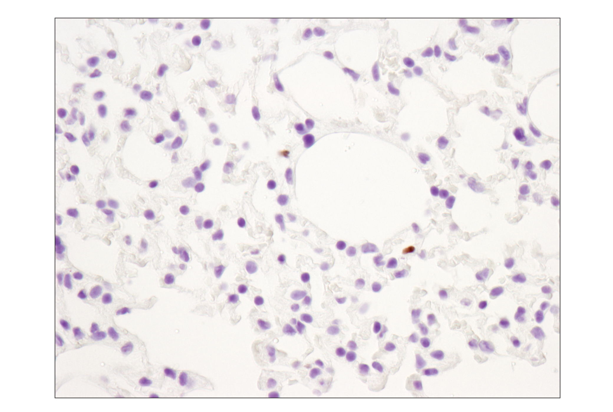
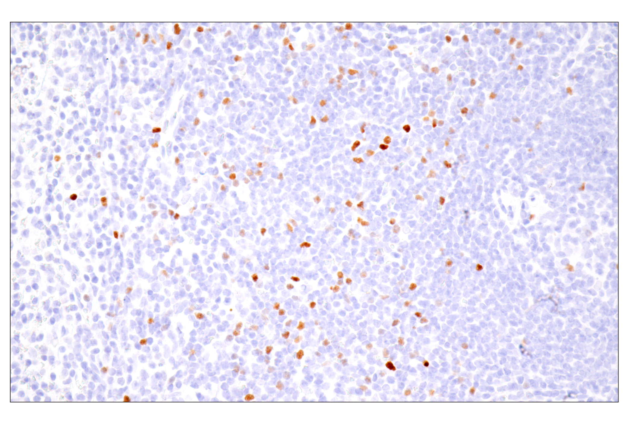
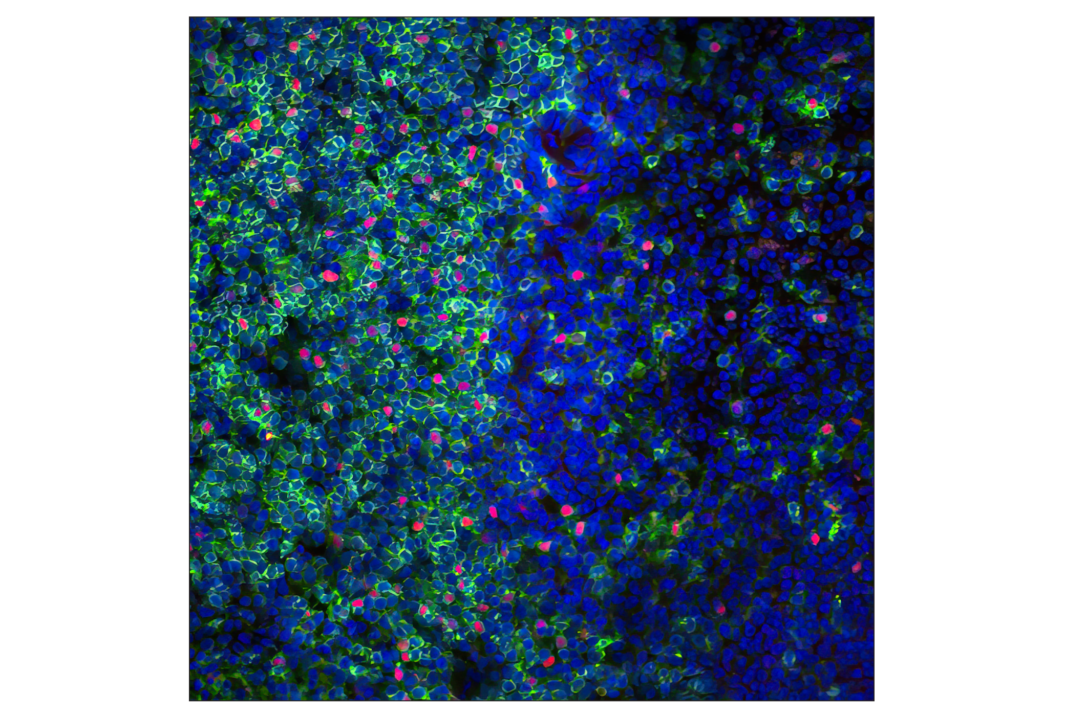
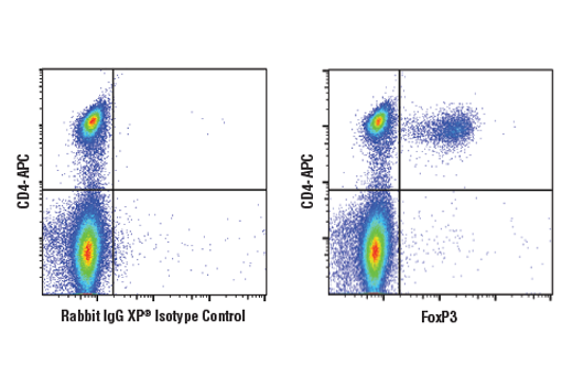
IHC-Bond, IHC-P, IF-F, FC-FP
M Mk
Endogenous
Rabbit IgG
#Q9BZS1
50943
Product Information
Product Usage Information
| Application | Dilution |
|---|---|
| IHC Leica Bond | 1:800 - 1:3200 |
| Immunohistochemistry (Paraffin) | 1:200 - 1:800 |
| Immunofluorescence (Frozen) | 1:400 - 1:800 |
| Flow Cytometry (Fixed/Permeabilized) | 1:50 - 1:200 |
Storage
For a carrier free (BSA and azide free) version of this product see product #72338.
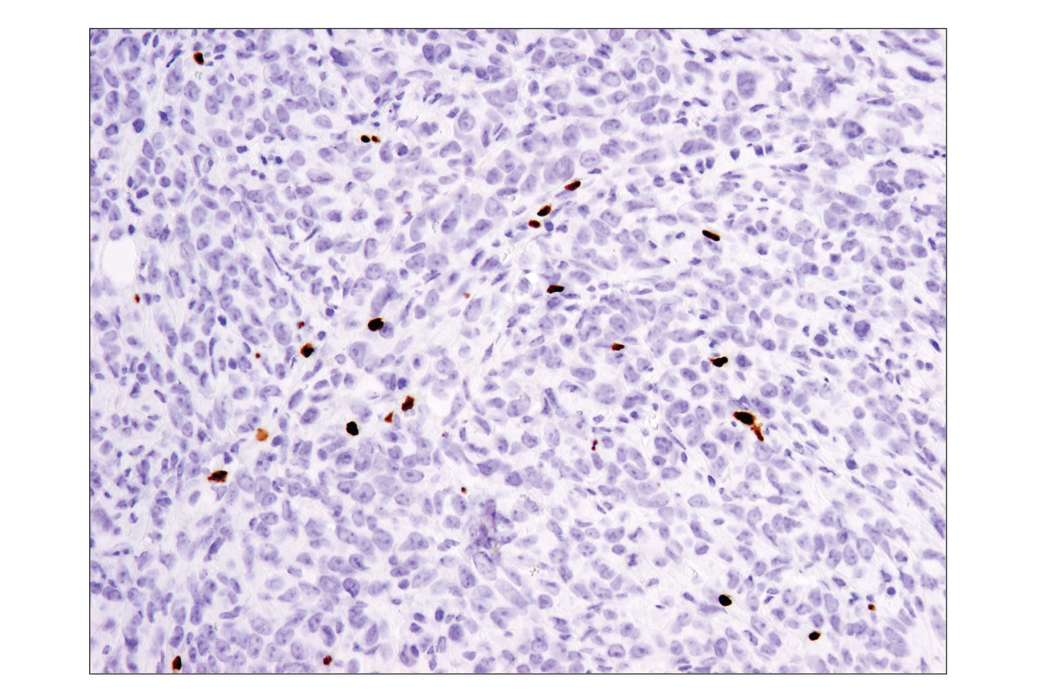
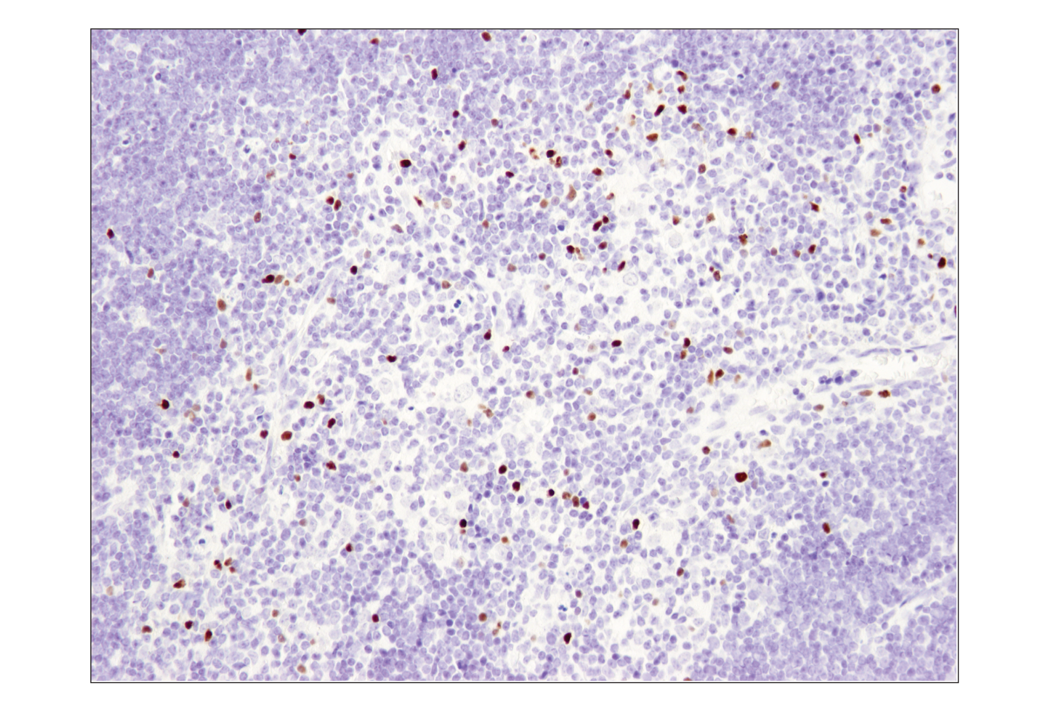
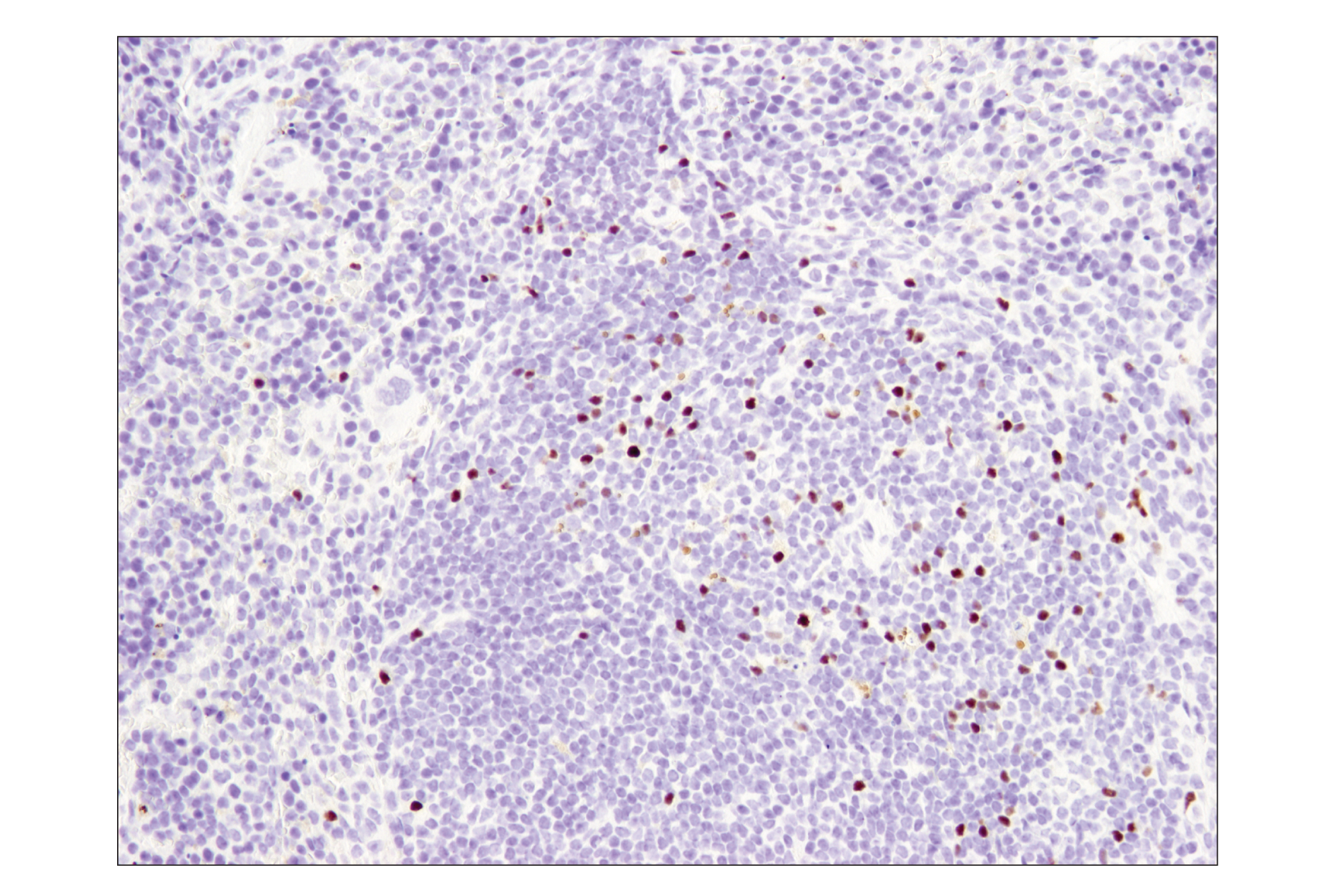
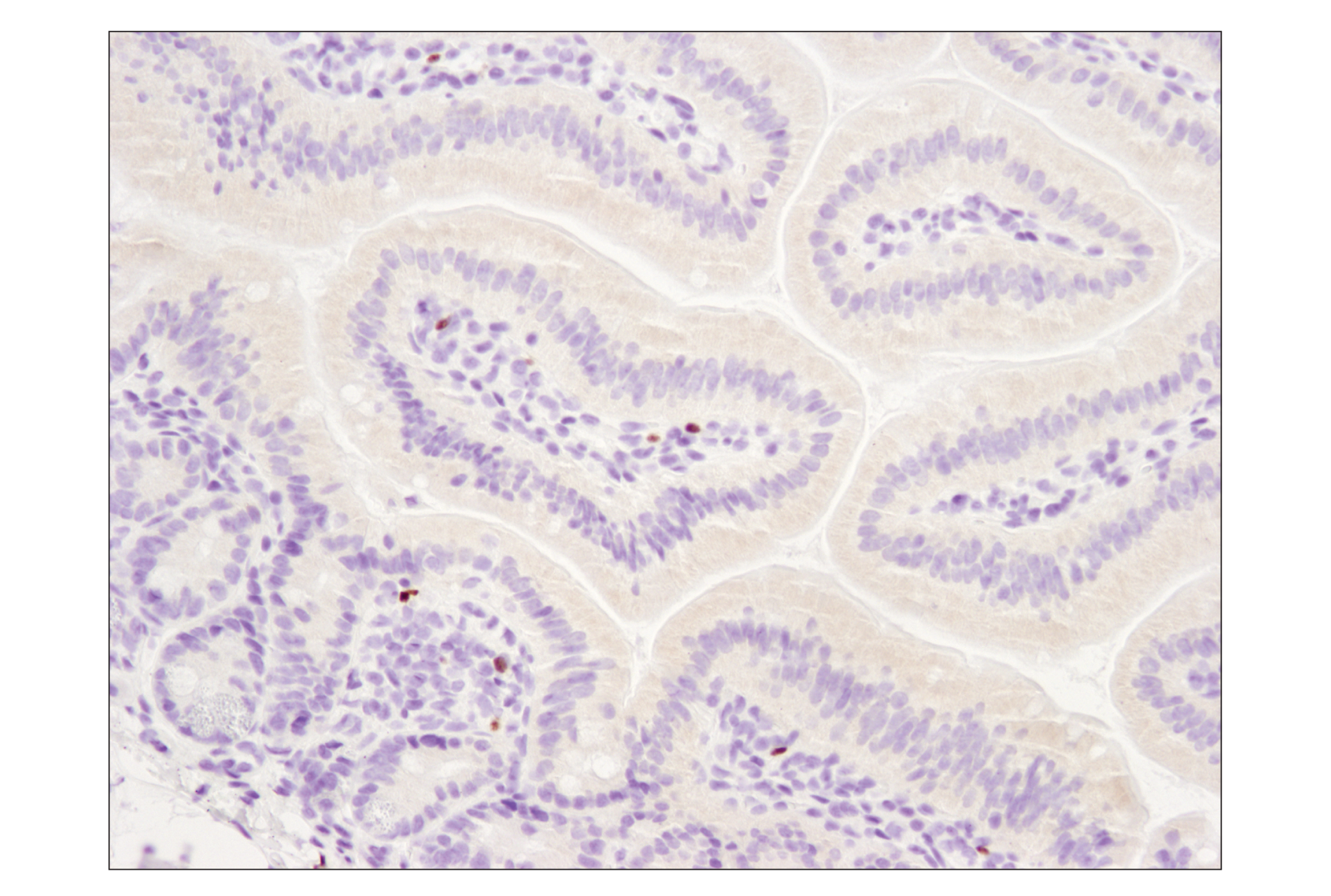
Specificity / Sensitivity
Species Reactivity:
Mouse, Monkey
Source / Purification
Monoclonal antibody is produced by immunizing animals with a synthetic peptide corresponding to residues surrounding Pro44 of mouse FoxP3 protein.
Background
Forkhead box (Fox) proteins are a family of evolutionarily conserved transcription factors containing a sequence known as Forkhead box or winged helix DNA binding domain (1). The human genome contains 43 Fox proteins that are divided into subfamilies. The FoxP subfamily has four members, FoxP1 - FoxP4, which are broadly expressed and play important roles in organ development, immune response, and cancer pathogenesis (2-4). The FoxP subfamily has several characteristics that are atypical among Fox proteins: their Forkhead domain is located at the carboxy-terminal region and they contain motifs that promote homo- and heterodimerization. FoxP proteins usually function as transcriptional repressors (4,5).
FoxP3 is crucial for the development of T cells with regulatory properties (Treg) (6). Mutations in FoxP3 are associated with immune dysregulation, polyendocrinopathy, enteropathy, and X-linked syndrome (IPEX) (7), while overexpression in mice causes severe immunodeficiency (8). Research studies have shown that FoxP3 functions as a tumor suppressor in several types of cancer (9-11).
- Myatt, S.S. and Lam, E.W. (2007) Nat Rev Cancer 7, 847-59.
- Shu, W. et al. (2001) J Biol Chem 276, 27488-97.
- Lu, M.M. et al. (2002) Gene Expr Patterns 2, 223-8.
- Koon, H.B. et al. (2007) Expert Opin Ther Targets 11, 955-65.
- Li, S. et al. (2004) Mol Cell Biol 24, 809-22.
- Ochs, H.D. et al. (2007) Immunol Res 38, 112-21.
- Bennett, C.L. et al. (2001) Nat Genet 27, 20-1.
- Kasprowicz, D.J. et al. (2003) J Immunol 171, 1216-23.
- Zuo, T. et al. (2007) Cell 129, 1275-86.
- Zuo, T. et al. (2007) J Clin Invest 117, 3765-73.
- Wang, L. et al. (2009) Cancer Cell 16, 336-46.
Species Reactivity
Species reactivity is determined by testing in at least one approved application (e.g., western blot).
Applications Key
IHC-Bond: IHC Leica Bond IHC-P: Immunohistochemistry (Paraffin) IF-F: Immunofluorescence (Frozen) FC-FP: Flow Cytometry (Fixed/Permeabilized)
Cross-Reactivity Key
H: human M: mouse R: rat Hm: hamster Mk: monkey Vir: virus Mi: mink C: chicken Dm: D. melanogaster X: Xenopus Z: zebrafish B: bovine Dg: dog Pg: pig Sc: S. cerevisiae Ce: C. elegans Hr: horse GP: Guinea Pig Rab: rabbit All: all species expected
Trademarks and Patents
Limited Uses
Except as otherwise expressly agreed in a writing signed by a legally authorized representative of CST, the following terms apply to Products provided by CST, its affiliates or its distributors. Any Customer's terms and conditions that are in addition to, or different from, those contained herein, unless separately accepted in writing by a legally authorized representative of CST, are rejected and are of no force or effect.
Products are labeled with For Research Use Only or a similar labeling statement and have not been approved, cleared, or licensed by the FDA or other regulatory foreign or domestic entity, for any purpose. Customer shall not use any Product for any diagnostic or therapeutic purpose, or otherwise in any manner that conflicts with its labeling statement. Products sold or licensed by CST are provided for Customer as the end-user and solely for research and development uses. Any use of Product for diagnostic, prophylactic or therapeutic purposes, or any purchase of Product for resale (alone or as a component) or other commercial purpose, requires a separate license from CST. Customer shall (a) not sell, license, loan, donate or otherwise transfer or make available any Product to any third party, whether alone or in combination with other materials, or use the Products to manufacture any commercial products, (b) not copy, modify, reverse engineer, decompile, disassemble or otherwise attempt to discover the underlying structure or technology of the Products, or use the Products for the purpose of developing any products or services that would compete with CST products or services, (c) not alter or remove from the Products any trademarks, trade names, logos, patent or copyright notices or markings, (d) use the Products solely in accordance with CST Product Terms of Sale and any applicable documentation, and (e) comply with any license, terms of service or similar agreement with respect to any third party products or services used by Customer in connection with the Products.