#P00519
25
Product Information
Storage
Keep enzymes on ice during use.
Avoid repeated freeze-thaw cycles.
Source / Purification
The GST-Kinase fusion protein was produced using a baculovirus expression system from a construct containing a human ABL1 cDNA (Pro118-Ser553) fragment amino-terminally fused to a GST-HIS6-Thrombin cleavage site. The protein was then purified by one-step affinity purification using glutathione-agarose.
The detection antibody, Phospho-Tyrosine Monoclonal Antibody (P-Tyr-100) #9411 was corresponding to mice immunized with phospho-tyrosine-containing peptides
Product Description
| Molecular Formula | Peptide Substrate, Biotin-Signal Transduction Protein (Tyr160): 1830 Daltons, GST-ABL1 Kinase domain: 74,140 Daltons |
Background
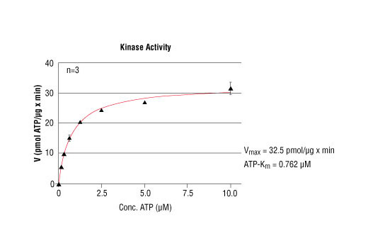
The c-Abl proto-oncogene encodes a nonreceptor protein tyrosine kinase that is ubiquitously expressed and highly conserved in metazoan evolution. c-Abl protein is distributed in both the nucleus and the cytoplasm of cells. It is implicated in regulating cell proliferation, differentiation, apoptosis, cell adhesion, and stress responses (1-3). c-Abl kinase activity is increased in vivo by diverse physiological stimuli including integrin activation; PDGF stimulation; and binding to c-Jun, Nck, and RFX1 (2,4). The in vivo mechanism for regulation of c-Abl kinase activity is not completely understood. Tyr245 is located in the linker region between the SH2 and catalytic domains. This positioning is conserved among Abl family members. Phosphorylation at Tyr245 is involved in the activation of c-Abl kinase (5). In addition, phosphorylation at Tyr412, which is located in the kinase activation loop of c-Abl, is required for kinase activity (6).
- Wang, J.Y. (2000) Oncogene 19, 5643-50.
- Van Etten, R.A. (1999) Trends Cell Biol 9, 179-86.
- Danial, N.N. and Rothman, P. (2000) Oncogene 19, 2523-31.
- Shaul, Y. (2000) Cell Death Differ 7, 10-6.
- Brasher, B.B. and Van Etten, R.A. (2000) J Biol Chem 275, 35631-7.
- Pluk, H. et al. (2002) Cell 108, 247-259.
Species Reactivity
Species reactivity is determined by testing in at least one approved application (e.g., western blot).
Cross-Reactivity Key
H: human M: mouse R: rat Hm: hamster Mk: monkey Vir: virus Mi: mink C: chicken Dm: D. melanogaster X: Xenopus Z: zebrafish B: bovine Dg: dog Pg: pig Sc: S. cerevisiae Ce: C. elegans Hr: horse GP: Guinea Pig Rab: rabbit All: all species expected
Trademarks and Patents
Limited Uses
Except as otherwise expressly agreed in a writing signed by a legally authorized representative of CST, the following terms apply to Products provided by CST, its affiliates or its distributors. Any Customer's terms and conditions that are in addition to, or different from, those contained herein, unless separately accepted in writing by a legally authorized representative of CST, are rejected and are of no force or effect.
Products are labeled with For Research Use Only or a similar labeling statement and have not been approved, cleared, or licensed by the FDA or other regulatory foreign or domestic entity, for any purpose. Customer shall not use any Product for any diagnostic or therapeutic purpose, or otherwise in any manner that conflicts with its labeling statement. Products sold or licensed by CST are provided for Customer as the end-user and solely for research and development uses. Any use of Product for diagnostic, prophylactic or therapeutic purposes, or any purchase of Product for resale (alone or as a component) or other commercial purpose, requires a separate license from CST. Customer shall (a) not sell, license, loan, donate or otherwise transfer or make available any Product to any third party, whether alone or in combination with other materials, or use the Products to manufacture any commercial products, (b) not copy, modify, reverse engineer, decompile, disassemble or otherwise attempt to discover the underlying structure or technology of the Products, or use the Products for the purpose of developing any products or services that would compete with CST products or services, (c) not alter or remove from the Products any trademarks, trade names, logos, patent or copyright notices or markings, (d) use the Products solely in accordance with CST Product Terms of Sale and any applicable documentation, and (e) comply with any license, terms of service or similar agreement with respect to any third party products or services used by Customer in connection with the Products.