| Product Includes | Product # | Quantity | Color | Storage Temp |
|---|---|---|---|---|
| M-CSFR Mouse mAb Coated Microwells | 27190 | 96 tests |
|
+4C |
| Phospho-M-CSFR (Tyr699) Rabbit Detection Ab | 62330 | 1 ea |
|
+4C |
| Anti-rabbit IgG, HRP-linked Antibody (ELISA Formulated) | 13272 | 1 ea |
|
+4C |
| Detection Antibody Diluent | 13339 | 11 ml |
|
+4C |
| HRP Diluent | 13515 | 11 ml |
|
+4C |
| TMB Substrate | 7004 | 11 ml |
|
+4C |
| STOP Solution | 7002 | 11 ml |
|
+4C |
| Sealing Tape | 54503 | 2 ea |
|
+4C |
| ELISA Wash Buffer (20X) | 9801 | 25 ml |
|
+4C |
| ELISA Sample Diluent | 11083 | 25 ml |
|
+4C |
| Cell Lysis Buffer (10X) | 9803 | 15 ml |
|
-20C |
*The microwell plate is supplied as 12 8-well modules - Each module is designed to break apart for 8 tests.
Description
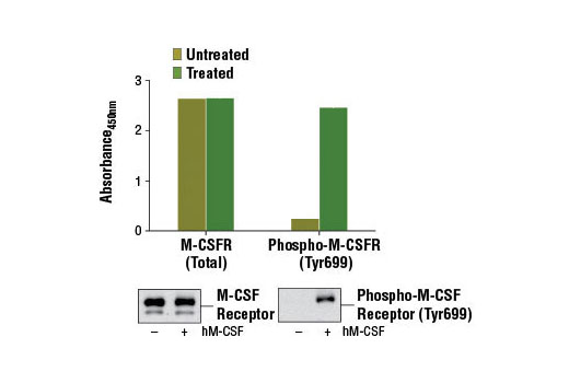
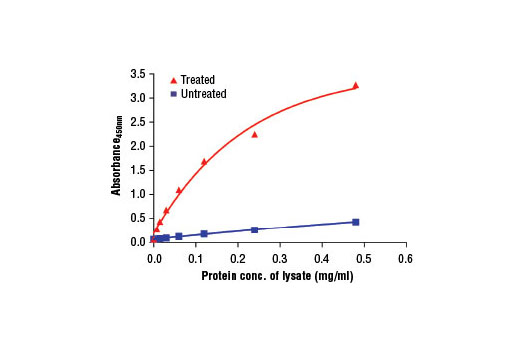
The PathScan® Phospho-M-CSF Receptor (Tyr699) Sandwich ELISA Kit is a solid phase sandwich enzyme-linked immunosorbent assay (ELISA) that detects endogenous levels of M-CSF receptor protein phosphorylated at Tyr699. A M-CSF receptor mouse mAb has been coated onto the microwells. After incubation with cell lysates, both phospho- and nonphospho-M-CSF receptor proteins are captured by the coated antibody. Following extensive washing, Phospho-M-CSF Receptor (Tyr699) Rabbit Detection mAb is added to detect the captured phospho-M-CSF receptor proteins. Anti-rabbit IgG, HRP-linked antibody is then used to recognize the bound detection antibody. HRP substrate, TMB, is added to develop color. The magnitude of optical density for this developed color is proportional to the quantity of M-CSF receptor protein phosphorylated at Tyr699.
Antibodies in kit are custom formulations specific to kit.
Specificity/Sensitivity
Background
Macrophage-colony stimulating factor (M-CSF, CSF-1) receptor is an integral membrane tyrosine kinase encoded by the c-fms proto-oncogene. M-CSF receptor is expressed in monocytes (macrophages and their progenitors) and drives growth and development of this blood cell lineage (1-3). Binding of M-CSF to its receptor induces receptor dimerization, activation, and autophosphorylation of cytoplasmic tyrosine residues used as docking sites for SH2-containing signaling proteins (4). There are at least five major tyrosine autophosphorylation sites. Tyr723 (Tyr721 in mouse) is located in the kinase insert (KI) region. Phosphorylated Tyr723 binds the p85 subunit of PI3 kinase as well as PLCγ2 (5). Phosphorylation of Tyr809 provides a docking site for Shc (5). Overactivation of this receptor can lead to a malignant phenotype in various cell systems (6). The activated M-CSF receptor has been shown to be a predictor of poor outcome in advanced epithelial ovarian carcinoma (7) and breast cancer (8).
Phosphorylation of M-CSF receptor at Tyr699 was identified at Cell Signaling Technology (CST) using PhosphoScan®, CST's LC-MS/MS platform for phosphorylation site discovery, and was cited in another publication (10). Autophosphorylation at Tyr699 within the kinase insert (KI) domain appears to provide a binding site for the Grb2 adaptor protein (9).
- Stanley, E.R. et al. (1978) Nature 274, 168-70.
- Byrne, P.V. et al. (1981) J Cell Biol 91, 848-53.
- Bourette, R.P. and Rohrschneider, L.R. (2000) Growth Factors 17, 155-66.
- Novak, U. et al. (1996) Oncogene 13, 2607-13.
- Bourette, R.P. et al. (1997) EMBO J 16, 5880-93.
- Morley, G.M. et al. (1999) Oncogene 18, 3076-84.
- Toy, E.P. et al. (2001) Gynecol Oncol 80, 194-200.
- Maher, M.G. et al. (1998) Clin Cancer Res 4, 1851-6.
- Hamilton, J.A. (1997) J Leukoc Biol 62, 145-55.
- Downing, J.R. et al. (1991) Mol Cell Biol 11, 2489-95.
Background References
Cross-Reactivity Key
H: human M: mouse R: rat Hm: hamster Mk: monkey Vir: virus Mi: mink C: chicken Dm: D. melanogaster X: Xenopus Z: zebrafish B: bovine Dg: dog Pg: pig Sc: S. cerevisiae Ce: C. elegans Hr: horse GP: Guinea Pig Rab: rabbit All: all species expected
Trademarks and Patents
Limited Uses
Except as otherwise expressly agreed in a writing signed by a legally authorized representative of CST, the following terms apply to Products provided by CST, its affiliates or its distributors. Any Customer's terms and conditions that are in addition to, or different from, those contained herein, unless separately accepted in writing by a legally authorized representative of CST, are rejected and are of no force or effect.
Products are labeled with For Research Use Only or a similar labeling statement and have not been approved, cleared, or licensed by the FDA or other regulatory foreign or domestic entity, for any purpose. Customer shall not use any Product for any diagnostic or therapeutic purpose, or otherwise in any manner that conflicts with its labeling statement. Products sold or licensed by CST are provided for Customer as the end-user and solely for research and development uses. Any use of Product for diagnostic, prophylactic or therapeutic purposes, or any purchase of Product for resale (alone or as a component) or other commercial purpose, requires a separate license from CST. Customer shall (a) not sell, license, loan, donate or otherwise transfer or make available any Product to any third party, whether alone or in combination with other materials, or use the Products to manufacture any commercial products, (b) not copy, modify, reverse engineer, decompile, disassemble or otherwise attempt to discover the underlying structure or technology of the Products, or use the Products for the purpose of developing any products or services that would compete with CST products or services, (c) not alter or remove from the Products any trademarks, trade names, logos, patent or copyright notices or markings, (d) use the Products solely in accordance with CST Product Terms of Sale and any applicable documentation, and (e) comply with any license, terms of service or similar agreement with respect to any third party products or services used by Customer in connection with the Products.