| Product Includes | Product # | Quantity | Color | Storage Temp |
|---|---|---|---|---|
| MAP Kinase Multi-Target | 79155 | 96 tests |
|
+4C |
| TMB Substrate | 7004 | 11 ml |
|
+4C |
| STOP Solution | 7002 | 11 ml |
|
+4C |
| Sealing Tape | 54503 | 2 ea |
|
+4C |
| ELISA Wash Buffer (20X) | 9801 | 25 ml |
|
+4C |
| ELISA Sample Diluent | 11083 | 25 ml |
|
+4C |
| Cell Lysis Buffer (10X) | 9803 | 15 ml |
|
-20C |
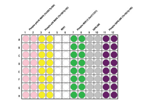
*The microwell plate is supplied as 12 8-well modules - Each module is designed to break apart for 8 tests.
Description
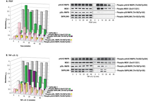
CST's PathScan® MAP Kinase Multi-Target Sandwich ELISA Kit is a solid phase sandwich enzyme-linked immunosorbent assay (ELISA) that combines the reagents necessary to detect endogenous levels of phospho-p44/42 MAPK (Thr202/Tyr204), phospho-p38 MAPK (Thr180/Tyr182), MEK1, phospho-MEK1 (Ser217/221), SAPK/JNK and phospho-SAPK/JNK (Thr183/Tyr185). These molecules represent convergence points and key regulatory proteins in signaling pathways controlling cellular events such as growth, differentiation and the response to stress and inflammation. Sixteen tests are provided for each target protein. Specific assay formulations for the indicated target proteins can be found in the datasheets associated with the individual PathScan® Sandwich ELISA Kits**. Briefly, a capture antibody has been coated onto the microwells. After incubation with cell lysates, the target protein is captured by the coated antibody. Following extensive washing, a detection antibody is added to detect the captured target protein. An HRP-linked secondary antibody is then used to recognize the bound detection antibody. HRP substrate, TMB, is added to develop color. The magnitude of absorbance for this developed color is proportional to the quantity of bound target protein. *Antibodies in kit are custom formulations specific to kit. **See companion products.
Specificity/Sensitivity
This kit detects proteins from the indicated species, as determined through in-house testing, but may also detect homologous proteins from other species.
Background
Both p44 and p42 MAP kinases (Erk1 and Erk2) function in a protein kinase cascade that plays a critical role in the regulation of cell growth and differentiation (1-6). MAP kinases are activated by a wide variety of extracellular signals including growth and neurotrophic factors, cytokines, hormones and neurotransmitters. Activation of MAP kinases occurs through phosphorylation of threonine and tyrosine (Thr202 and Tyrr204 of human MAP kinase or Thr183 and Tyr185 of rat MAP kinase) at the sequence T*EY* by a single upstream MAP kinase kinase (MEK) (7,8).
MEK1 and MEK2 are dual-specificity protein kinases that function in a mitogen activated protein kinase cascade controlling cell growth and differentiation. Activation of MEK1 and MEK2 occurs through phosphorylation of Ser217 and Ser221 by Raf-like molecules. MEK activates p44 and p42 MAP kinase (1,9,10).
p38 MAP kinase (MAPK) participates in a signaling cascade controlling the cellular response to pro-inflammatory cytokines and a variety of cellular stresses. MKK3, MKK6 and SEK (MKK4) activate p38 MAP kinase by phosphorylation at Thr180 and Tyr182 (11-14).
The stress-activated protein kinase/Jun-amino-terminal kinase SAPK/JNK is activated by a variety of environmental stresses, including UV and gamma radiation, ceramides, inflammatory cytokines and in some instances, by growth factors and GPCR agonists (15-20). As with the other MAPKs, the core-signaling unit is composed of a MAPKKK, typically MEKK1-4, or by a mixed lineage kinase (MLK), which phosphorylates and activates MKK4-7, which then phosphorylates Thr183 and Tyr185 to activate the SAPK/JNK kinase (16). Stress signals are delivered to this cascade by small GTPases of the Rho family (Rac, Rho, cdc42) (17). Both Rac1 and cdc42 mediate the stimulation of MEKKs and MLKs (17). Alternatively, MKK4-7 can be activated by a pathway independent of small GTPases via stimulation of a member of the germinal center kinase (GCK) family (18).
- McKay, M.M. and Morrison, D.K. (2007) Oncogene 26, 3113-21.
- Pearson, G. et al. (2001) Endocr Rev 22, 153-83.
- Marshall, C.J. (1995) Cell 80, 179-85.
- Hunter, T. (1995) Cell 80, 225-36.
- Hill, C.S. and Treisman, R. (1995) Cell 80, 199-211.
- Cowley, S. et al. (1994) Cell 77, 841-52.
- Sturgill, T.W. et al. (1988) Nature 334, 715-8.
- Payne, D.M. et al. (1991) EMBO J 10, 885-92.
- Alessi, D.R. et al. (1994) EMBO J 13, 1610-9.
- Pearson, G. et al. (2001) Endocr Rev 22, 153-83.
- Raingeaud, J. et al. (1995) J Biol Chem 270, 7420-6.
- Raman, M. et al. (2007) Oncogene 26, 3100-12.
- Zarubin, T. and Han, J. (2005) Cell Res 15, 11-8.
- Roux, P.P. and Blenis, J. (2004) Microbiol Mol Biol Rev 68, 320-44.
- Davis, R.J. (1999) Biochem Soc Symp 64, 1-12.
- Ichijo, H. (1999) Oncogene 18, 6087-93.
- Kyriakis, J.M. and Avruch, J. (2001) Physiol Rev 81, 807-69.
- Kyriakis, J.M. (1999) J Biol Chem 274, 5259-62.
- Leppä, S. and Bohmann, D. (1999) Oncogene 18, 6158-62.
- Whitmarsh, A.J. and Davis, R.J. (1998) Trends Biochem Sci 23, 481-5.
Background References
Cross-Reactivity Key
H: human M: mouse R: rat Hm: hamster Mk: monkey Vir: virus Mi: mink C: chicken Dm: D. melanogaster X: Xenopus Z: zebrafish B: bovine Dg: dog Pg: pig Sc: S. cerevisiae Ce: C. elegans Hr: horse GP: Guinea Pig Rab: rabbit All: all species expected
Trademarks and Patents
Limited Uses
Except as otherwise expressly agreed in a writing signed by a legally authorized representative of CST, the following terms apply to Products provided by CST, its affiliates or its distributors. Any Customer's terms and conditions that are in addition to, or different from, those contained herein, unless separately accepted in writing by a legally authorized representative of CST, are rejected and are of no force or effect.
Products are labeled with For Research Use Only or a similar labeling statement and have not been approved, cleared, or licensed by the FDA or other regulatory foreign or domestic entity, for any purpose. Customer shall not use any Product for any diagnostic or therapeutic purpose, or otherwise in any manner that conflicts with its labeling statement. Products sold or licensed by CST are provided for Customer as the end-user and solely for research and development uses. Any use of Product for diagnostic, prophylactic or therapeutic purposes, or any purchase of Product for resale (alone or as a component) or other commercial purpose, requires a separate license from CST. Customer shall (a) not sell, license, loan, donate or otherwise transfer or make available any Product to any third party, whether alone or in combination with other materials, or use the Products to manufacture any commercial products, (b) not copy, modify, reverse engineer, decompile, disassemble or otherwise attempt to discover the underlying structure or technology of the Products, or use the Products for the purpose of developing any products or services that would compete with CST products or services, (c) not alter or remove from the Products any trademarks, trade names, logos, patent or copyright notices or markings, (d) use the Products solely in accordance with CST Product Terms of Sale and any applicable documentation, and (e) comply with any license, terms of service or similar agreement with respect to any third party products or services used by Customer in connection with the Products.