| Product Includes | Product # | Quantity | Color | Storage Temp |
|---|---|---|---|---|
| Apoptosis Multi-Target | 43136 | 96 tests |
|
+4C |
| TMB Substrate | 7004 | 11 ml |
|
+4C |
| STOP Solution | 7002 | 11 ml |
|
+4C |
| Sealing Tape | 54503 | 2 ea |
|
+4C |
| ELISA Wash Buffer (20X) | 9801 | 25 ml |
|
+4C |
| ELISA Sample Diluent | 11083 | 25 ml |
|
+4C |
| Cell Lysis Buffer (10X) | 9803 | 15 ml |
|
-20C |
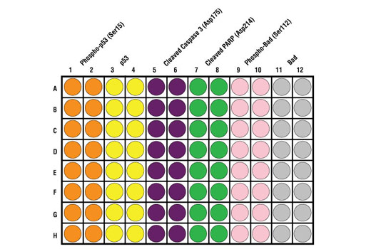
*The microwell plate is supplied as 12 8-well modules - Each module is designed to break apart for 8 tests.
Description
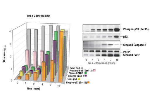
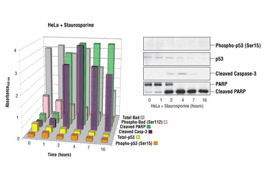
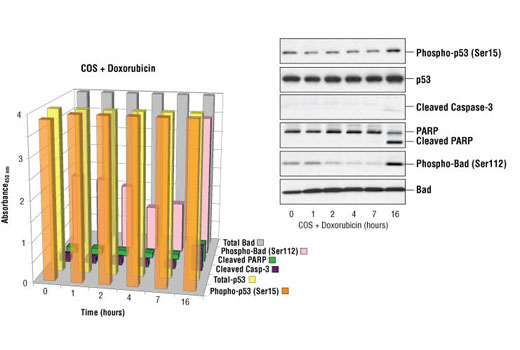
CST’s PathScan® Apoptosis Multi-Target Sandwich ELISA Kit is a solid phase sandwich enzyme-linked immunosorbent assay (ELISA) that combines the reagents necessary to detect endogenous levels of p53 protein, phospho-p53 protein (Ser15), Bad, phospho-Bad (Ser112), Cleaved Caspase-3 (Asp175) and Cleaved PARP (Asp214). These molecules represent key signaling proteins in pathways controlling survival and apoptosis. Sixteen assays are provided for each target protein. Specific assay formulations for the indicated target proteins can be found in the datasheets associated with the individual sandwich ELISA kits*. Briefly, a capture antibody** has been coated onto the microwells. After incubation with cell lysates, the target protein is captured by the coated antibody. Following extensive washing, a detection antibody** is added to detect the captured target protein. An HRP-linked secondary antibody is then used to recognize the bound detection antibody. HRP substrate, TMB, is added to develop color. The magnitude of absorbance for this developed color is proportional to the quantity of bound target protein.
*See companion products.
**Antibodies in kit are custom formulations specific to kit.
Specificity/Sensitivity
*See companion products.
This kit detects proteins from the indicated species, as determined through in-house testing, but may also detect homologous proteins from other species.
Background
Apoptosis is a regulated physiological process leading to cell death. Caspases, a family of cysteine acid proteases, are central regulators of apoptosis. Initiator caspases (including 8, 9, 10, and 12) are closely coupled to proapoptotic signals. Once activated, these caspases cleave and activate downstream effector caspases (including 3, 6, and 7), which in turn cleave cytoskeletal and nuclear proteins like PARP, α-fodrin, DFF, and lamin A and induce apoptosis. Cytochrome c released from mitochondria is coupled to the activation of caspase-9, a key initiator caspase (1). Proapoptotic stimuli include FasL, TNF-α, DNA damage and ER stress. Fas and TNFR activate caspase-8 and -10 (2), DNA damage leads to the activation of caspase-9 and ER stress leads to the calcium-mediated activation of caspase-12 (3). The inhibitor of apoptosis protein (IAP) family includes XIAP and survivin and functions by binding and inhibiting several caspases (4,5). Smac/Diablo, a mitochondrial protein, is released into the cytosol upon mitochondrial stress and competes with caspases for binding of IAPs. The interaction of Smac/Diablo with IAPs relieves the inhibitory effects of IAPs on caspases (6).
- Baker, S.J. and Reddy, E.P. (1998) Oncogene 17, 3261-3270.
- Budihardjo, I. et al. (1999) Annu. Rev. Cell Dev. Biol. 15, 269-290.
- Nakagawa, T. et al. (2000) Nature 403, 98-103.
- Deveraux, Q. L. et al. (1998) EMBO J. 17, 2215-2223.
- Li, F. et al. (1998) Nature 396, 580-584.
- Du, C. et al. (2000) Cell 102, 33-42.
Background References
Cross-Reactivity Key
H: human M: mouse R: rat Hm: hamster Mk: monkey Vir: virus Mi: mink C: chicken Dm: D. melanogaster X: Xenopus Z: zebrafish B: bovine Dg: dog Pg: pig Sc: S. cerevisiae Ce: C. elegans Hr: horse GP: Guinea Pig Rab: rabbit All: all species expected
Trademarks and Patents
Limited Uses
Except as otherwise expressly agreed in a writing signed by a legally authorized representative of CST, the following terms apply to Products provided by CST, its affiliates or its distributors. Any Customer's terms and conditions that are in addition to, or different from, those contained herein, unless separately accepted in writing by a legally authorized representative of CST, are rejected and are of no force or effect.
Products are labeled with For Research Use Only or a similar labeling statement and have not been approved, cleared, or licensed by the FDA or other regulatory foreign or domestic entity, for any purpose. Customer shall not use any Product for any diagnostic or therapeutic purpose, or otherwise in any manner that conflicts with its labeling statement. Products sold or licensed by CST are provided for Customer as the end-user and solely for research and development uses. Any use of Product for diagnostic, prophylactic or therapeutic purposes, or any purchase of Product for resale (alone or as a component) or other commercial purpose, requires a separate license from CST. Customer shall (a) not sell, license, loan, donate or otherwise transfer or make available any Product to any third party, whether alone or in combination with other materials, or use the Products to manufacture any commercial products, (b) not copy, modify, reverse engineer, decompile, disassemble or otherwise attempt to discover the underlying structure or technology of the Products, or use the Products for the purpose of developing any products or services that would compete with CST products or services, (c) not alter or remove from the Products any trademarks, trade names, logos, patent or copyright notices or markings, (d) use the Products solely in accordance with CST Product Terms of Sale and any applicable documentation, and (e) comply with any license, terms of service or similar agreement with respect to any third party products or services used by Customer in connection with the Products.