| Product Includes | Product # | Quantity | Color | Storage Temp |
|---|---|---|---|---|
| Chk1 Mouse mAb Coated Microwells | 32550 | 96 tests |
|
+4C |
| Chk1 Detection Ab | 37897 | 11 ml |
|
+4C |
| Anti-rabbit IgG, HRP-linked Antibody | 93699 | 11 ml |
|
+4C |
| TMB Substrate | 7004 | 11 ml |
|
+4C |
| STOP Solution | 7002 | 11 ml |
|
+4C |
| Sealing Tape | 54503 | 2 ea |
|
+4C |
| ELISA Wash Buffer (20X) | 9801 | 25 ml |
|
+4C |
| ELISA Sample Diluent | 11083 | 25 ml |
|
+4C |
| Cell Lysis Buffer (10X) | 9803 | 15 ml |
|
-20C |
*The microwell plate is supplied as 12 8-well modules - Each module is designed to break apart for 8 tests.
Description
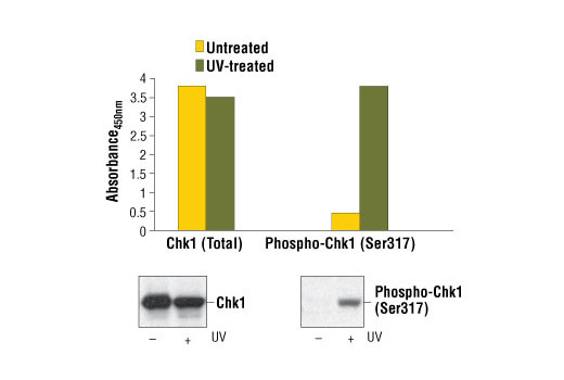
The PathScan® Total Chk1 Sandwich ELISA Kit is a solid phase sandwich enzyme-linked immunosorbent assay (ELISA) that detects endogenous levels of Chk1. A Chk1 Mouse Antibody has been coated onto the microwells. After incubation with cell lysates, Chk1 (phospho and nonphospho) is captured by the coated antibody. Following extensive washing, a Chk1 Rabbit Detection Antibody is added to the captured phospho and nonphospho Chk1 protein. Anti-rabbit IgG, HRP-linked Antibody is then used to recognize the bound detection antibody. HRP substrate, TMB, is added to develop color. The magnitude of the absorbance for this developed color is proportional to the quantity of total Chk1.
Antibodies in kit are custom formulations specific to kit.
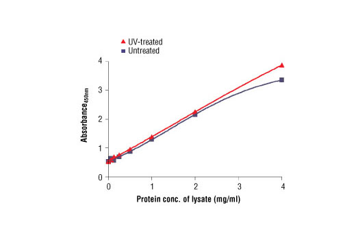
Specificity/Sensitivity
Background
Chk1 kinase acts downstream of ATM/ATR kinase and plays an important role in DNA damage checkpoint control, embryonic development, and tumor suppression (1). Activation of Chk1 involves phosphorylation at Ser317 and Ser345 by ATM/ATR, followed by autophosphorylation of Ser296. Activation occurs in response to blocked DNA replication and certain forms of genotoxic stress (2). While phosphorylation at Ser345 serves to localize Chk1 to the nucleus following checkpoint activation (3), phosphorylation at Ser317 along with site-specific phosphorylation of PTEN allows for re-entry into the cell cycle following stalled DNA replication (4). Chk1 exerts its checkpoint mechanism on the cell cycle, in part, by regulating the cdc25 family of phosphatases. Chk1 phosphorylation of cdc25A targets it for proteolysis and inhibits its activity through 14-3-3 binding (5). Activated Chk1 can inactivate cdc25C via phosphorylation at Ser216, blocking the activation of cdc2 and transition into mitosis (6). Centrosomal Chk1 has been shown to phosphorylate cdc25B and inhibit its activation of CDK1-cyclin B1, thereby abrogating mitotic spindle formation and chromatin condensation (7). Furthermore, Chk1 plays a role in spindle checkpoint function through regulation of aurora B and BubR1 (8). Research studies have implicated Chk1 as a drug target for cancer therapy as its inhibition leads to cell death in many cancer cell lines (9).
- Liu, Q. et al. (2000) Genes Dev 14, 1448-59.
- Zhao, H. and Piwnica-Worms, H. (2001) Mol Cell Biol 21, 4129-39.
- Jiang, K. et al. (2003) J Biol Chem 278, 25207-17.
- Martin, S.A. and Ouchi, T. (2008) Mol Cancer Ther 7, 2509-16.
- Chen, M.S. et al. (2003) Mol Cell Biol 23, 7488-97.
- Zeng, Y. et al. (1998) Nature 395, 507-10.
- Löffler, H. et al. (2006) Cell Cycle 5, 2543-7.
- Zachos, G. et al. (2007) Dev Cell 12, 247-60.
- Garber, K. (2005) J Natl Cancer Inst 97, 1026-8.
Background References
Cross-Reactivity Key
H: human M: mouse R: rat Hm: hamster Mk: monkey Vir: virus Mi: mink C: chicken Dm: D. melanogaster X: Xenopus Z: zebrafish B: bovine Dg: dog Pg: pig Sc: S. cerevisiae Ce: C. elegans Hr: horse GP: Guinea Pig Rab: rabbit All: all species expected
Trademarks and Patents
Limited Uses
Except as otherwise expressly agreed in a writing signed by a legally authorized representative of CST, the following terms apply to Products provided by CST, its affiliates or its distributors. Any Customer's terms and conditions that are in addition to, or different from, those contained herein, unless separately accepted in writing by a legally authorized representative of CST, are rejected and are of no force or effect.
Products are labeled with For Research Use Only or a similar labeling statement and have not been approved, cleared, or licensed by the FDA or other regulatory foreign or domestic entity, for any purpose. Customer shall not use any Product for any diagnostic or therapeutic purpose, or otherwise in any manner that conflicts with its labeling statement. Products sold or licensed by CST are provided for Customer as the end-user and solely for research and development uses. Any use of Product for diagnostic, prophylactic or therapeutic purposes, or any purchase of Product for resale (alone or as a component) or other commercial purpose, requires a separate license from CST. Customer shall (a) not sell, license, loan, donate or otherwise transfer or make available any Product to any third party, whether alone or in combination with other materials, or use the Products to manufacture any commercial products, (b) not copy, modify, reverse engineer, decompile, disassemble or otherwise attempt to discover the underlying structure or technology of the Products, or use the Products for the purpose of developing any products or services that would compete with CST products or services, (c) not alter or remove from the Products any trademarks, trade names, logos, patent or copyright notices or markings, (d) use the Products solely in accordance with CST Product Terms of Sale and any applicable documentation, and (e) comply with any license, terms of service or similar agreement with respect to any third party products or services used by Customer in connection with the Products.