| Product Includes | Product # | Quantity | Color | Storage Temp |
|---|---|---|---|---|
| Luminol/Enhancer Solution | 84850 | 3 ml |
|
RT |
| Stable Peroxide Buffer | 42552 | 3 ml |
|
RT |
| Sealing Tape | 54503 | 2 ea |
|
+4C |
| ELISA Wash Buffer (20X) | 9801 | 25 ml |
|
+4C |
| Cell Lysis Buffer (10X) | 9803 | 15 ml |
|
-20C |
*The microwell plate is supplied as 12 8-well modules - Each module is designed to break apart for 8 tests.
Description
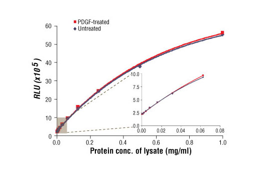
The PathScan® Total MEK1 Chemiluminescent Sandwich ELISA Kit is a solid phase sandwich enzyme-linked immunosorbent assay (ELISA) that detects endogenous levels of total MEK1 protein with a chemiluminescent readout. Chemiluminescent ELISAs often have a wider dynamic range and higher sensitivity than conventional chromogenic detection. This chemiluminescent ELISA, which is offered in low volume microplates, shows increased signal and sensitivity while using smaller sample size. A MEK1 mouse mAb has been coated on the microwells. After incubation with cell lysates, the MEK1 protein is captured by the coated antibody. Following extensive washing, a MEK1/2 rabbit antibody is added to detect the captured total MEK1 protein. Anti-rabbit IgG, HRP-linked antibody is then used to recognize the bound detection antibody. Chemiluminescent reagent is added for signal development. The magnitude of light emission, measured in relative light units (RLU), is proportional to the quantity of total MEK1 protein.
Specificity/Sensitivity
Background
MEK1 and MEK2, also called MAPK or Erk kinases, are dual-specificity protein kinases that function in a mitogen activated protein kinase cascade controlling cell growth and differentiation (1-3). Activation of MEK1 and MEK2 occurs through phosphorylation of two serine residues at positions 217 and 221, located in the activation loop of subdomain VIII, by Raf-like molecules. MEK1/2 is activated by a wide variety of growth factors and cytokines and also by membrane depolarization and calcium influx (1-4). Constitutively active forms of MEK1/2 are sufficient for the transformation of NIH/3T3 cells or the differentiation of PC-12 cells (4). MEK activates p44 and p42 MAP kinase by phosphorylating both threonine and tyrosine residues at sites located within the activation loop of kinase subdomain VIII.
Background References
Cross-Reactivity Key
H: human M: mouse R: rat Hm: hamster Mk: monkey Vir: virus Mi: mink C: chicken Dm: D. melanogaster X: Xenopus Z: zebrafish B: bovine Dg: dog Pg: pig Sc: S. cerevisiae Ce: C. elegans Hr: horse GP: Guinea Pig Rab: rabbit All: all species expected
Trademarks and Patents
Limited Uses
Except as otherwise expressly agreed in a writing signed by a legally authorized representative of CST, the following terms apply to Products provided by CST, its affiliates or its distributors. Any Customer's terms and conditions that are in addition to, or different from, those contained herein, unless separately accepted in writing by a legally authorized representative of CST, are rejected and are of no force or effect.
Products are labeled with For Research Use Only or a similar labeling statement and have not been approved, cleared, or licensed by the FDA or other regulatory foreign or domestic entity, for any purpose. Customer shall not use any Product for any diagnostic or therapeutic purpose, or otherwise in any manner that conflicts with its labeling statement. Products sold or licensed by CST are provided for Customer as the end-user and solely for research and development uses. Any use of Product for diagnostic, prophylactic or therapeutic purposes, or any purchase of Product for resale (alone or as a component) or other commercial purpose, requires a separate license from CST. Customer shall (a) not sell, license, loan, donate or otherwise transfer or make available any Product to any third party, whether alone or in combination with other materials, or use the Products to manufacture any commercial products, (b) not copy, modify, reverse engineer, decompile, disassemble or otherwise attempt to discover the underlying structure or technology of the Products, or use the Products for the purpose of developing any products or services that would compete with CST products or services, (c) not alter or remove from the Products any trademarks, trade names, logos, patent or copyright notices or markings, (d) use the Products solely in accordance with CST Product Terms of Sale and any applicable documentation, and (e) comply with any license, terms of service or similar agreement with respect to any third party products or services used by Customer in connection with the Products.