| Product Includes | Product # | Quantity | Color | Storage Temp |
|---|---|---|---|---|
| Tyro3 Rabbit mAb Coated Microwells | 29196 | 96 tests |
|
+4C |
| Tyro3 Mouse Detection mAb | 23487 | 1 ea |
|
+4C |
| Anti-mouse IgG, HRP-linked Antibody (ELISA Formulated) | 13304 | 1 ea |
|
+4C |
| Detection Antibody Diluent | 13339 | 11 ml |
|
+4C |
| HRP Diluent | 13515 | 11 ml |
|
+4C |
| TMB Substrate | 7004 | 11 ml |
|
+4C |
| STOP Solution | 7002 | 11 ml |
|
+4C |
| Sealing Tape | 54503 | 2 ea |
|
+4C |
| ELISA Wash Buffer (20X) | 9801 | 25 ml |
|
+4C |
| ELISA Sample Diluent | 11083 | 25 ml |
|
+4C |
| Cell Lysis Buffer (10X) | 9803 | 15 ml |
|
-20C |
*The microwell plate is supplied as 12 8-well modules - Each module is designed to break apart for 8 tests.
Description
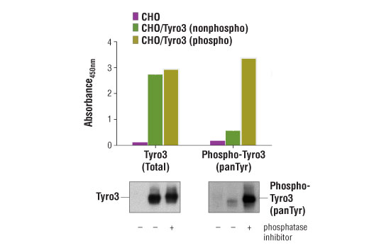
PathScan® Total Tyro3 Sandwich ELISA Kit is a solid phase sandwich enzyme-linked immunosorbent assay (ELISA) that detects endogenous levels of Tyro3 protein. A Tyro3 Rabbit mAb has been coated onto the microwells. After incubation with cell lysates, Tyro3 protein (phospho and nonphospho) is captured by the coated antibody. Following extensive washing, a Tyro3 Mouse Detection mAb is added to detect the captured Tyro3 proteins. Anti-mouse IgG, HRP-linked antibody is then used to recognize the bound detection antibody. HRP substrate, TMB, is added to develop color. The magnitude of optical density for this developed color is proportional to the quantity of total tyro3 protein.
*Antibodies in this kit are custom formulations specific to kit.
Specificity/Sensitivity
Background
Tyro3 is a receptor tyrosine kinase belonging to the TAM subfamily (Tyro3, Axl and Mer). All three members have similar domain structure composed of an extracellular region with 2 Ig-like domains, followed by 2 FNII-like domains, a single transmembrane region, and a cytoplasmic tyrosine kinase domain (1). The natural ligand for Tyro3, as well as Axl and Mer, is Gas6 (growth arrest-specific gene 6) (1,2). Expression pattern and target knockout data indicate an important role of Tyro3 in apoptotic cell phagocytosis of dendritic cells and macrophages (3), NK cell differentiation (4), reproductive neuron survival and migration (5), osteoclast stimulation (6,7), as well as cortical and hippocampal neuron function (8). Both MAPK and PI3K pathways have been suggested as downstream targets of Tyro3 activation (7,8). Tyro3 has also been shown to be correlated to melanoma tumorigenesis, likely through its reglulatory role in the expression of oncogenic microphthalmia-associated transcription factor (MITF) (9).
- Binder, M.D. and Kilpatrick, T.J. (2009) Neurosignals 17, 277-87.
- Nagata, K. et al. (1996) J Biol Chem 271, 30022-7.
- Seitz, H.M. et al. (2007) J Immunol 178, 5635-42.
- Caraux, A. et al. (2006) Nat Immunol 7, 747-54.
- Pierce, A. et al. (2008) Mol Endocrinol 22, 2481-95.
- Nakamura, Y.S. et al. (1998) Stem Cells 16, 229-38.
- Katagiri, M. et al. (2001) J Biol Chem 276, 7376-82.
- Prieto, A.L. et al. (2007) Neuroscience 150, 319-34.
- Zhu, S. et al. (2009) Proc Natl Acad Sci U S A 106, 17025-30.
Background References
Cross-Reactivity Key
H: human M: mouse R: rat Hm: hamster Mk: monkey Vir: virus Mi: mink C: chicken Dm: D. melanogaster X: Xenopus Z: zebrafish B: bovine Dg: dog Pg: pig Sc: S. cerevisiae Ce: C. elegans Hr: horse GP: Guinea Pig Rab: rabbit All: all species expected
Trademarks and Patents
Limited Uses
Except as otherwise expressly agreed in a writing signed by a legally authorized representative of CST, the following terms apply to Products provided by CST, its affiliates or its distributors. Any Customer's terms and conditions that are in addition to, or different from, those contained herein, unless separately accepted in writing by a legally authorized representative of CST, are rejected and are of no force or effect.
Products are labeled with For Research Use Only or a similar labeling statement and have not been approved, cleared, or licensed by the FDA or other regulatory foreign or domestic entity, for any purpose. Customer shall not use any Product for any diagnostic or therapeutic purpose, or otherwise in any manner that conflicts with its labeling statement. Products sold or licensed by CST are provided for Customer as the end-user and solely for research and development uses. Any use of Product for diagnostic, prophylactic or therapeutic purposes, or any purchase of Product for resale (alone or as a component) or other commercial purpose, requires a separate license from CST. Customer shall (a) not sell, license, loan, donate or otherwise transfer or make available any Product to any third party, whether alone or in combination with other materials, or use the Products to manufacture any commercial products, (b) not copy, modify, reverse engineer, decompile, disassemble or otherwise attempt to discover the underlying structure or technology of the Products, or use the Products for the purpose of developing any products or services that would compete with CST products or services, (c) not alter or remove from the Products any trademarks, trade names, logos, patent or copyright notices or markings, (d) use the Products solely in accordance with CST Product Terms of Sale and any applicable documentation, and (e) comply with any license, terms of service or similar agreement with respect to any third party products or services used by Customer in connection with the Products.