| Product Includes | Product # | Quantity | Color | Storage Temp |
|---|---|---|---|---|
| Histone H3 Rabbit mAb Coated Microwells | 45940 | 96 tests |
|
+4C |
| Tri-Methyl-Histone H3 (Lys27) Rabbit Detection mAb (Biotinylated) | 13765 | 1 ea |
|
+4C |
| HRP-Linked Streptavidin (ELISA Formulated) | 11805 | 1 ea |
|
+4C |
| Detection Antibody Diluent | 13339 | 11 ml |
|
+4C |
| HRP Diluent | 13515 | 11 ml |
|
+4C |
| TMB Substrate | 7004 | 11 ml |
|
+4C |
| STOP Solution | 7002 | 11 ml |
|
+4C |
| ELISA Wash Buffer (20X) | 9801 | 25 ml |
|
+4C |
| ELISA Sample Diluent | 11083 | 25 ml |
|
+4C |
| Sealing Tape | 54503 | 2 ea |
|
+4C |
| Cell Lysis Buffer (10X) | 9803 | 15 ml |
|
-20C |
*The microwell plate is supplied as 12 8-well modules - Each module is designed to break apart for 8 tests.
Description
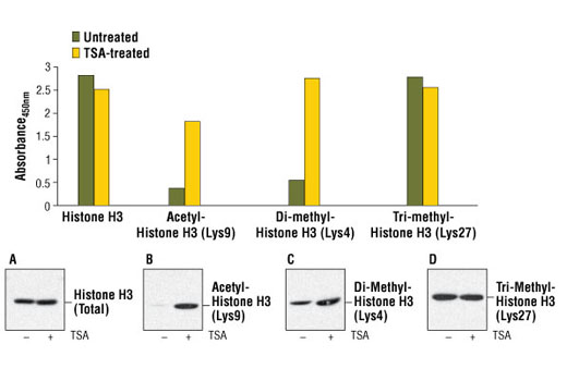
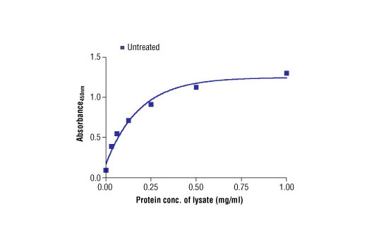
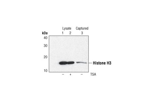
The PathScan® Tri-Methyl-Histone H3 (Lys27) Sandwich ELISA Kit is a solid phase sandwich enzyme-linked immunosorbent assay (ELISA) that detects endogenous levels of histone H3 when tri-methylated at Lys27. A Histone H3 Rabbit mAb has been coated onto the microwells. After incubation with cell lysates, histone H3 is captured by the coated antibody. Following extensive washing, biotinylated Tri-Methyl Histone H3 (Lys27) Rabbit Antibody is added to detect the captured histone H3 protein. HRP-linked streptavidin is then used to recognize the bound detection antibody. HRP substrate, TMB, is added to develop color. The magnitude of the absorbance for this developed color is proportional to the quantity of histone H3 tri-methylated at Lys27.
Antibodies in kit are custom formulations specific to kit.
Specificity/Sensitivity
Background
Modulation of chromatin structure plays an important role in the regulation of transcription in eukaryotes. The nucleosome, made up of DNA wound around eight core histone proteins (two each of H2A, H2B, H3, and H4), is the primary building block of chromatin (1). The amino-terminal tails of core histones undergo various posttranslational modifications, including acetylation, phosphorylation, methylation, and ubiquitination (2-5). These modifications occur in response to various stimuli and have a direct effect on the accessibility of chromatin to transcription factors and, therefore, gene expression (6). In most species, histone H2B is primarily acetylated at Lys5, 12, 15, and 20 (4,7). Histone H3 is primarily acetylated at Lys9, 14, 18, 23, 27, and 56. Acetylation of H3 at Lys9 appears to have a dominant role in histone deposition and chromatin assembly in some organisms (2,3). Phosphorylation at Ser10, Ser28, and Thr11 of histone H3 is tightly correlated with chromosome condensation during both mitosis and meiosis (8-10). Phosphorylation at Thr3 of histone H3 is highly conserved among many species and is catalyzed by the kinase haspin. Immunostaining with phospho-specific antibodies in mammalian cells reveals mitotic phosphorylation at Thr3 of H3 in prophase and its dephosphorylation during anaphase (11).
- Workman, J.L. and Kingston, R.E. (1998) Annu Rev Biochem 67, 545-79.
- Hansen, J.C. et al. (1998) Biochemistry 37, 17637-41.
- Strahl, B.D. and Allis, C.D. (2000) Nature 403, 41-5.
- Cheung, P. et al. (2000) Cell 103, 263-71.
- Bernstein, B.E. and Schreiber, S.L. (2002) Chem Biol 9, 1167-73.
- Jaskelioff, M. and Peterson, C.L. (2003) Nat Cell Biol 5, 395-9.
- Thorne, A.W. et al. (1990) Eur J Biochem 193, 701-13.
- Hendzel, M.J. et al. (1997) Chromosoma 106, 348-60.
- Goto, H. et al. (1999) J Biol Chem 274, 25543-9.
- Preuss, U. et al. (2003) Nucleic Acids Res 31, 878-85.
- Dai, J. et al. (2005) Genes Dev 19, 472-88.
Background References
Cross-Reactivity Key
H: human M: mouse R: rat Hm: hamster Mk: monkey Vir: virus Mi: mink C: chicken Dm: D. melanogaster X: Xenopus Z: zebrafish B: bovine Dg: dog Pg: pig Sc: S. cerevisiae Ce: C. elegans Hr: horse GP: Guinea Pig Rab: rabbit All: all species expected
Trademarks and Patents
Limited Uses
Except as otherwise expressly agreed in a writing signed by a legally authorized representative of CST, the following terms apply to Products provided by CST, its affiliates or its distributors. Any Customer's terms and conditions that are in addition to, or different from, those contained herein, unless separately accepted in writing by a legally authorized representative of CST, are rejected and are of no force or effect.
Products are labeled with For Research Use Only or a similar labeling statement and have not been approved, cleared, or licensed by the FDA or other regulatory foreign or domestic entity, for any purpose. Customer shall not use any Product for any diagnostic or therapeutic purpose, or otherwise in any manner that conflicts with its labeling statement. Products sold or licensed by CST are provided for Customer as the end-user and solely for research and development uses. Any use of Product for diagnostic, prophylactic or therapeutic purposes, or any purchase of Product for resale (alone or as a component) or other commercial purpose, requires a separate license from CST. Customer shall (a) not sell, license, loan, donate or otherwise transfer or make available any Product to any third party, whether alone or in combination with other materials, or use the Products to manufacture any commercial products, (b) not copy, modify, reverse engineer, decompile, disassemble or otherwise attempt to discover the underlying structure or technology of the Products, or use the Products for the purpose of developing any products or services that would compete with CST products or services, (c) not alter or remove from the Products any trademarks, trade names, logos, patent or copyright notices or markings, (d) use the Products solely in accordance with CST Product Terms of Sale and any applicable documentation, and (e) comply with any license, terms of service or similar agreement with respect to any third party products or services used by Customer in connection with the Products.