FC-FP
#Q02242, #P36402, #P10144, #P22646, #P11942, #P01731
18566, 6932, 3002, 12501, 12502, 12525
Product Information
Product Usage Information
This panel can be combined with peptide-MHC tetramer staining to identify antigen-specific cells. Tetramer staining is often performed on live cells. If tetramer staining is desired, we recommend comparing activity of the reagent in live cell staining vs. the recommended protocol for the panel. If there is not a significant difference between the two conditions, the tetramer can be included in the antibody panel staining mix. If there is significant loss of activity following fixation and permeabilization, tetramer staining should be done on live cells prior to fixation and permeabilization.
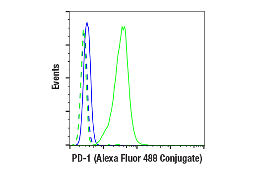
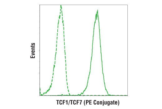
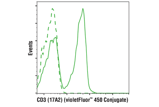
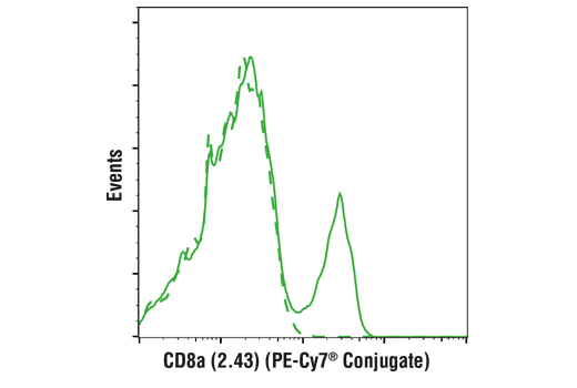
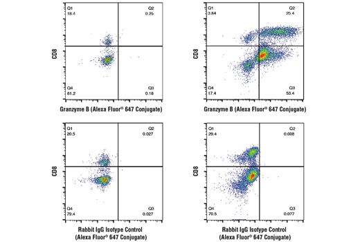
Gating strategy for observing progenitor exhausted CD8+ T cells:
If a fixable viability dye was used, first gate on viable cells. Next, gate on lymphocytes based on forward scatter and side scatter. Cytotoxic T cells are the CD3+CD8+ cells within the lymphocyte gate. Next, observe TCF1 and PD-1 expression on the cytotoxic T cells. Cytotoxic T cells that are TCF1+PD-1+ are exhausted progenitor cells and are expected to be mostly negative for Granzyme B expression. Cytotoxic T cells that are TCF1-PD-1+ are differentiated cells with effector potential and are expected to be mostly positive for expression of Granzyme B.
Storage
Store at 4oC. Do not aliquot the antibodies. Protect from light. Do not freeze.
All components in this kit are stable in accordance with the date printed on the outer packaging label when stored at the recommended temperature. Please refer to product labels, datasheets, or web pages for specific “Best By” dates for each individual component.
Specificity / Sensitivity
Species Reactivity:
Mouse
Source / Purification
Monoclonal antibodies were purified from tissue culture supernatant via affinity chromatography. The purified antibodies were conjugated under optimal conditions, with unreacted dye removed from the preparation.
Product Description
Cytotoxic T cells are identified by co-expression of CD3 and CD8α. Among these cells, the exhausted progenitor cells are TCF1+PD-1+ cells and are mostly Granzyme B-; the differentiated cells with effector potential are TCF1-PD-1+ and are mostly GranzymeB+. In response to immunotherapy, these progenitor TCF1+PD-1+CD8+ cells can expand and differentiate into TCF1-PD-1+CD8+ effector cells.
Species Reactivity
Species reactivity is determined by testing in at least one approved application (e.g., western blot).
Applications Key
FC-FP: Flow Cytometry (Fixed/Permeabilized)
Cross-Reactivity Key
H: human M: mouse R: rat Hm: hamster Mk: monkey Vir: virus Mi: mink C: chicken Dm: D. melanogaster X: Xenopus Z: zebrafish B: bovine Dg: dog Pg: pig Sc: S. cerevisiae Ce: C. elegans Hr: horse GP: Guinea Pig Rab: rabbit All: all species expected
Trademarks and Patents
Limited Uses
Except as otherwise expressly agreed in a writing signed by a legally authorized representative of CST, the following terms apply to Products provided by CST, its affiliates or its distributors. Any Customer's terms and conditions that are in addition to, or different from, those contained herein, unless separately accepted in writing by a legally authorized representative of CST, are rejected and are of no force or effect.
Products are labeled with For Research Use Only or a similar labeling statement and have not been approved, cleared, or licensed by the FDA or other regulatory foreign or domestic entity, for any purpose. Customer shall not use any Product for any diagnostic or therapeutic purpose, or otherwise in any manner that conflicts with its labeling statement. Products sold or licensed by CST are provided for Customer as the end-user and solely for research and development uses. Any use of Product for diagnostic, prophylactic or therapeutic purposes, or any purchase of Product for resale (alone or as a component) or other commercial purpose, requires a separate license from CST. Customer shall (a) not sell, license, loan, donate or otherwise transfer or make available any Product to any third party, whether alone or in combination with other materials, or use the Products to manufacture any commercial products, (b) not copy, modify, reverse engineer, decompile, disassemble or otherwise attempt to discover the underlying structure or technology of the Products, or use the Products for the purpose of developing any products or services that would compete with CST products or services, (c) not alter or remove from the Products any trademarks, trade names, logos, patent or copyright notices or markings, (d) use the Products solely in accordance with CST Product Terms of Sale and any applicable documentation, and (e) comply with any license, terms of service or similar agreement with respect to any third party products or services used by Customer in connection with the Products.