| Product Includes | Product # | Quantity | Mol. Wt | Isotype/Source |
|---|---|---|---|---|
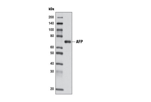
| AFP (D12C1) Rabbit mAb | 4448 | 40 µl | 65 kDa | Rabbit IgG |
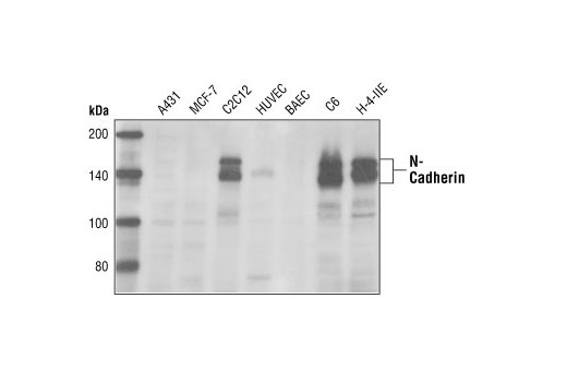
| N-Cadherin Antibody | 4061 | 40 µl | 140 kDa | Rabbit |
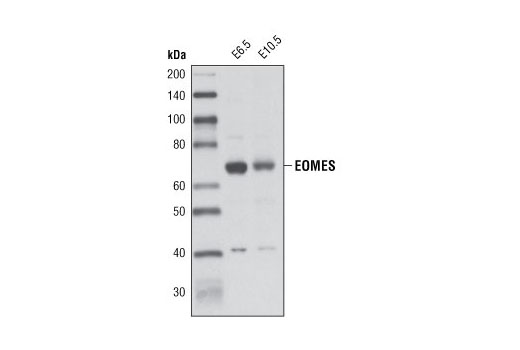
| EOMES Antibody | 4540 | 40 µl | 70 kDa | Rabbit |
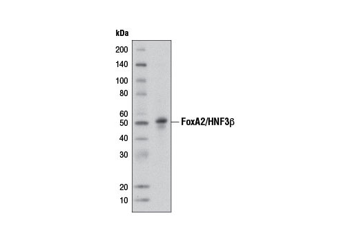
| FoxA2/HNF3β (D56D6) XP® Rabbit mAb | 8186 | 40 µl | 50 kDa | Rabbit IgG |
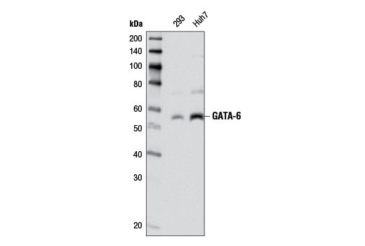
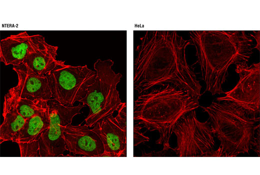
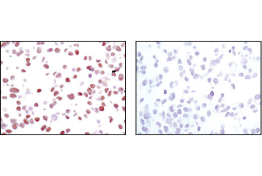
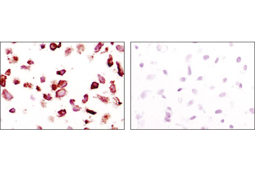
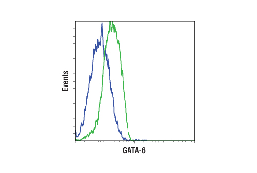
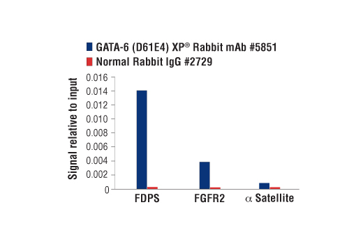
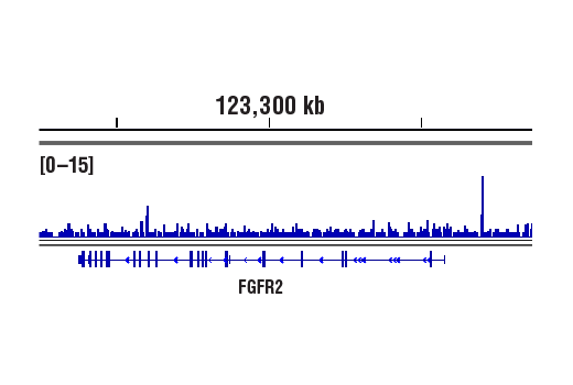
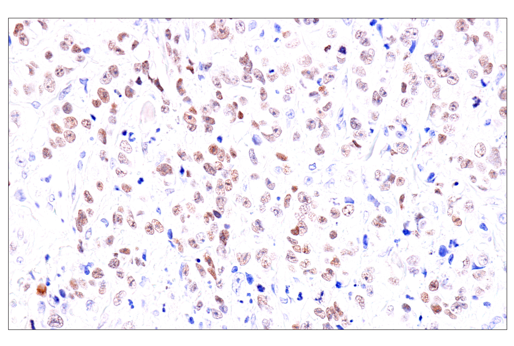
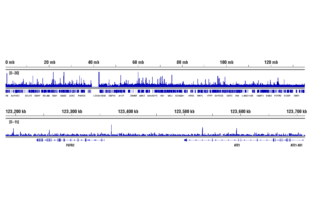
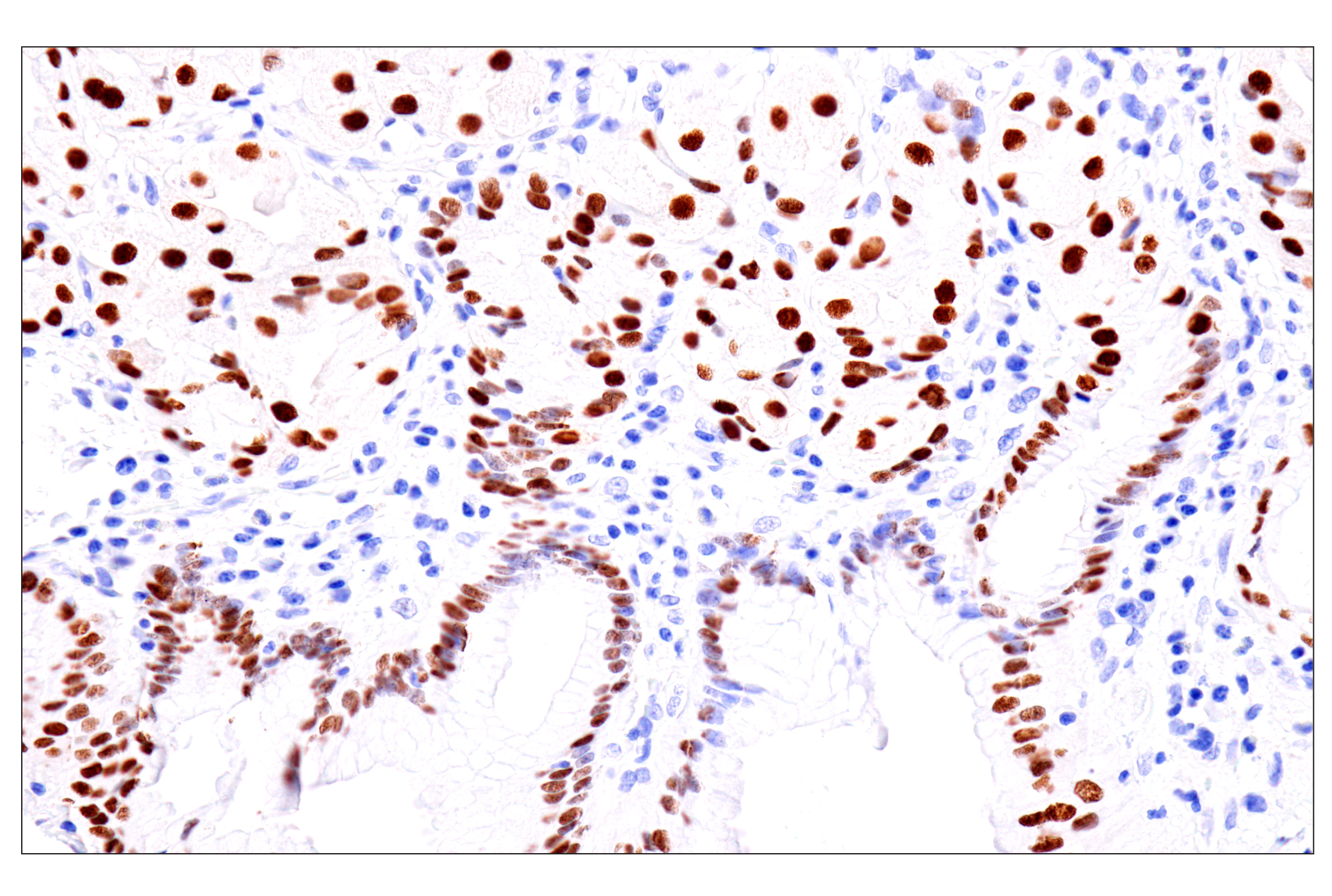
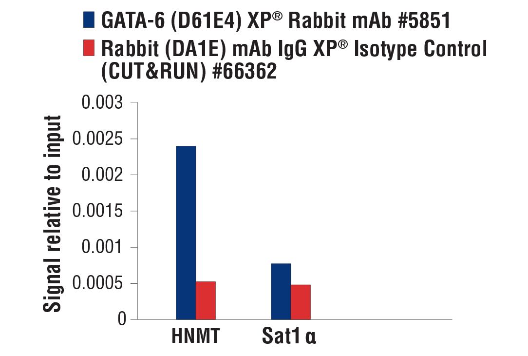
| GATA-6 (D61E4) XP® Rabbit mAb | 5851 | 40 µl | 55 kDa | Rabbit IgG |
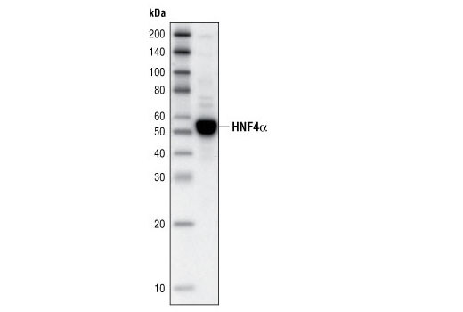
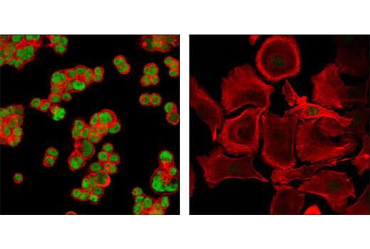
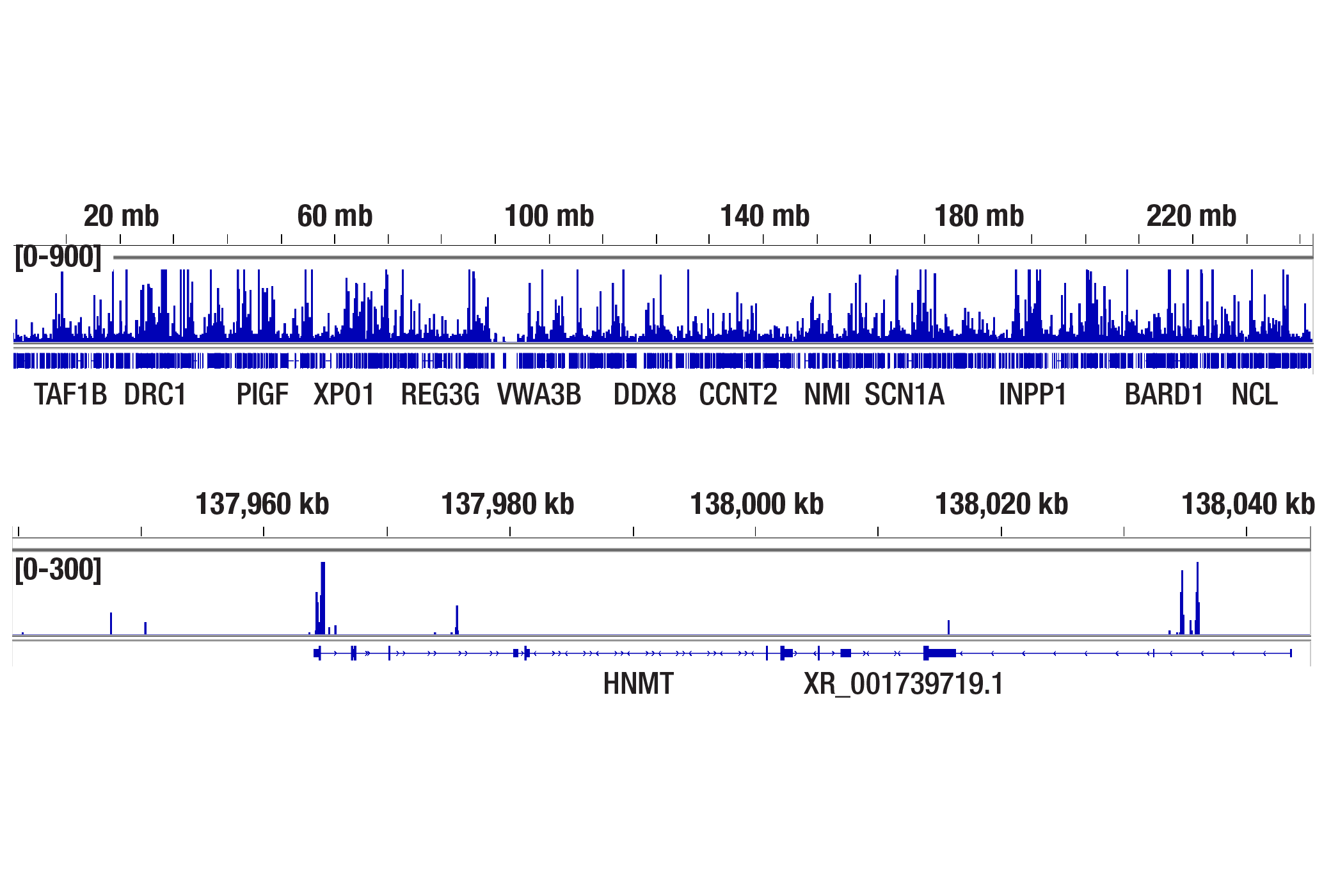
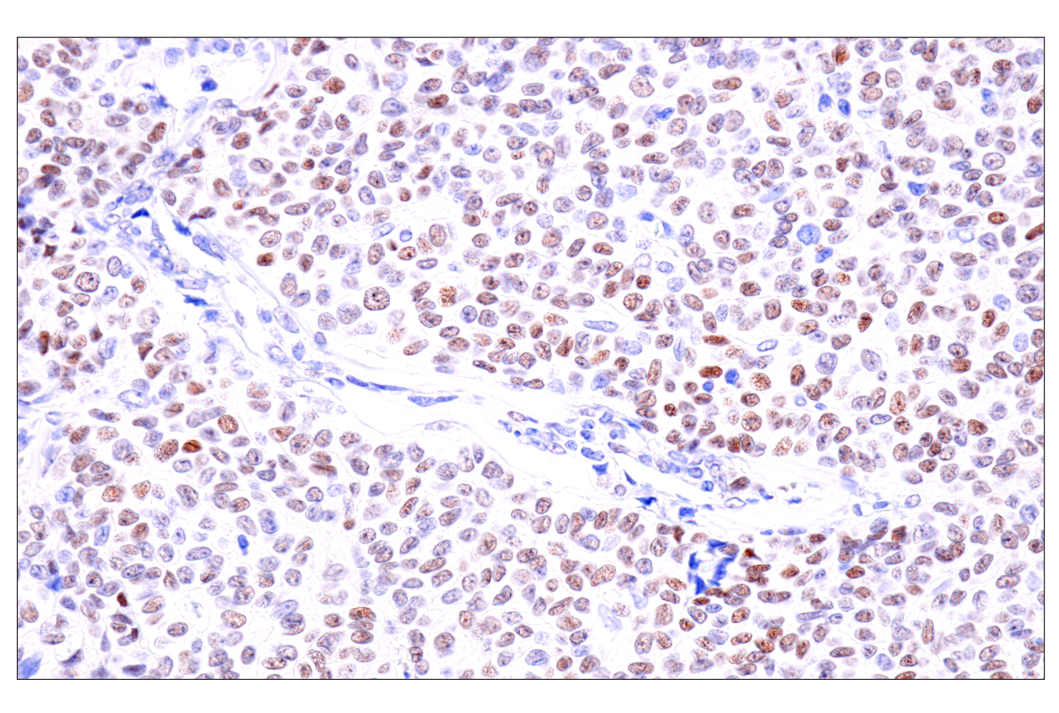
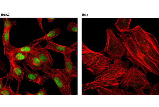
| HNF4α (C11F12) Rabbit mAb | 3113 | 40 µl | 52 kDa | Rabbit IgG |
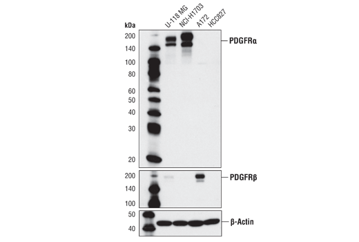
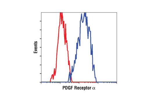
| PDGF Receptor α (D13C6) XP® Rabbit mAb | 5241 | 40 µl | 190 kDa | Rabbit IgG |
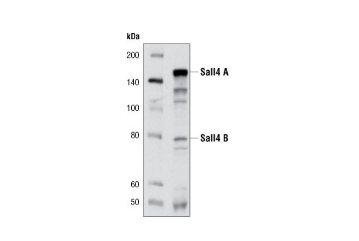
| Sall4 (D16H12) Rabbit mAb | 8459 | 40 µl | 80, 142 kDa | Rabbit IgG |
| Anti-rabbit IgG, HRP-linked Antibody | 7074 | 100 µl | Goat |
Please visit cellsignal.com for individual component applications, species cross-reactivity, dilutions, protocols, and additional product information.
Description
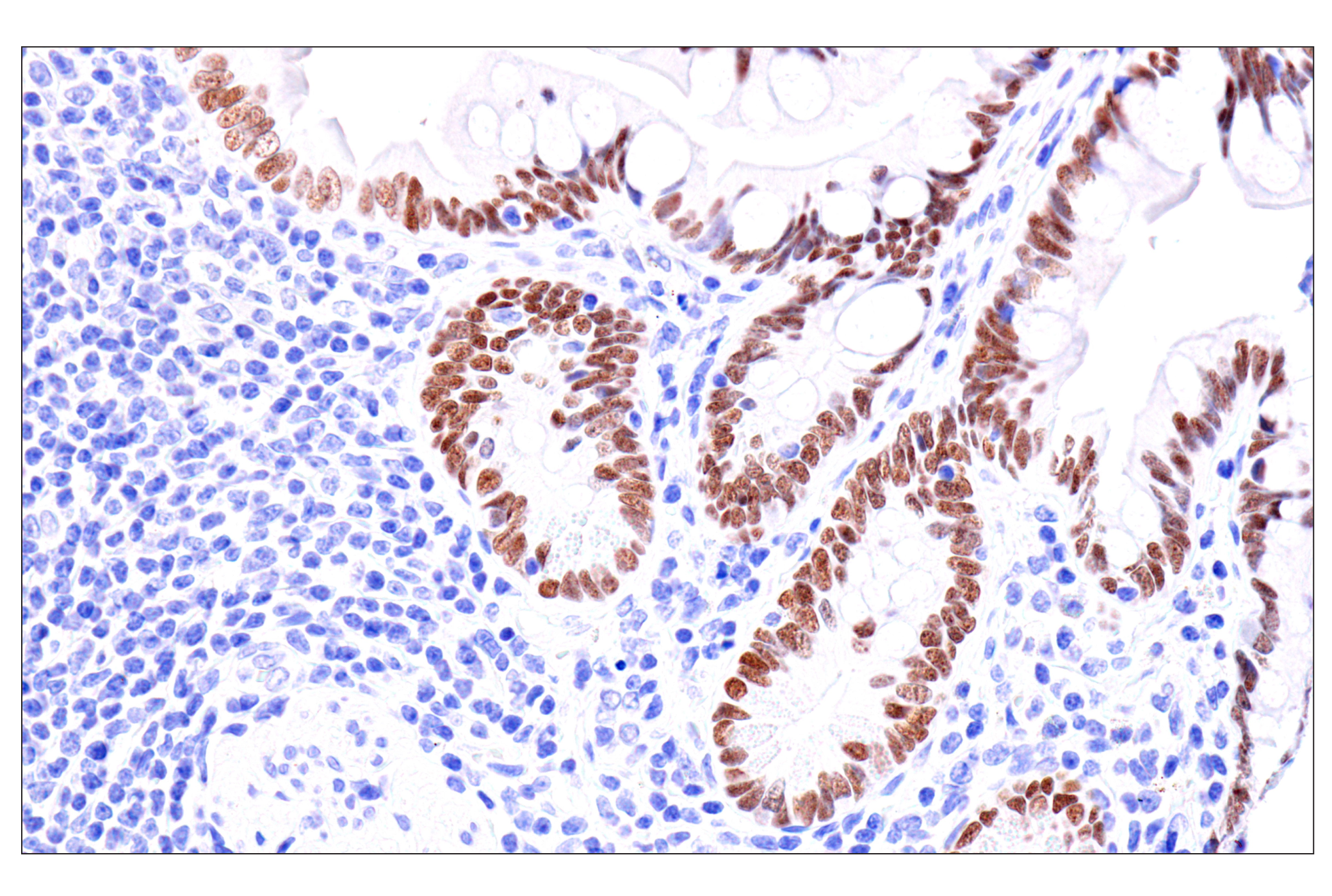
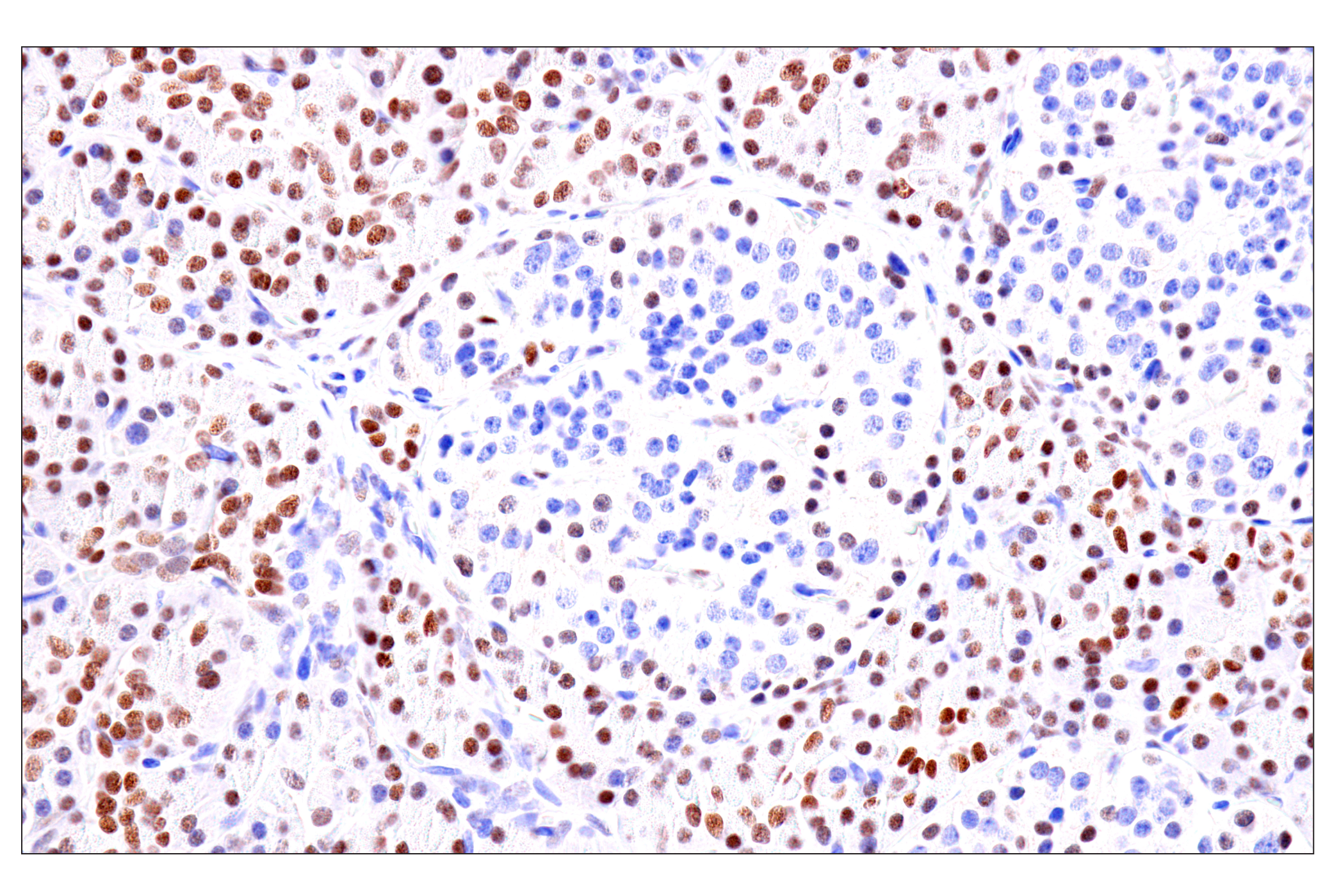
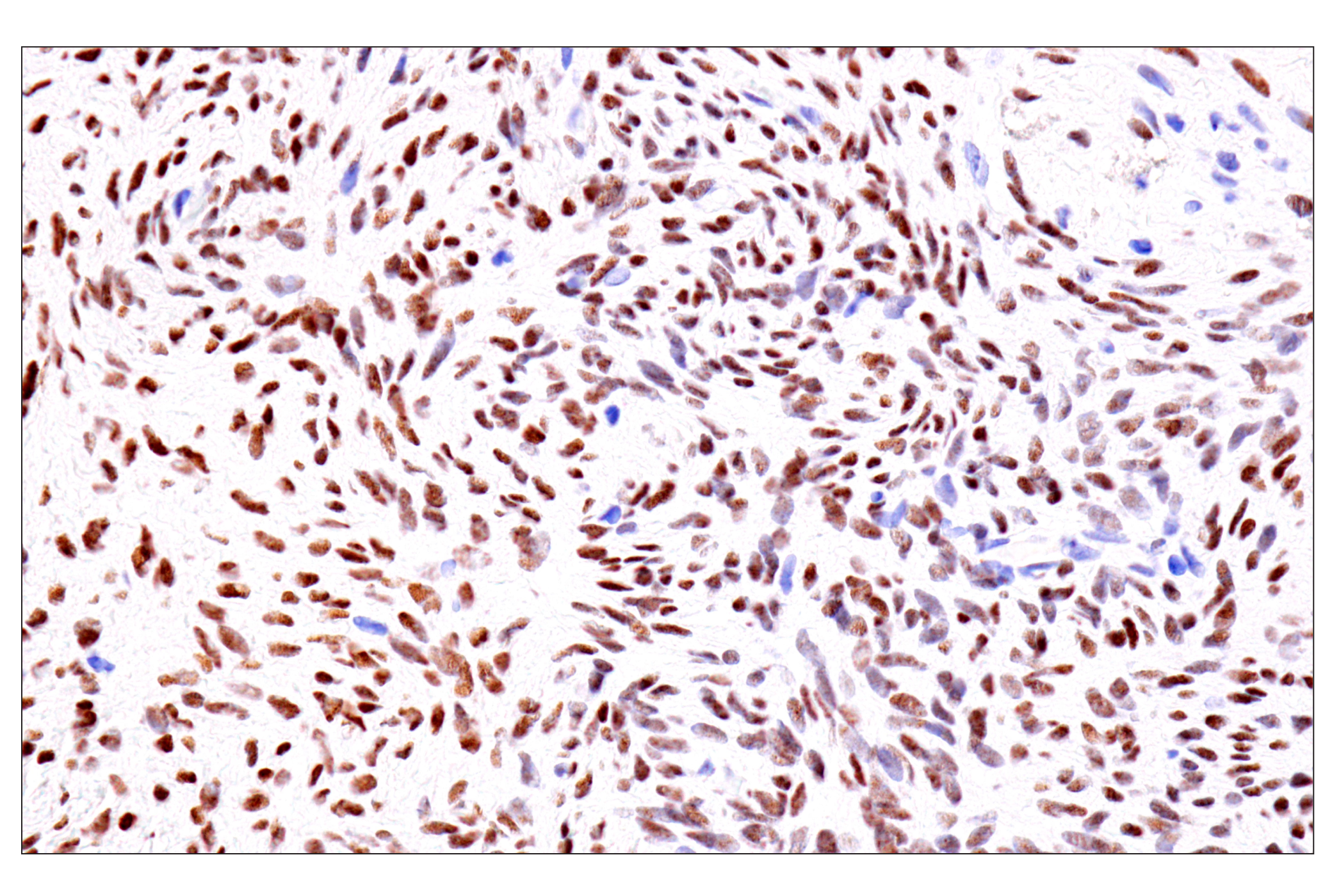
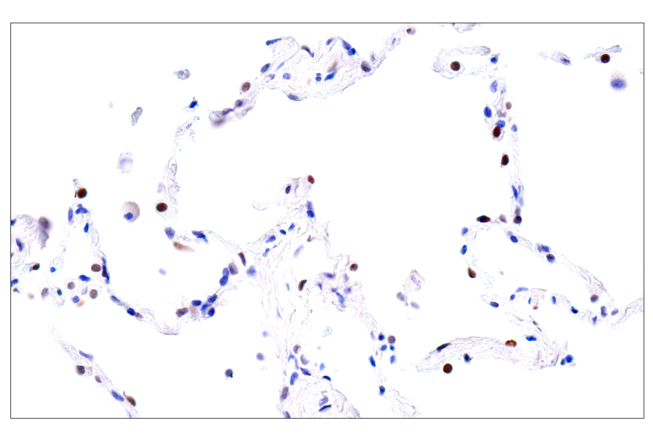
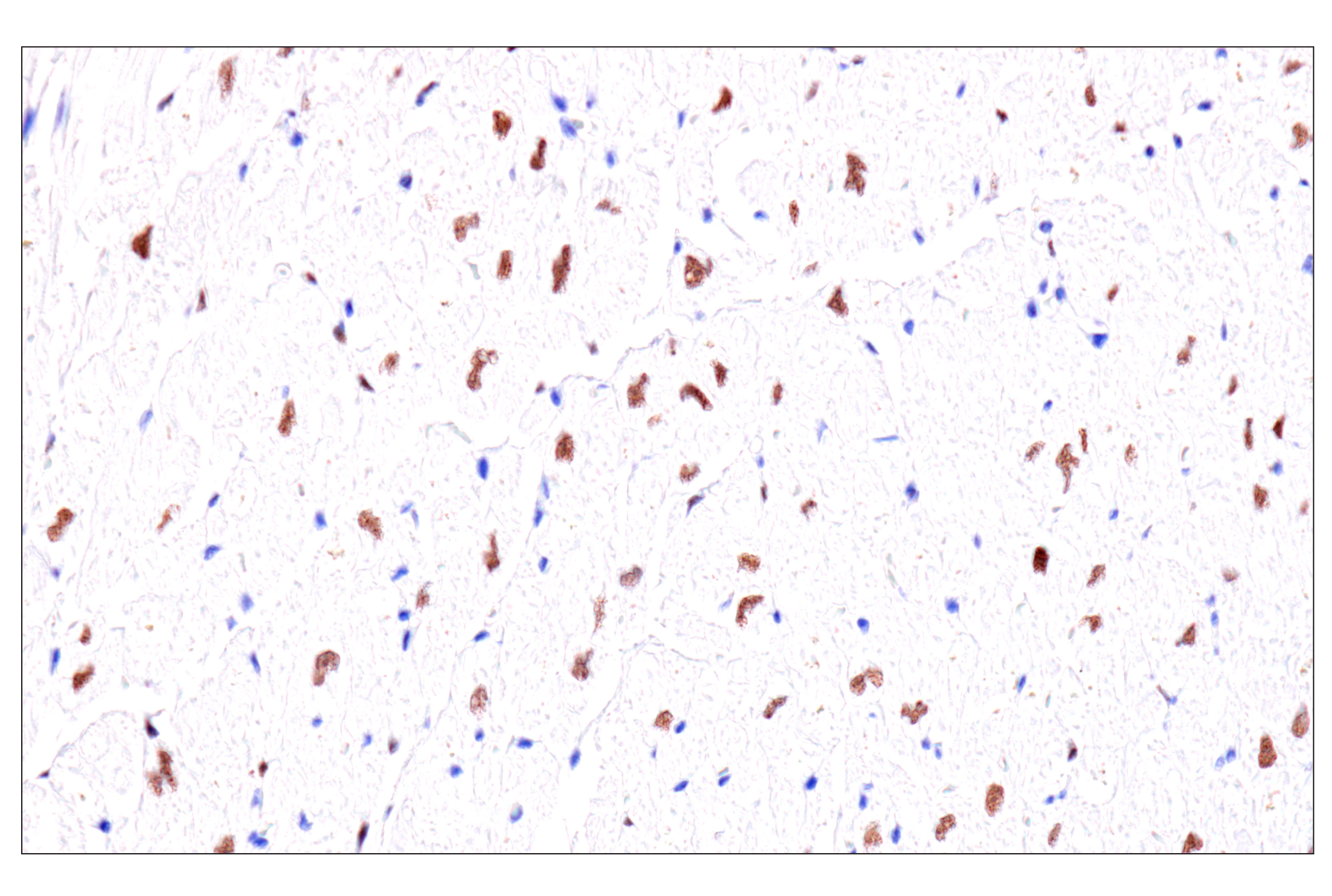
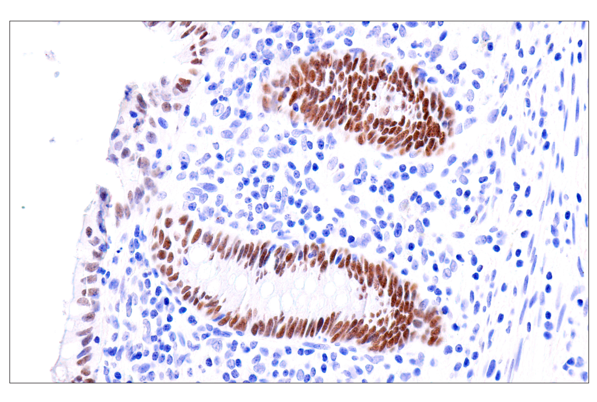
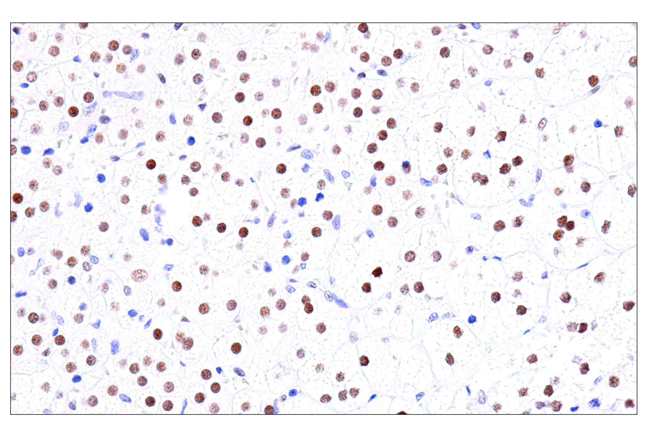
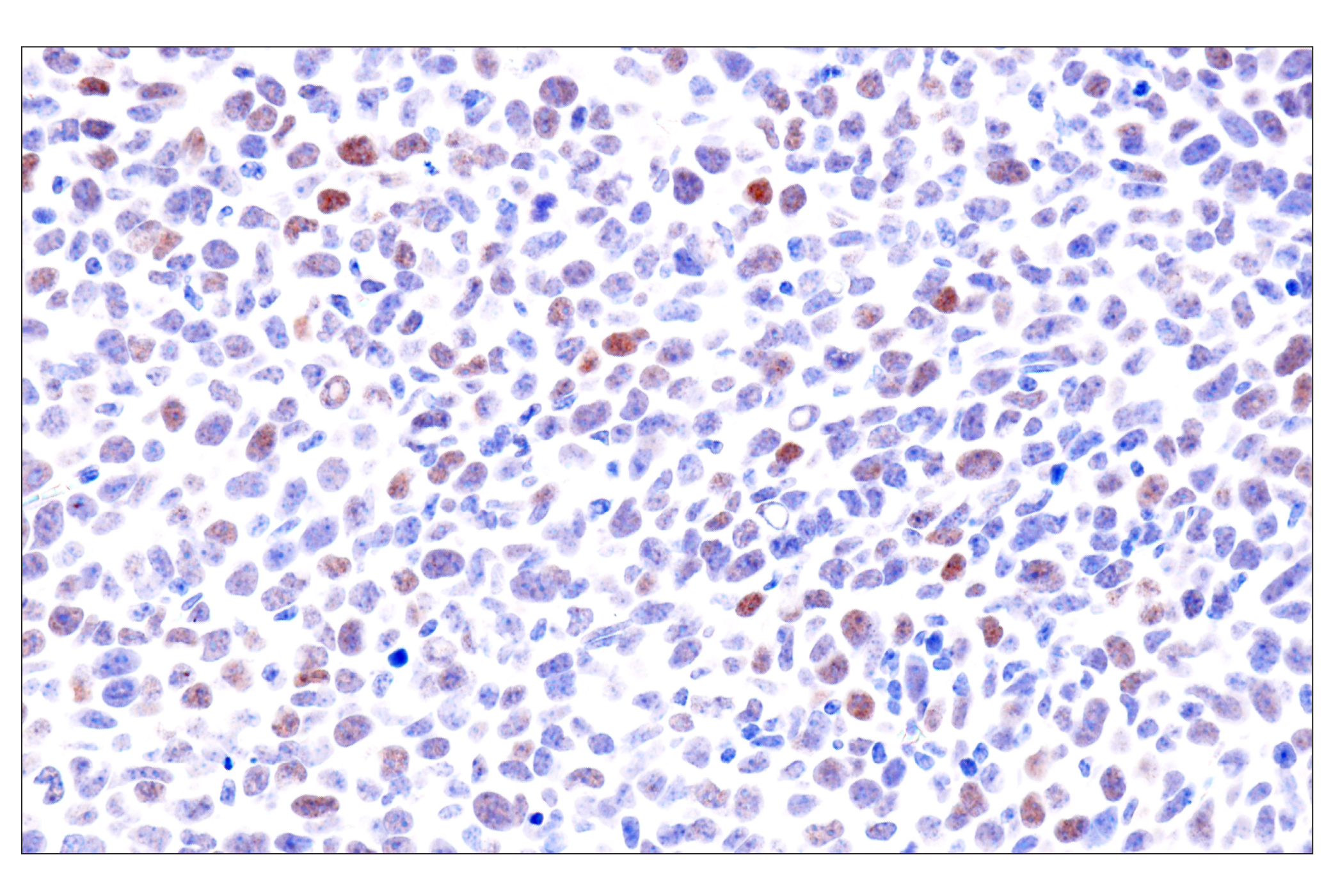
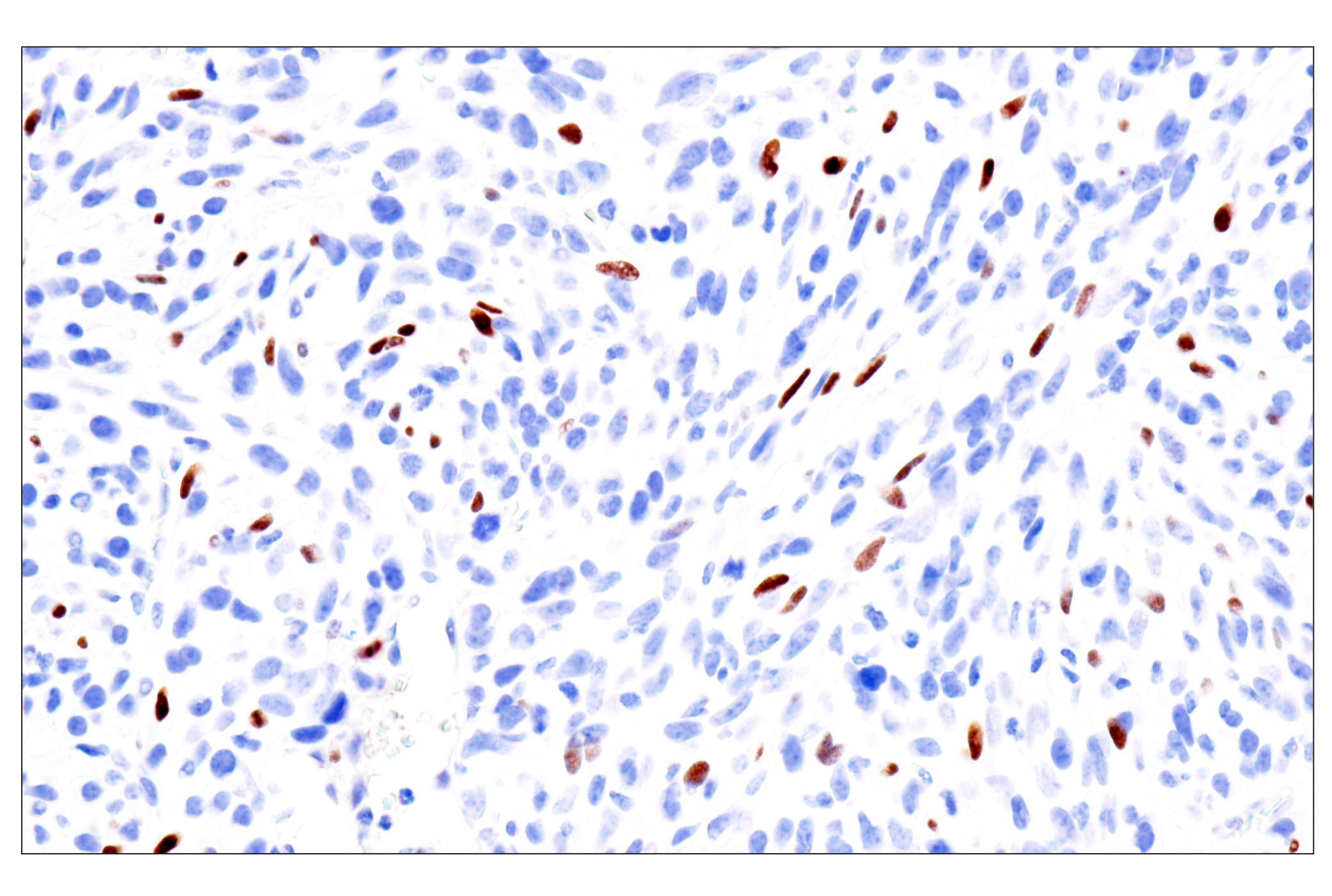
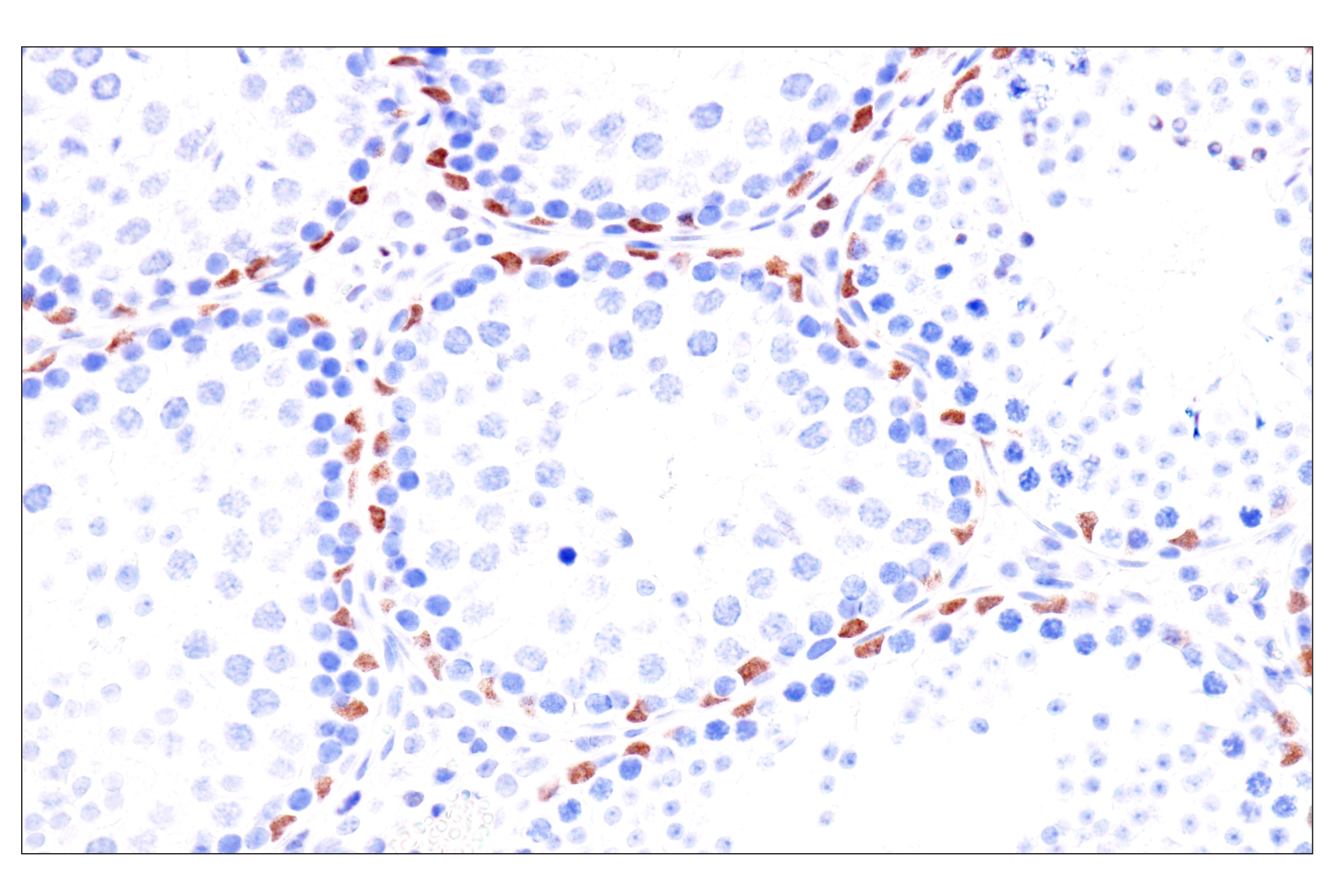
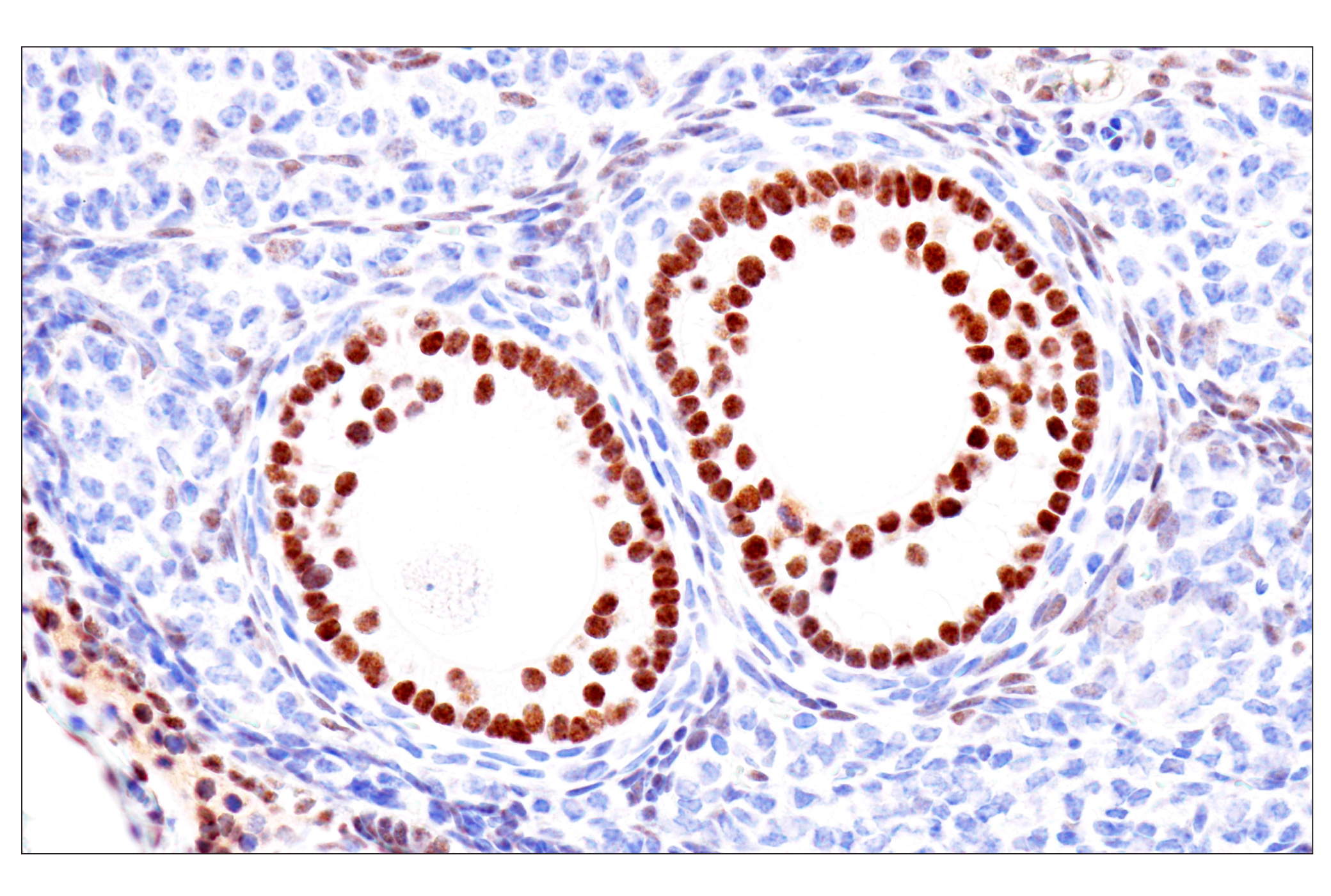
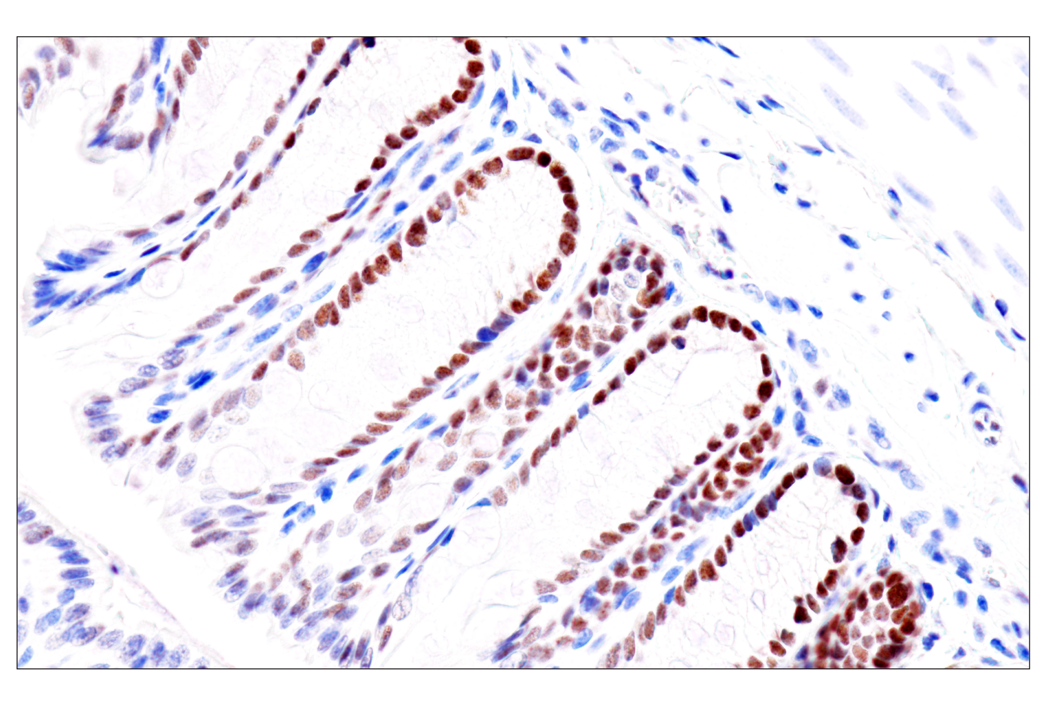
The Endodermal Lineage Marker Antibody Sampler Kit provides an economical means of evaluating proteins expressed during endoderm development. This kit contains enough antibody to perform four western blot experiments per primary antibody.
Storage
Background
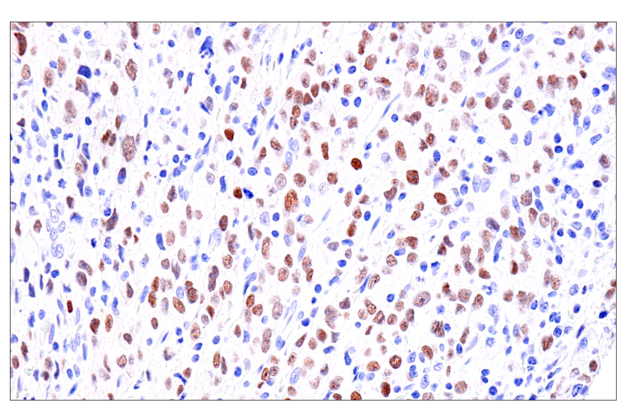
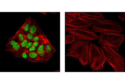
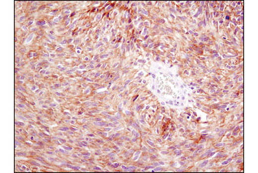
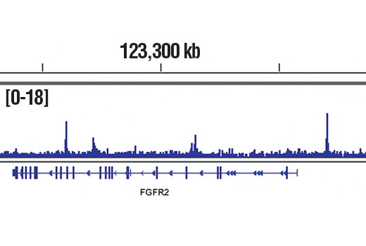
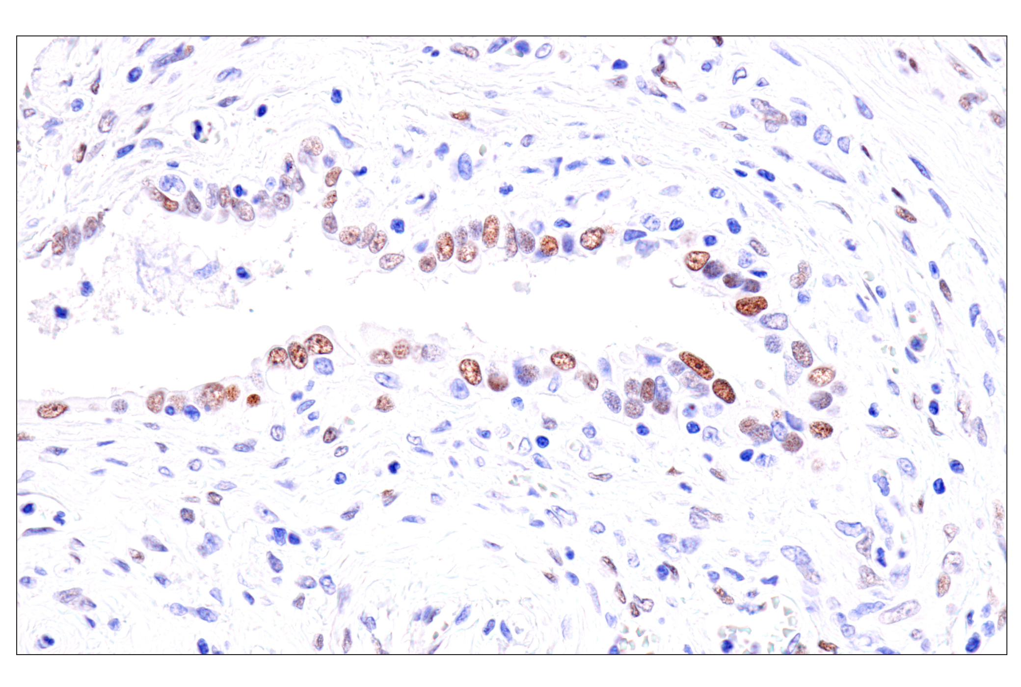
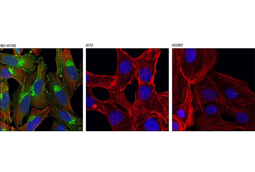
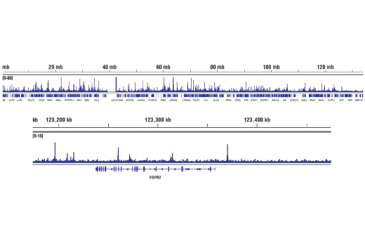
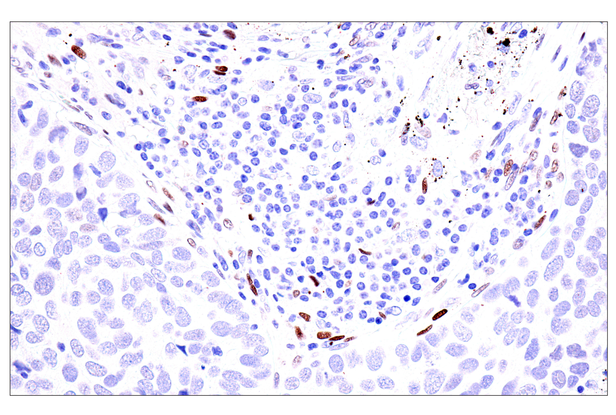
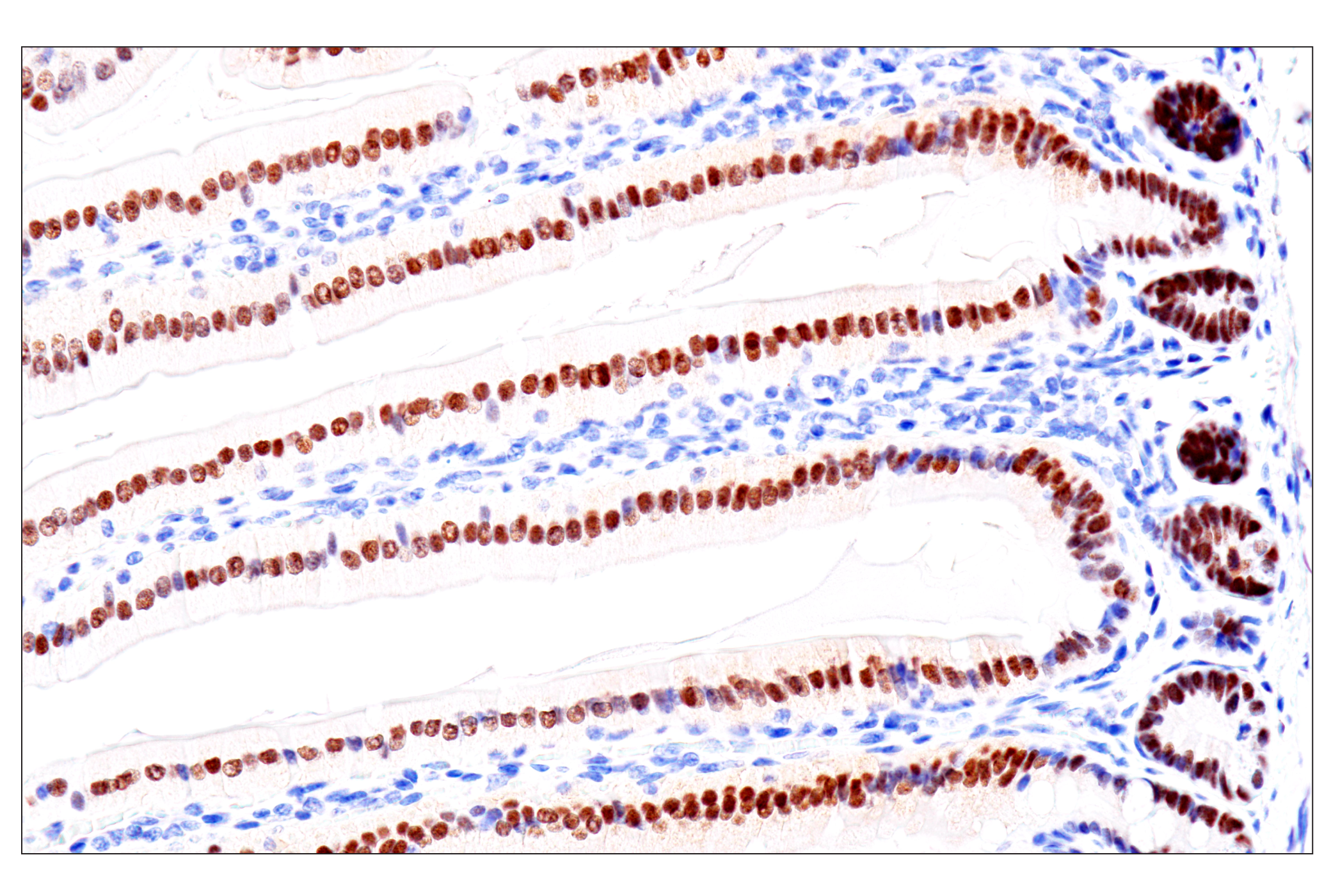
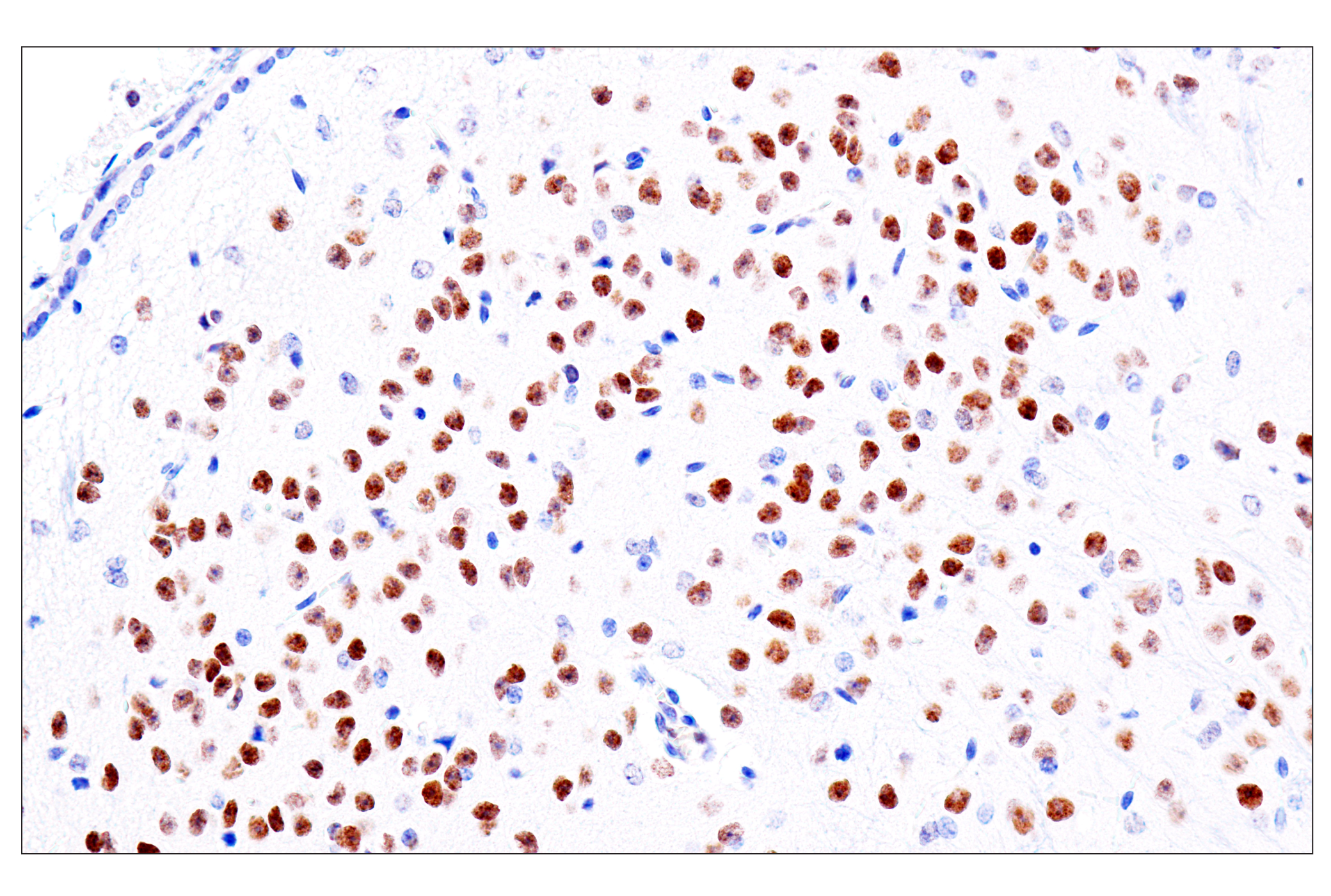
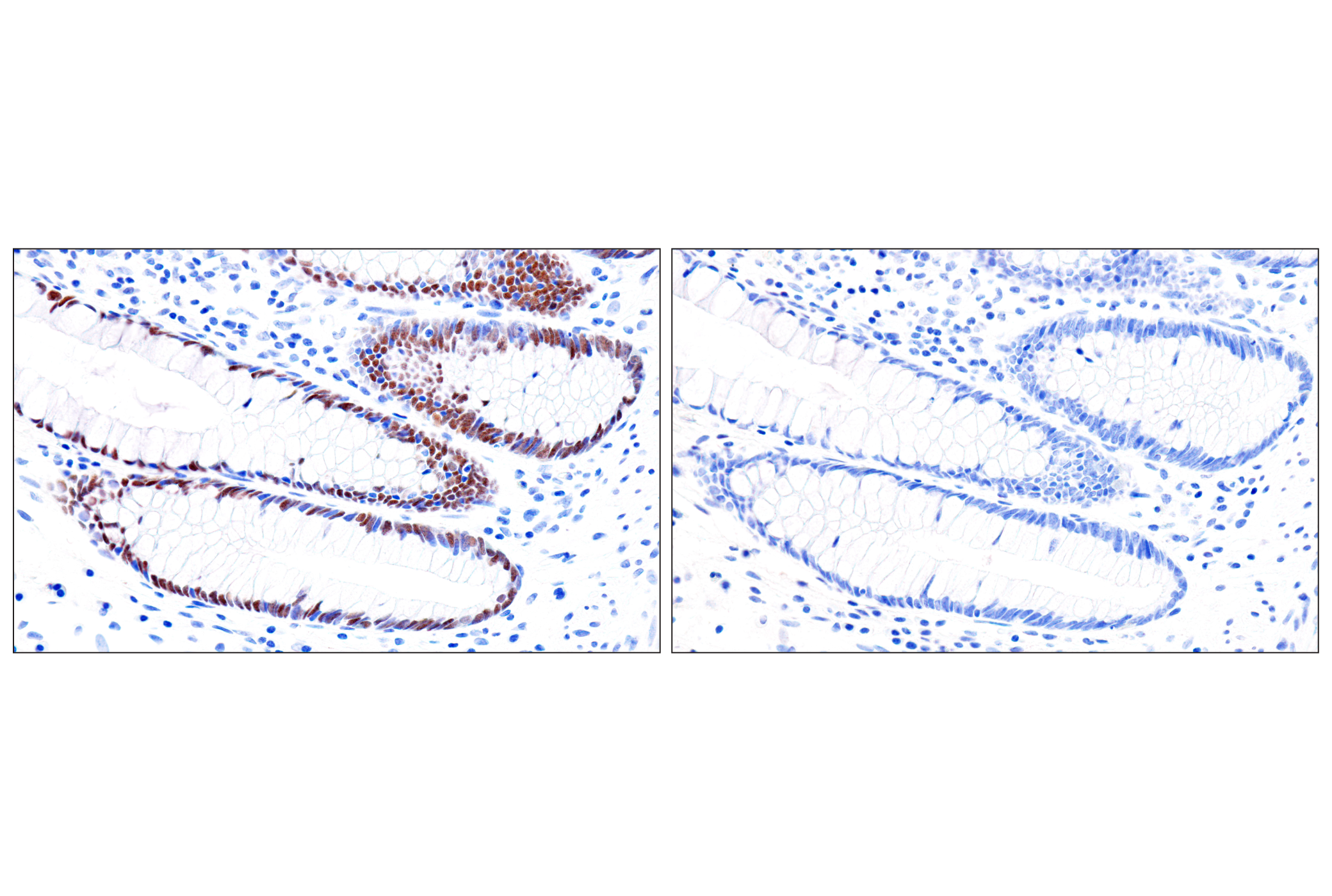
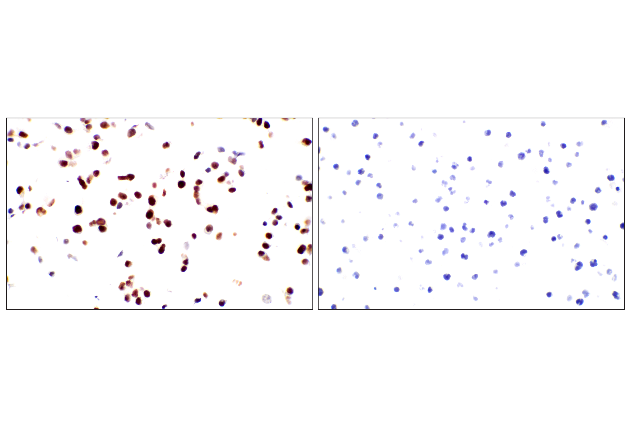
Two endodermal lineages develop during mammalian embryogenesis, the primitive endoderm of the blastocyst stage embryo and the definitive endoderm at gastrulation. The primitive endoderm gives rise to extra-embryonic lineages encompassing the visceral and the parietal endoderm. The definitive endoderm contributes to the respiratory and gastrointestinal tracts by forming the epithelial lining of the trachea, esophagus, lungs, stomach and intestines, and is a major component of many glands, including thyroid, thymus, pancreas and liver (1). Understanding molecular mechanisms that regulate early endodermal fates is seminal for the advance of stem cell research as they connect the transition from pluripotency to endoderm specification during mammalian development and contribute to the generation of clinically relevant cell types. FoxA2/HNF3β is a transcription factor essential for development of the endoderm and midline structures in mouse embryos (2,3). EOMES acts during gastrulation to promote the specification of the definitive endoderm (4). Markers of hepatic differentiation in the endoderm include expression of α-fetoprotein (AFP) and N-cadherin (5,6). HNF4α is involved in the differentiation of the visceral endoderm. GATA-6 lies upstream of HNF4 in a transcriptional cascade that regulates differentiation of the visceral endoderm and is also required for the establishment of the endodermally derived bronchial epithelium (7). Sall4 is required for the formation of the primitive endoderm from inner cell mass. It has been reported that extra-embryonic stem cell lines cannot be formed in Sall4-deficient blastocysts (8). PDGF receptor α is expressed in primitive endoderm derivatives throughout embryogenesis (9).
- Wells, J.M. and Melton, D.A. (1999) Annu Rev Cell Dev Biol 15, 393-410.
- Weinstein, D.C. et al. (1994) Cell 78, 575-88.
- Ang, S.L. and Rossant, J. (1994) Cell 78, 561-74.
- Costello, I. et al. (2011) Nat Cell Biol 13, 1084-91.
- Zhao, D. et al. (2009) PLoS One 4, e6468.
- Meier, V. et al. (2006) Comp Hepatol 5, 2.
- Morrisey, E.E. et al. (1998) Genes Dev 12, 3579-90.
- Elling, U. et al. (2006) Proc Natl Acad Sci U S A 103, 16319-24.
- Orr-Urtreger, A. et al. (1992) Development 115, 289-303.
Background References
Trademarks and Patents
Limited Uses
Except as otherwise expressly agreed in a writing signed by a legally authorized representative of CST, the following terms apply to Products provided by CST, its affiliates or its distributors. Any Customer's terms and conditions that are in addition to, or different from, those contained herein, unless separately accepted in writing by a legally authorized representative of CST, are rejected and are of no force or effect.
Products are labeled with For Research Use Only or a similar labeling statement and have not been approved, cleared, or licensed by the FDA or other regulatory foreign or domestic entity, for any purpose. Customer shall not use any Product for any diagnostic or therapeutic purpose, or otherwise in any manner that conflicts with its labeling statement. Products sold or licensed by CST are provided for Customer as the end-user and solely for research and development uses. Any use of Product for diagnostic, prophylactic or therapeutic purposes, or any purchase of Product for resale (alone or as a component) or other commercial purpose, requires a separate license from CST. Customer shall (a) not sell, license, loan, donate or otherwise transfer or make available any Product to any third party, whether alone or in combination with other materials, or use the Products to manufacture any commercial products, (b) not copy, modify, reverse engineer, decompile, disassemble or otherwise attempt to discover the underlying structure or technology of the Products, or use the Products for the purpose of developing any products or services that would compete with CST products or services, (c) not alter or remove from the Products any trademarks, trade names, logos, patent or copyright notices or markings, (d) use the Products solely in accordance with CST Product Terms of Sale and any applicable documentation, and (e) comply with any license, terms of service or similar agreement with respect to any third party products or services used by Customer in connection with the Products.