| Product Includes | Product # | Quantity | Mol. Wt | Isotype/Source |
|---|---|---|---|---|
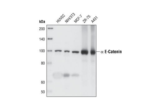
| α-E-Catenin (23B2) Rabbit mAb | 3240 | 40 µl | 100 kDa | Rabbit IgG |
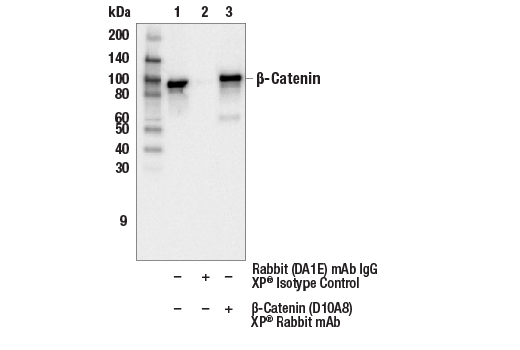
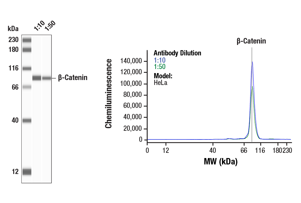
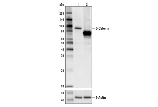
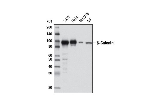
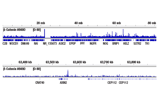
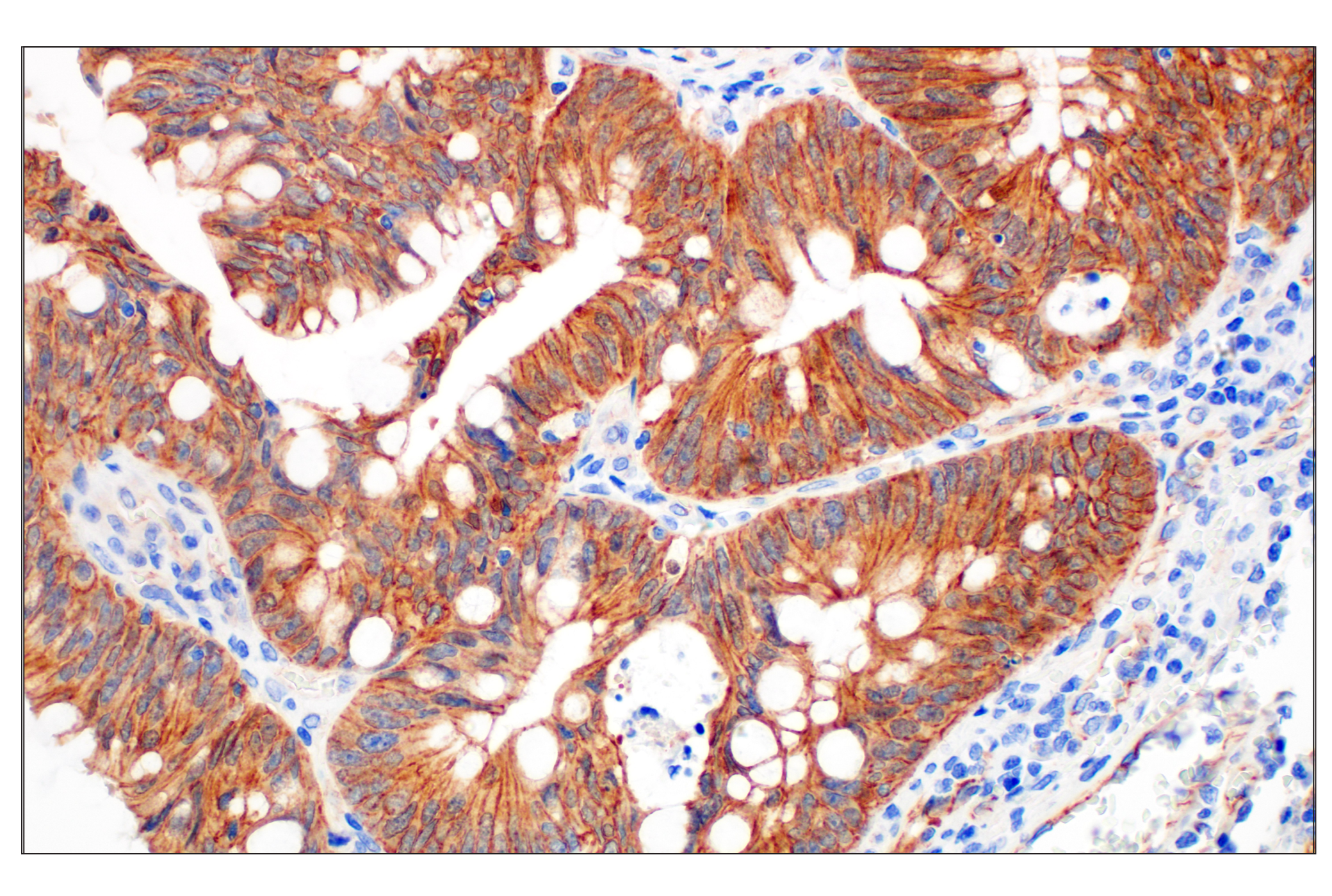
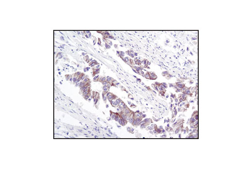
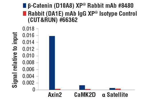
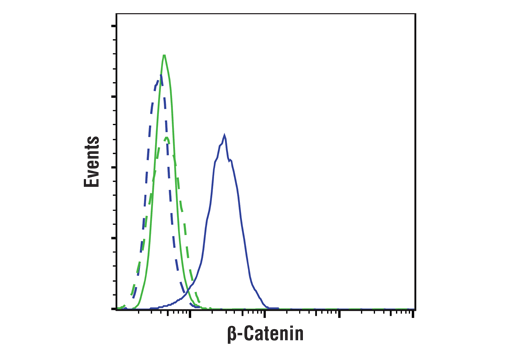
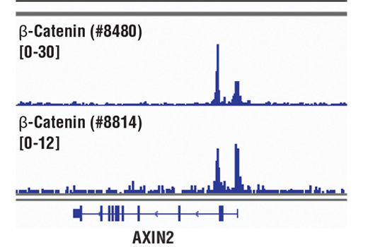
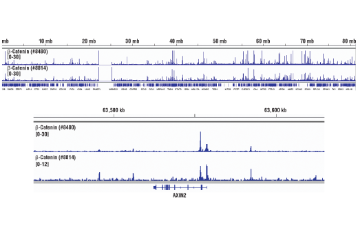
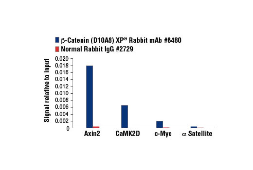
| β-Catenin (D10A8) XP® Rabbit mAb | 8480 | 40 µl | 92 kDa | Rabbit IgG |
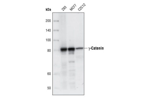
| γ-Catenin Antibody | 2309 | 40 µl | 83 kDa | Rabbit |
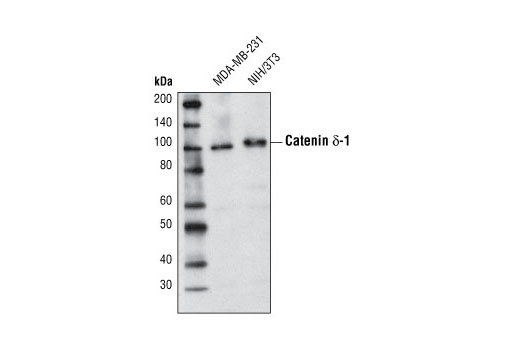
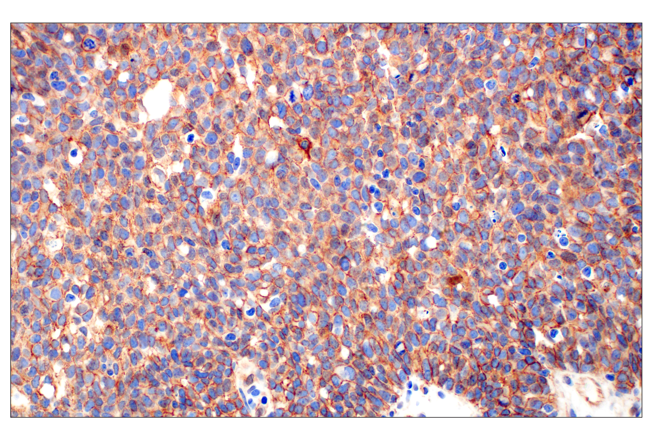
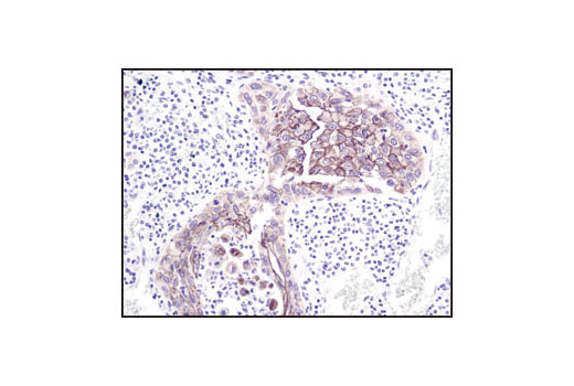
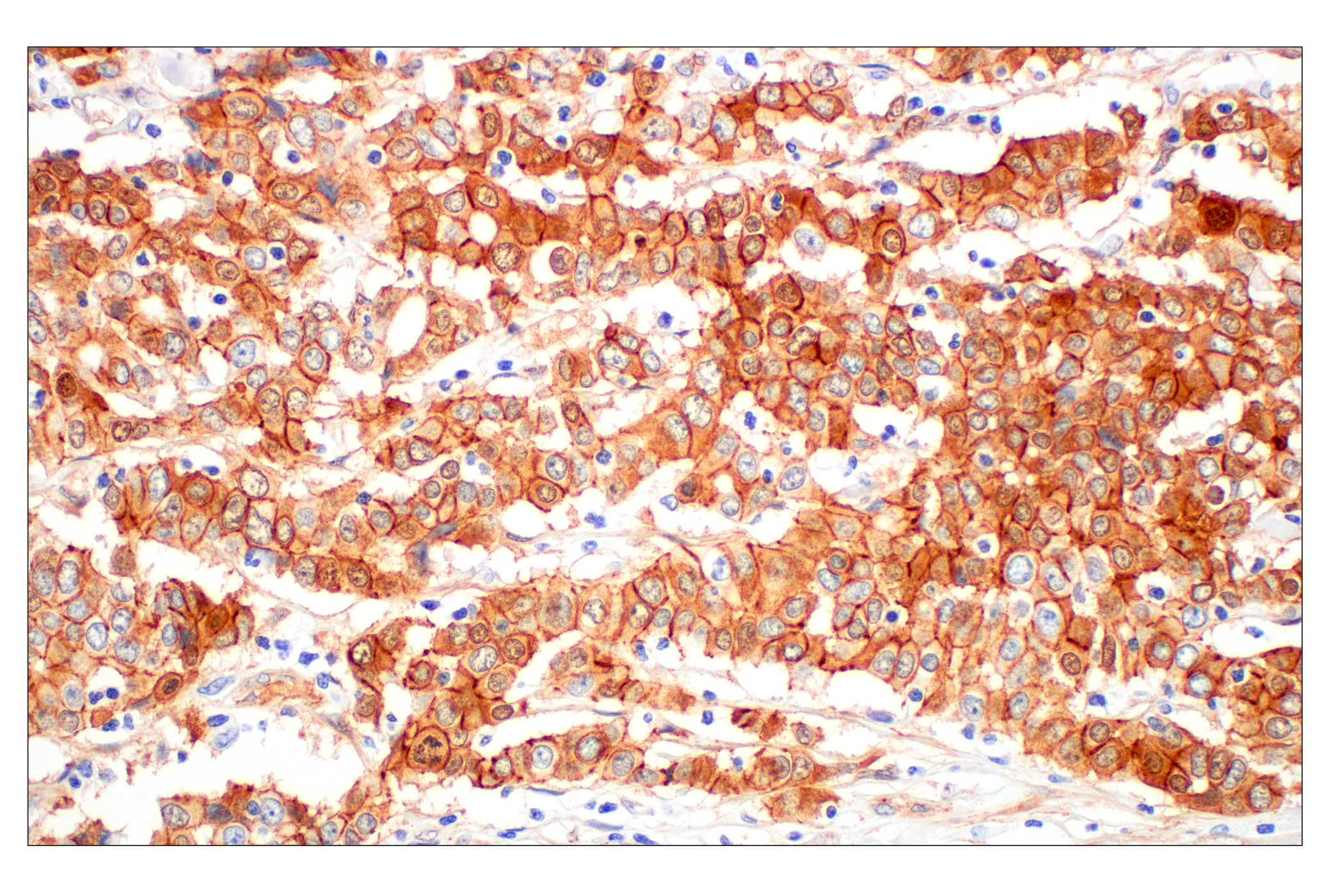
| Catenin δ-1 Antibody | 4989 | 40 µl | 100 kDa | Rabbit |
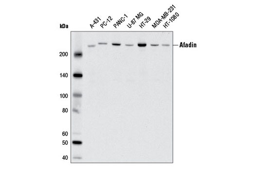
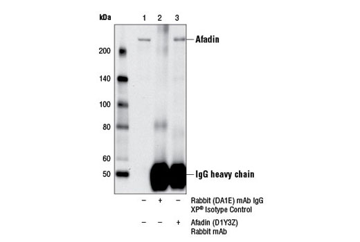
| Afadin (D1Y3Z) Rabbit mAb | 13531 | 40 µl | 205 kDa | Rabbit IgG |
| Anti-rabbit IgG, HRP-linked Antibody | 7074 | 100 µl | Goat |
Please visit cellsignal.com for individual component applications, species cross-reactivity, dilutions, protocols, and additional product information.
Description
The Adherens Junction Antibody Sampler Kit provides an economical means of detecting the protein components of adherens junctions. The kit includes enough antibody to perform four western blot experiments per primary antibody.
Storage
Background
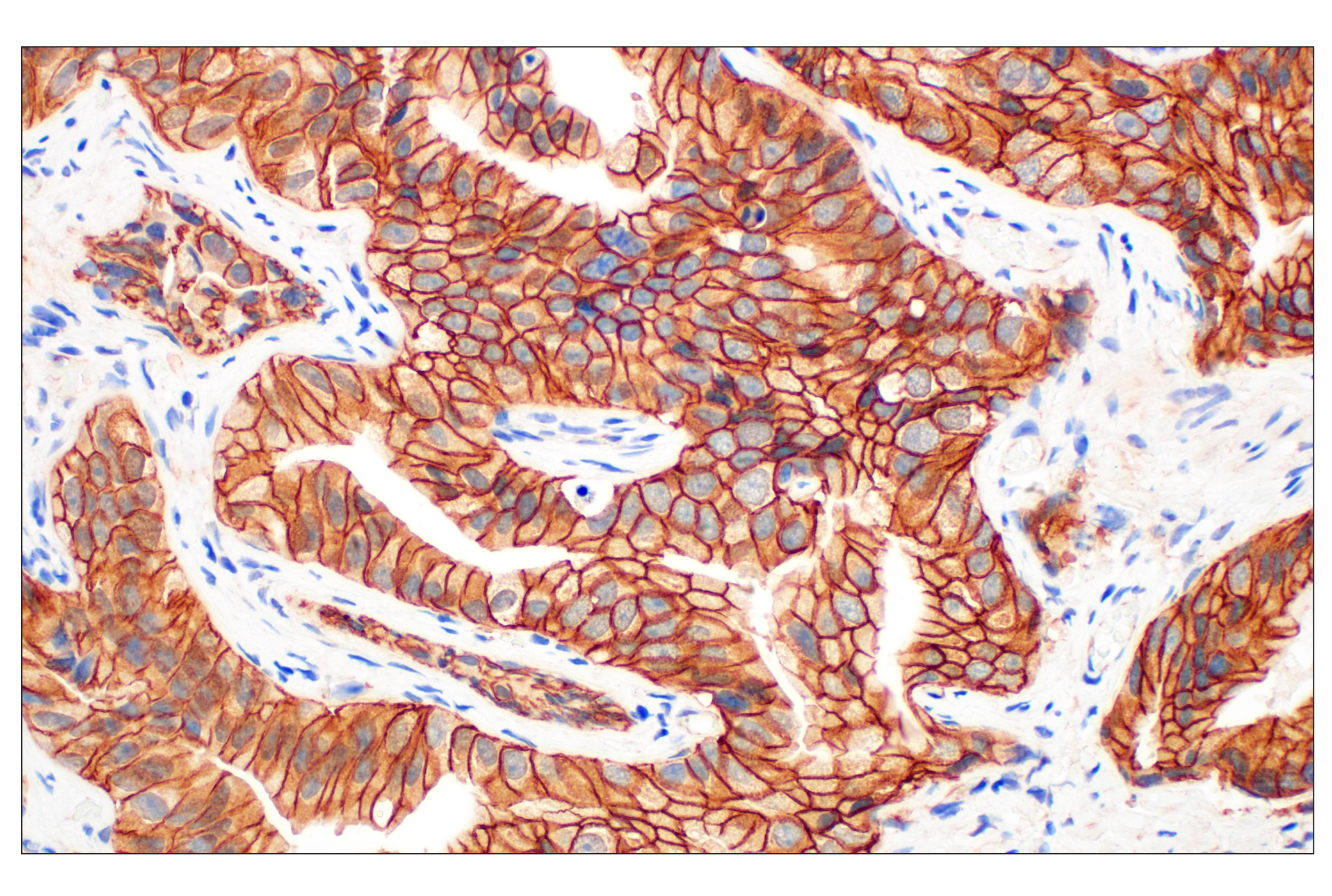
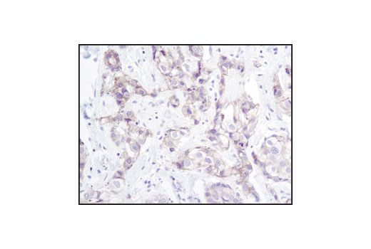
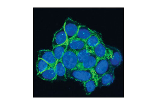
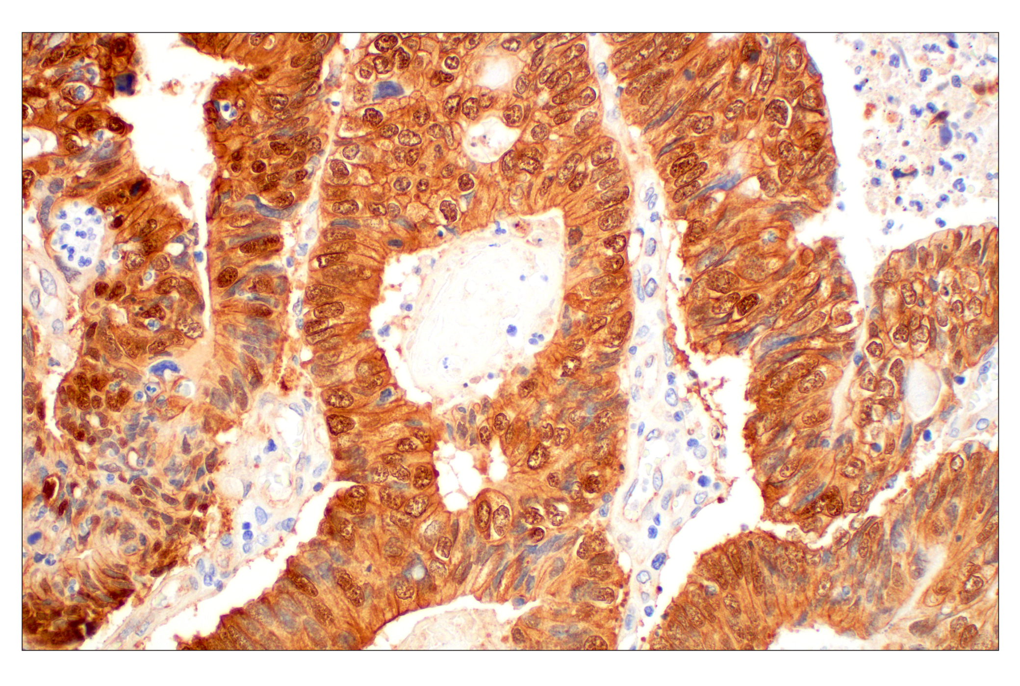
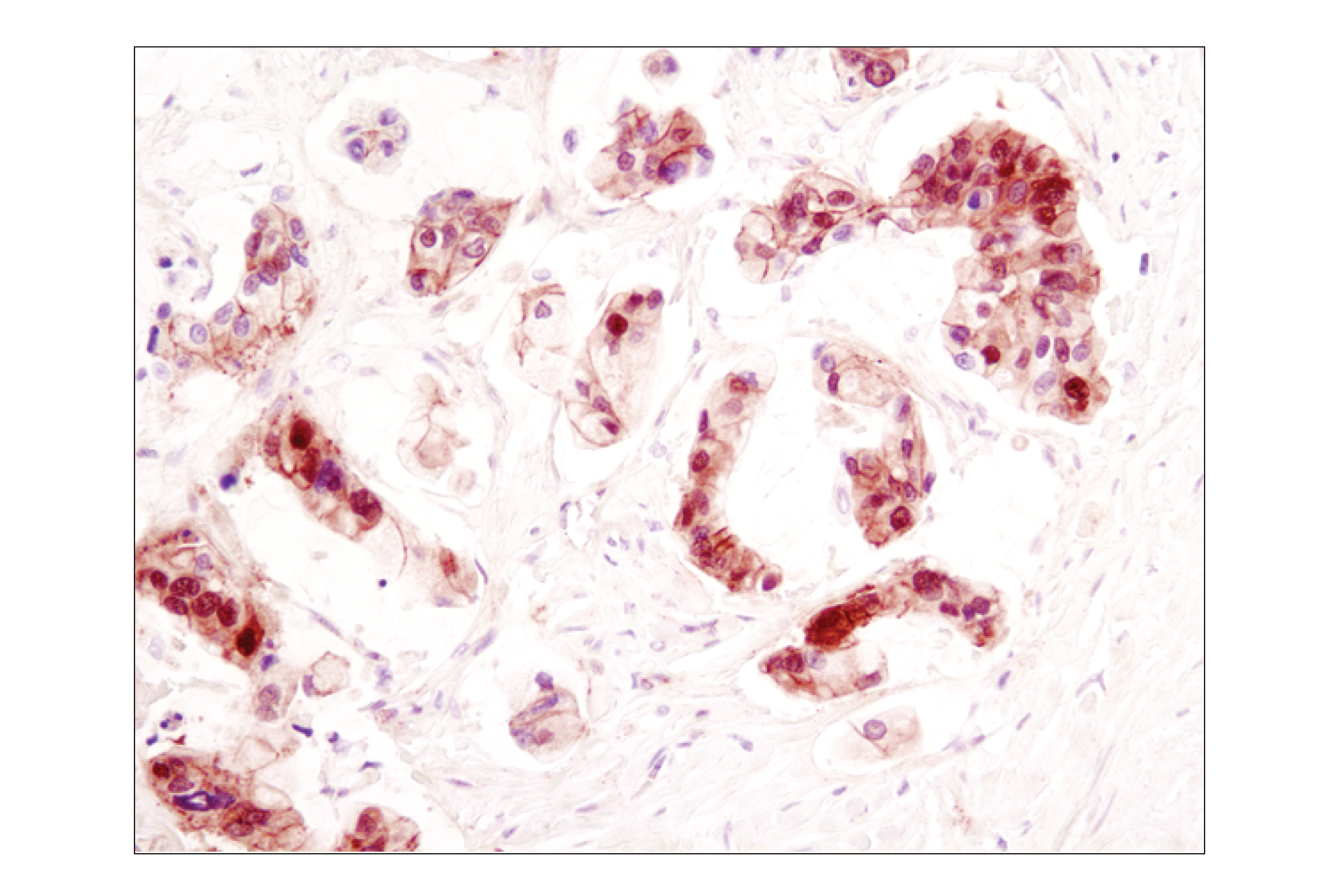
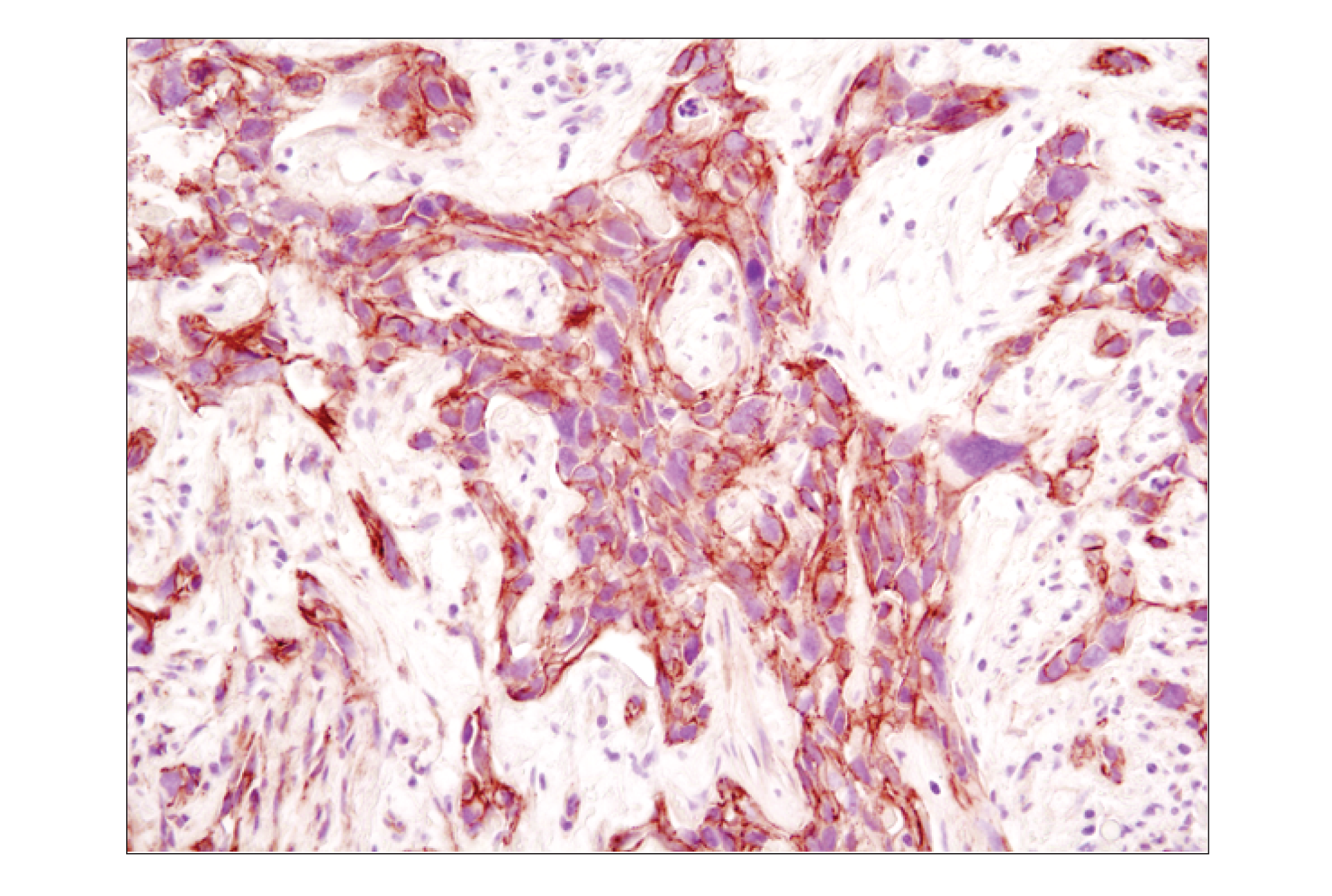
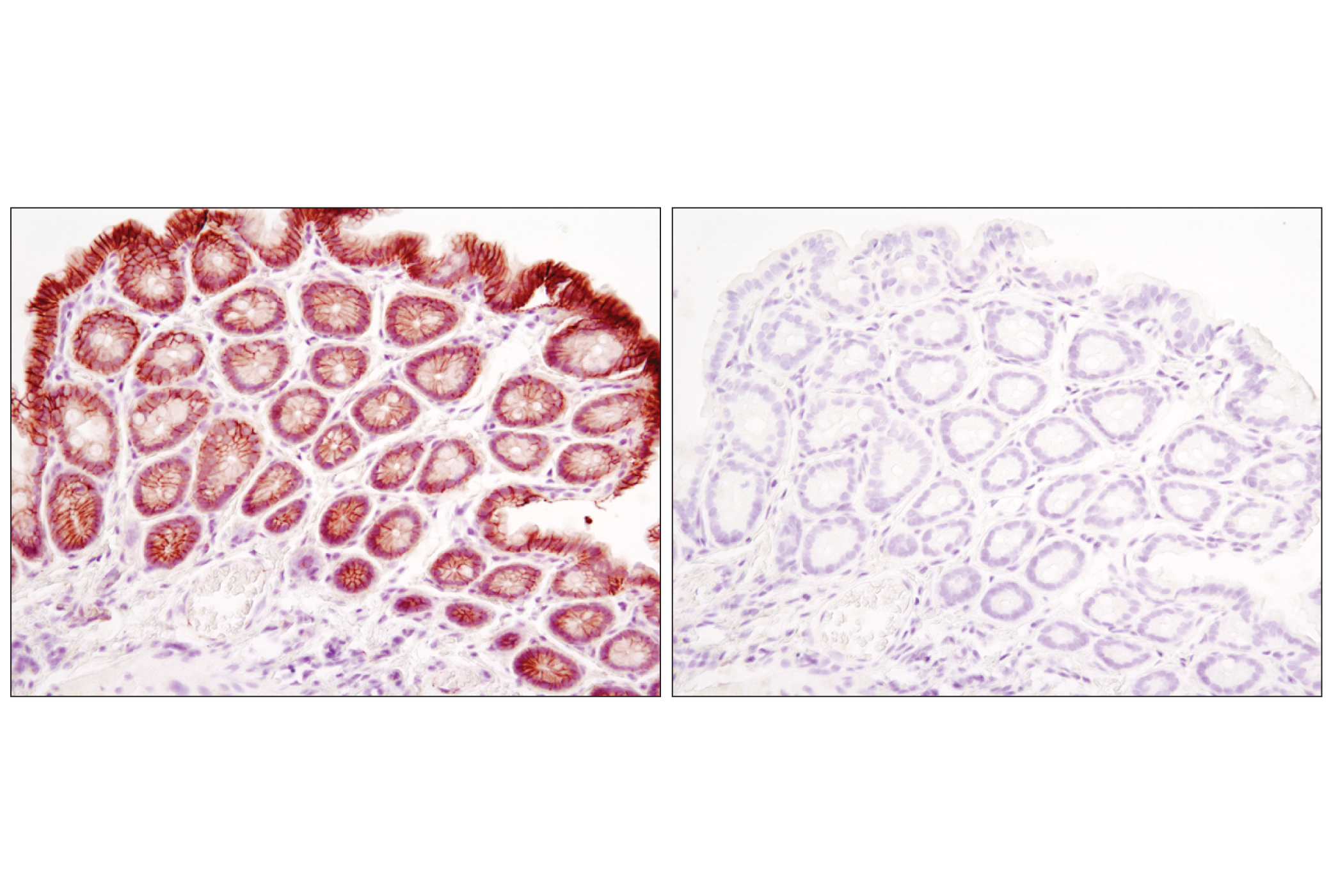
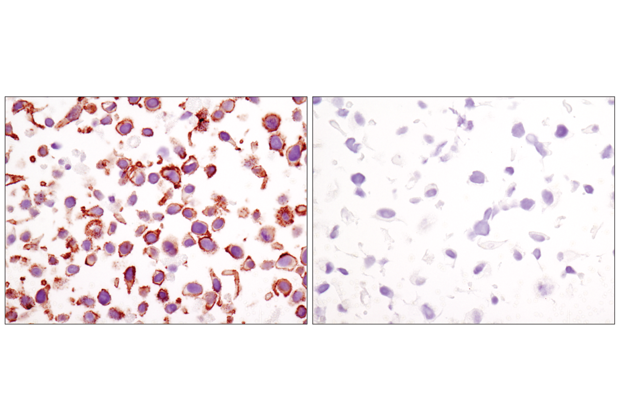
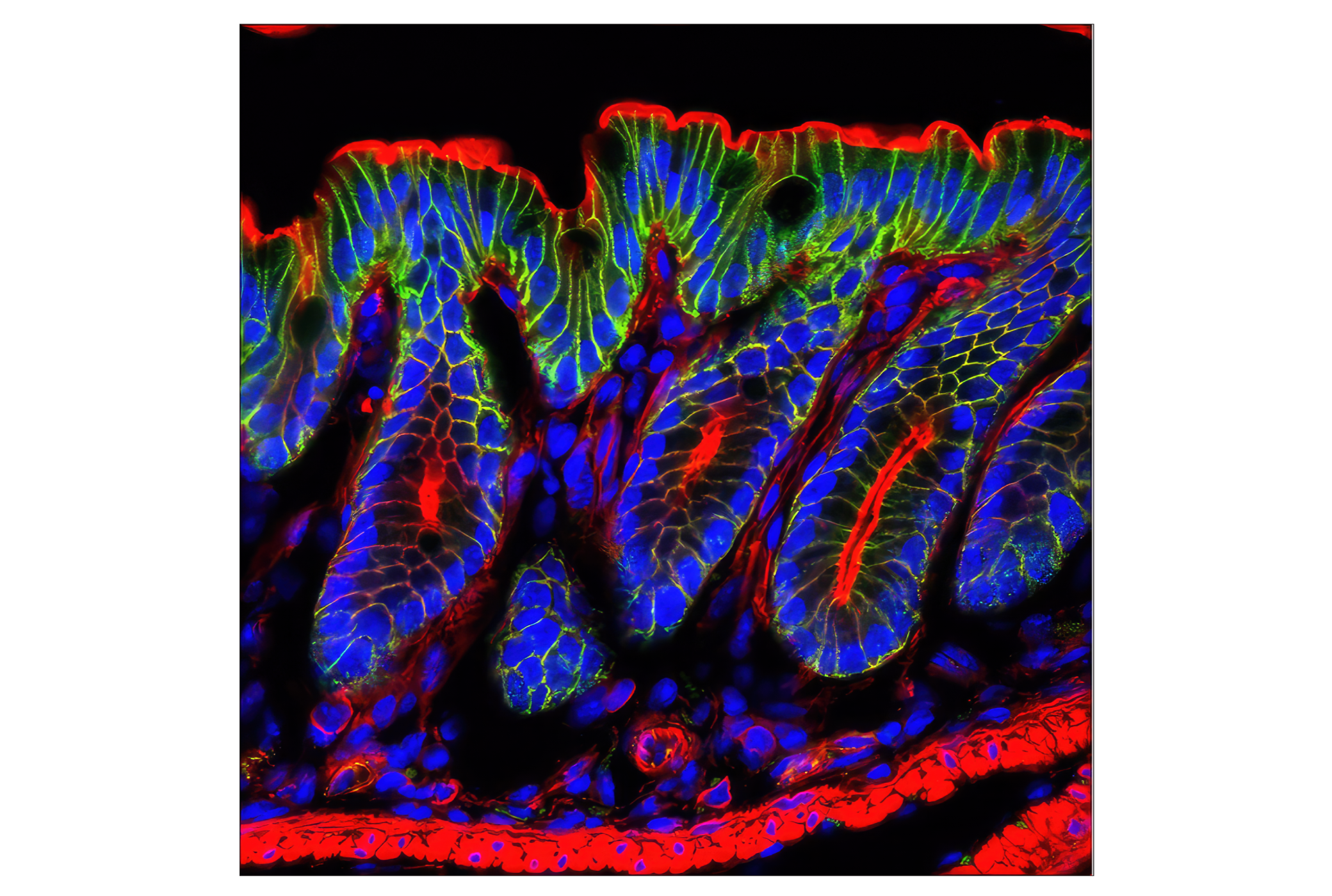
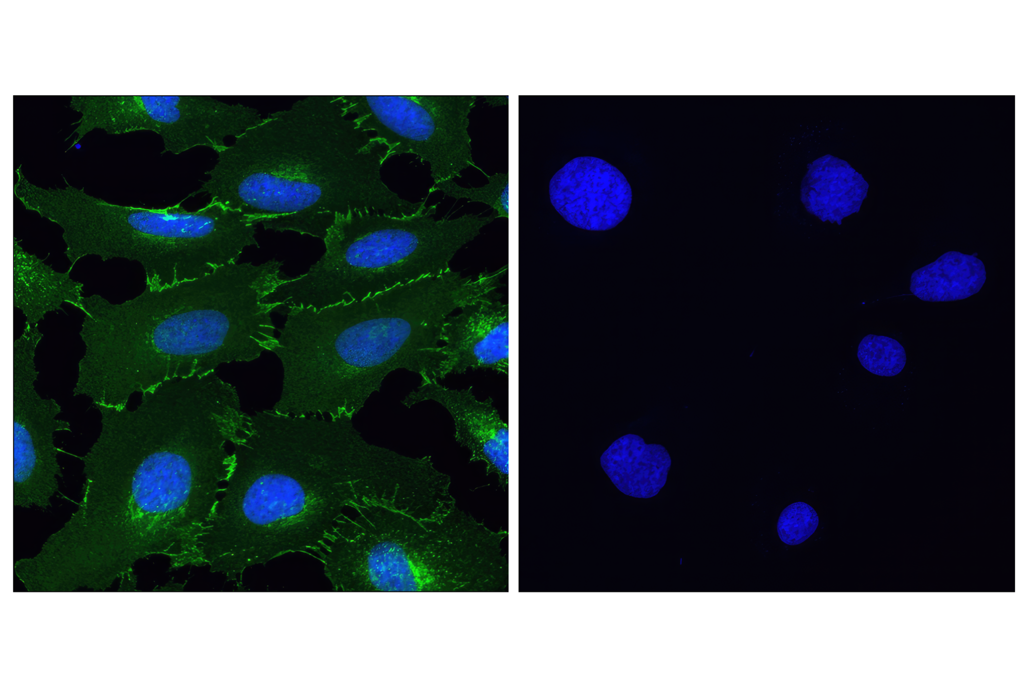
Adherens junctions are dynamic structures that form cell-cell contacts and are important in development, differentiation, tissue integrity, morphology and cell polarity. They are composed of the transmembrane proteins, cadherins, which bind cadherins on adjacent cells in a calcium-dependent manner. On the cytoplasmic side of adherens junctions, the classic model states that cadherins are linked to the cytoskeleton through β- and α-catenin. α-E-catenin is ubiquitously expressed, α-N-catenin is expressed in neuronal tissue, and α-T-catenin is primarily expressed in heart tissue. Research studies have demonstrated that loss of E-cadherin and α-E-catenin occurs during the progression of several human cancers, indicating that the breakdown of adherens junctions is important in cancer progression (reviewed in 1).
Research studies also suggest that, rather than acting as a static link between cadherins and actin, α-catenin regulates actin dynamics directly, possibly by competing with the actin nucleating arp2/3 complex (2,3). α-catenin also plays a role in regulating β-catenin-dependent transcriptional activity, affecting differentiation and response to Wnt signaling. α-catenin binds to β-catenin in the nucleus, preventing it from regulating transcription, and levels of both proteins appear to be regulated via proteasome-dependent degradation (4).
Afadin has two splice variants: l-afadin, which is ubiquitously expressed, and s-afadin, which is expressed predominantly in neural tissue. s-afadin is a shorter form lacking one of the three proline-rich regions found in l-afadin, as well as the carboxyl-terminal F-actin binding region (5). Human s-afadin is identical to AF-6, the ALL-1 fusion partner involved in acute myeloid leukemias (6). Recent research has also shown that afadin is involved in controlling the directionality of cell movement when it is localized at the leading edge of moving cells (7,8).
- Kobielak, A. and Fuchs, E. (2004) Nat Rev Mol Cell Biol 5, 614-25.
- Yamada, S. et al. (2005) Cell 123, 889-901.
- Drees, F. et al. (2005) Cell 123, 903-15.
- Hwang, S.G. et al. (2005) J Biol Chem 280, 12758-65.
- Mandai, K. et al. (1997) J Cell Biol 139, 517-28.
- Prasad, R. et al. (1993) Cancer Res 53, 5624-8.
- Miyata, M. et al. (2009) J Cell Sci 122, 4319-29.
- Miyata, M. et al. (2009) J Biol Chem 284, 24595-609.
Background References
Trademarks and Patents
Limited Uses
Except as otherwise expressly agreed in a writing signed by a legally authorized representative of CST, the following terms apply to Products provided by CST, its affiliates or its distributors. Any Customer's terms and conditions that are in addition to, or different from, those contained herein, unless separately accepted in writing by a legally authorized representative of CST, are rejected and are of no force or effect.
Products are labeled with For Research Use Only or a similar labeling statement and have not been approved, cleared, or licensed by the FDA or other regulatory foreign or domestic entity, for any purpose. Customer shall not use any Product for any diagnostic or therapeutic purpose, or otherwise in any manner that conflicts with its labeling statement. Products sold or licensed by CST are provided for Customer as the end-user and solely for research and development uses. Any use of Product for diagnostic, prophylactic or therapeutic purposes, or any purchase of Product for resale (alone or as a component) or other commercial purpose, requires a separate license from CST. Customer shall (a) not sell, license, loan, donate or otherwise transfer or make available any Product to any third party, whether alone or in combination with other materials, or use the Products to manufacture any commercial products, (b) not copy, modify, reverse engineer, decompile, disassemble or otherwise attempt to discover the underlying structure or technology of the Products, or use the Products for the purpose of developing any products or services that would compete with CST products or services, (c) not alter or remove from the Products any trademarks, trade names, logos, patent or copyright notices or markings, (d) use the Products solely in accordance with CST Product Terms of Sale and any applicable documentation, and (e) comply with any license, terms of service or similar agreement with respect to any third party products or services used by Customer in connection with the Products.