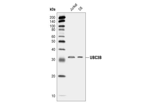
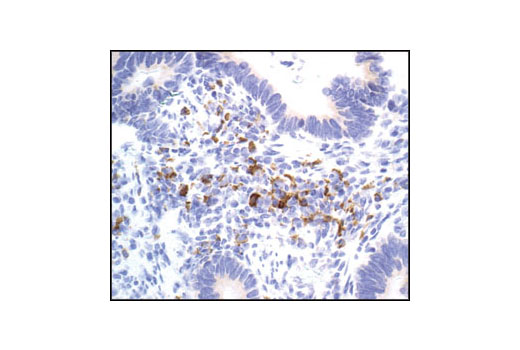
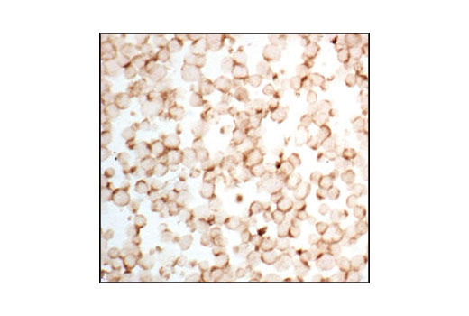
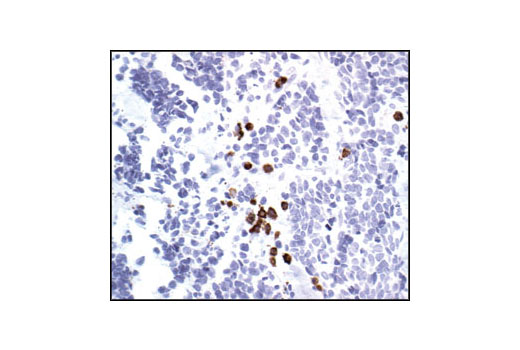
WB, IHC-P, FC-FP
H M R
Endogenous
32
Rabbit
#Q712K3
54926
Product Information
Product Usage Information
| Application | Dilution |
|---|---|
| Western Blotting | 1:1000 |
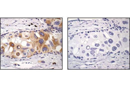
| Immunohistochemistry (Paraffin) | 1:50 |
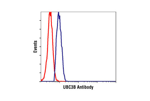
| Flow Cytometry (Fixed/Permeabilized) | 1:25 |
Storage
Specificity / Sensitivity
Species Reactivity:
Human, Mouse, Rat
Source / Purification
Polyclonal antibodies are produced by immunizing animals with a synthetic peptide corresponding to the sequence of human UBC3B. Antibodies are purified by protein A and peptide affinity chromatography.
Background
Ubiquitin can be covalently linked to many cellular proteins by the ubiquitination process, which targets proteins for degradation by the 26S proteasome. Three components are involved in the target protein-ubiquitin conjugation process. Ubiquitin is first activated by forming a thiolester complex with the activation component E1; the activated ubiquitin is subsequently transferred to the ubiquitin-carrier protein E2, and then from E2 to ubiquitin ligase E3 for final delivery to the epsilon-NH2 of the target protein lysine residue (1-3). Combinatorial interactions of different E2 and E3 proteins result in substrate specificity (4). Recent data suggest that activated E2 associates transiently with E3, and that the dissociation is a critical step for ubiqitination (5). UBC3, the mammalian orthologue of yeast Cdc34, and UBC3B, a UBC3 family member, are E2 ubiquitin-carrier proteins. These proteins contain a conserved core domain containing a cysteine residue, which forms the thioester bond with ubiquitin (6). UBC3 in concert with the SCFSkp2 (Skp1, Cullin and F-box protein/Skp2) complex mediates cell cycle progression from G1 to S phase by targeting the CDK inhibitor p27 for proteolysis (7). UBC3B in concert with the SCFb-Trcp (Skp1, Cullin and F-box protein/b-Trcp) complex mediates degradation of b-catenin (6).
- Ciechanover, A. (1998) EMBO J. 17, 7151-7160.
- Hochstrasser, M. (2000) Nat. Cell Biol. 2, E153-E157.
- Hochstrasser, M. (2000) Science 289, 563-564.
- DeSalle, L.M. and Pagano, M. (2001) FEBS Lett. 490, 179-189.
- Deffenbaugh, A. E. et al. (2003) Cell 114, 611-622.
- Semplici, F. et al. (2002) Oncogene 21 , 3978-3987.
- Pagano, M. et al. (1995) Science 269, 682-685.
Species Reactivity
Species reactivity is determined by testing in at least one approved application (e.g., western blot).
Western Blot Buffer
IMPORTANT: For western blots, incubate membrane with diluted primary antibody in 5% w/v BSA, 1X TBS, 0.1% Tween® 20 at 4°C with gentle shaking, overnight.
Applications Key
WB: Western Blotting IHC-P: Immunohistochemistry (Paraffin) FC-FP: Flow Cytometry (Fixed/Permeabilized)
Cross-Reactivity Key
H: human M: mouse R: rat Hm: hamster Mk: monkey Vir: virus Mi: mink C: chicken Dm: D. melanogaster X: Xenopus Z: zebrafish B: bovine Dg: dog Pg: pig Sc: S. cerevisiae Ce: C. elegans Hr: horse GP: Guinea Pig Rab: rabbit All: all species expected
Trademarks and Patents
Limited Uses
Except as otherwise expressly agreed in a writing signed by a legally authorized representative of CST, the following terms apply to Products provided by CST, its affiliates or its distributors. Any Customer's terms and conditions that are in addition to, or different from, those contained herein, unless separately accepted in writing by a legally authorized representative of CST, are rejected and are of no force or effect.
Products are labeled with For Research Use Only or a similar labeling statement and have not been approved, cleared, or licensed by the FDA or other regulatory foreign or domestic entity, for any purpose. Customer shall not use any Product for any diagnostic or therapeutic purpose, or otherwise in any manner that conflicts with its labeling statement. Products sold or licensed by CST are provided for Customer as the end-user and solely for research and development uses. Any use of Product for diagnostic, prophylactic or therapeutic purposes, or any purchase of Product for resale (alone or as a component) or other commercial purpose, requires a separate license from CST. Customer shall (a) not sell, license, loan, donate or otherwise transfer or make available any Product to any third party, whether alone or in combination with other materials, or use the Products to manufacture any commercial products, (b) not copy, modify, reverse engineer, decompile, disassemble or otherwise attempt to discover the underlying structure or technology of the Products, or use the Products for the purpose of developing any products or services that would compete with CST products or services, (c) not alter or remove from the Products any trademarks, trade names, logos, patent or copyright notices or markings, (d) use the Products solely in accordance with CST Product Terms of Sale and any applicable documentation, and (e) comply with any license, terms of service or similar agreement with respect to any third party products or services used by Customer in connection with the Products.