| Product Includes | Product # | Quantity | Mol. Wt | Isotype/Source |
|---|---|---|---|---|
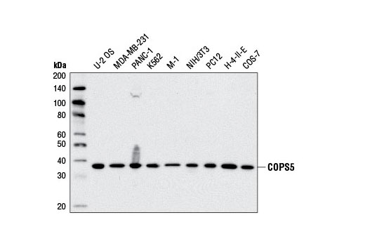
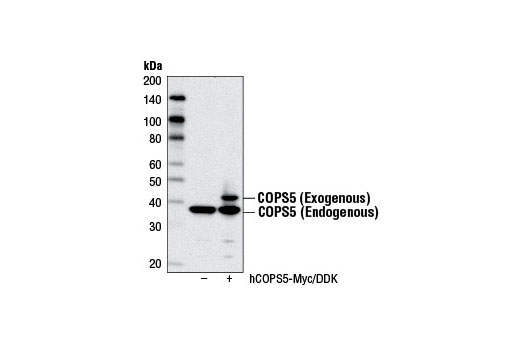
| COPS5 Antibody | 6895 | 40 µl | 37 kDa | Rabbit |
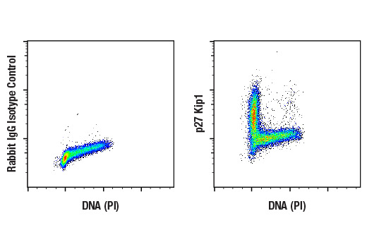
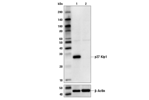
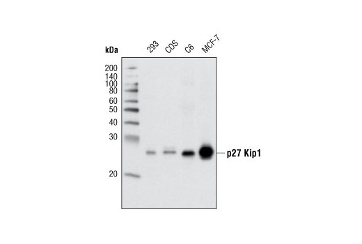
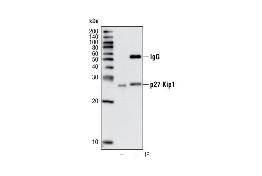
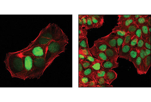
| p27 Kip1 (D69C12) XP® Rabbit mAb | 3686 | 40 µl | 27 kDa | Rabbit IgG |
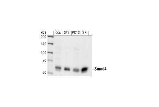
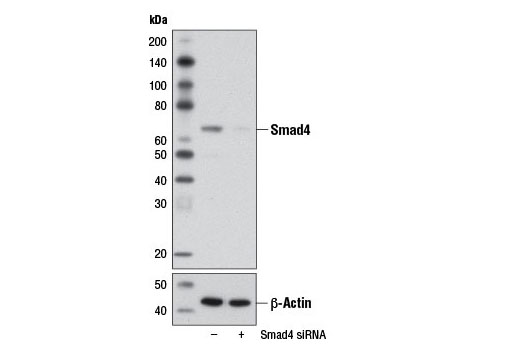
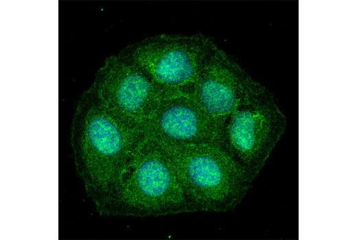
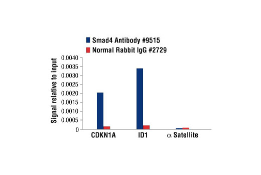
| Smad4 Antibody | 9515 | 40 µl | 70 kDa | Rabbit |
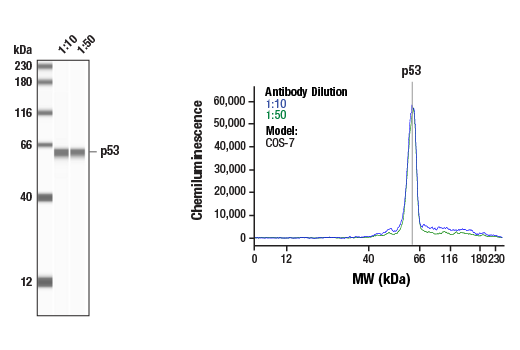
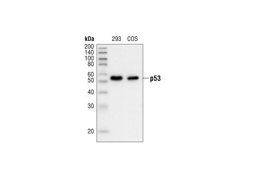
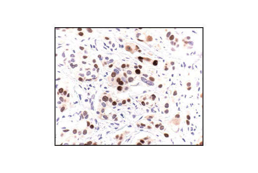
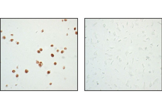
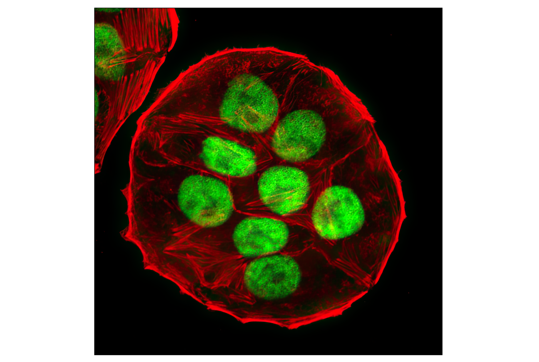
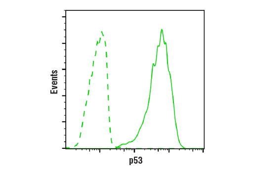
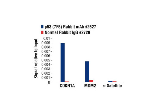
| p53 (7F5) Rabbit mAb | 2527 | 40 µl | 53 kDa | Rabbit IgG |
| Anti-rabbit IgG, HRP-linked Antibody | 7074 | 100 µl | Goat |
Please visit cellsignal.com for individual component applications, species cross-reactivity, dilutions, protocols, and additional product information.
Description
The Tumor Suppressor Inactivation Antibody Sampler Kit provides a fast and economical means of evaluating the role of COPS5 in the inhibition of the tumor suppressors p27 Kip1, p53, and Smad4. The kit contains enough primary antibody to perform four western blot experiments with each primary antibody.
Storage
Background
COPS5/CSN5/Jab1 (c-Jun activation domain-binding protein-1) was originally identified as a transcriptional coactivator of c-Jun and subsequently discovered to be a fifth component and integral part of the CSN (1). As the catalytic center of the CSN, COPS5 is able to integrate multiple functions of the CSN complex such as cell cycle control, transcription, and DNA damage response by regulating the activity of CRLs through deneddylation of cullins (2). Indeed, COPS5 harbors an Mpr1-Pad1-N-terminal (MPN) domain with an embedded Jab1/CSN5 MPN domain metalloenzyme (JAMM) motif that is essential for the CSN isopeptidase activity responsible for deneddylation of CRLs. COPS5 is an evolutionarily conserved 38 kDa protein in humans, mice, fission yeast, and plants, which suggests that it is critical to cell survival and proliferation. A role for COPS5 as a positive regulator of cellular proliferation is supported by evidence that it functionally inactivates several key tumor suppressors such as p53, RUNX3, Smad4, and p27 Kip1 through altered subcellular localization, degradation, and deneddylation (3-7). These findings are underscored by the observation that COPS5 overexpression has been identified in a number of different tumor types and has been implicated in the initiation and progression of several types of cancer (8). Moreover, COPS5 deficient mice display an embryonically lethal phenotype highlighted by elevated expression of COPS5 targets such as p53 and p27 (9,10).
- Claret, F.X. et al. (1996) Nature 383, 453-7.
- Wei, N. et al. (2008) Trends Biochem Sci 33, 592-600.
- Bech-Otschir, D. et al. (2001) EMBO J 20, 1630-9.
- Oh, W. et al. (2006) J Biol Chem 281, 17457-65.
- Wan, M. et al. (2002) EMBO Rep 3, 171-6.
- Tomoda, K. et al. (2002) J Biol Chem 277, 2302-10.
- Kim, J.H. et al. (2009) J Cell Biochem 107, 557-65.
- Shackleford, T.J. and Claret, F.X. (2010) Cell Div 5, 26.
- Tian, L. et al. (2010) Oncogene 29, 6125-37.
- Tomoda, K. et al. (2004) J Biol Chem 279, 43013-8.
Background References
Trademarks and Patents
Limited Uses
Except as otherwise expressly agreed in a writing signed by a legally authorized representative of CST, the following terms apply to Products provided by CST, its affiliates or its distributors. Any Customer's terms and conditions that are in addition to, or different from, those contained herein, unless separately accepted in writing by a legally authorized representative of CST, are rejected and are of no force or effect.
Products are labeled with For Research Use Only or a similar labeling statement and have not been approved, cleared, or licensed by the FDA or other regulatory foreign or domestic entity, for any purpose. Customer shall not use any Product for any diagnostic or therapeutic purpose, or otherwise in any manner that conflicts with its labeling statement. Products sold or licensed by CST are provided for Customer as the end-user and solely for research and development uses. Any use of Product for diagnostic, prophylactic or therapeutic purposes, or any purchase of Product for resale (alone or as a component) or other commercial purpose, requires a separate license from CST. Customer shall (a) not sell, license, loan, donate or otherwise transfer or make available any Product to any third party, whether alone or in combination with other materials, or use the Products to manufacture any commercial products, (b) not copy, modify, reverse engineer, decompile, disassemble or otherwise attempt to discover the underlying structure or technology of the Products, or use the Products for the purpose of developing any products or services that would compete with CST products or services, (c) not alter or remove from the Products any trademarks, trade names, logos, patent or copyright notices or markings, (d) use the Products solely in accordance with CST Product Terms of Sale and any applicable documentation, and (e) comply with any license, terms of service or similar agreement with respect to any third party products or services used by Customer in connection with the Products.