| Product Includes | Product # | Quantity | Mol. Wt | Isotype/Source |
|---|---|---|---|---|
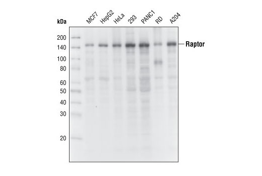
| Raptor (24C12) Rabbit mAb | 2280 | 40 µl | 150 kDa | Rabbit |
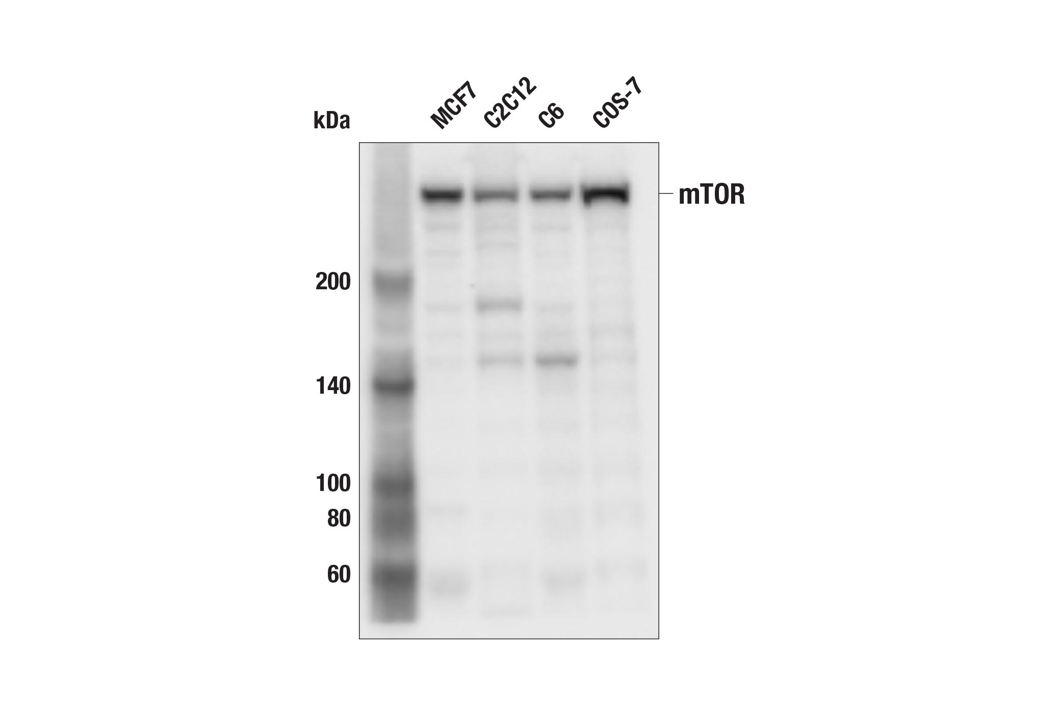
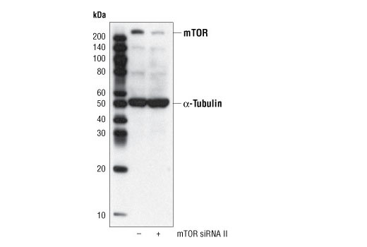
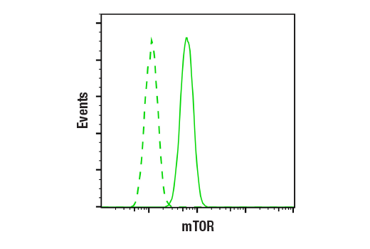
| mTOR (7C10) Rabbit mAb | 2983 | 40 µl | 289 kDa | Rabbit IgG |
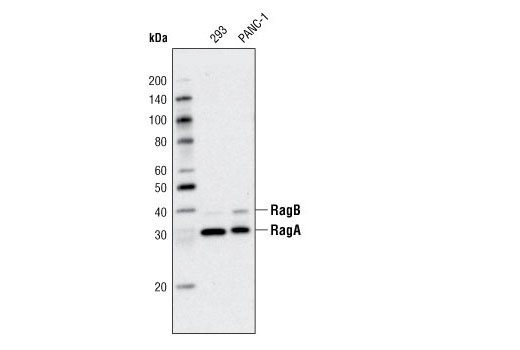
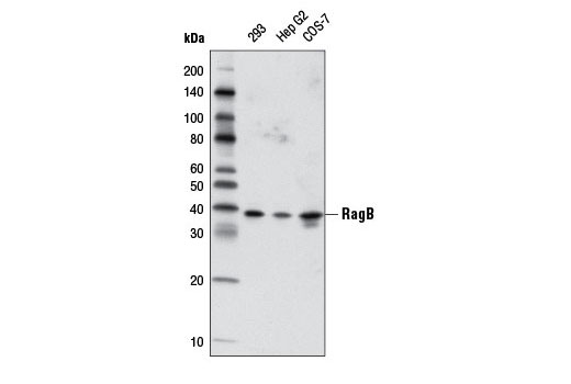
| RagB (D18F3) Rabbit mAb | 8150 | 40 µl | 40 kDa | Rabbit IgG |
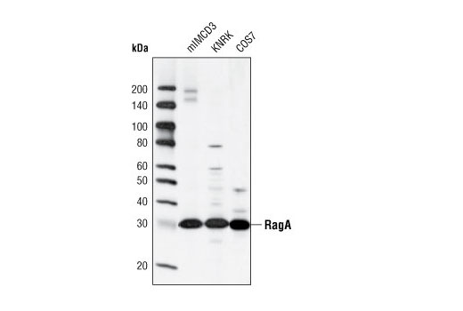
| RagA (D8B5) Rabbit mAb | 4357 | 40 µl | 30 kDa | Rabbit IgG |
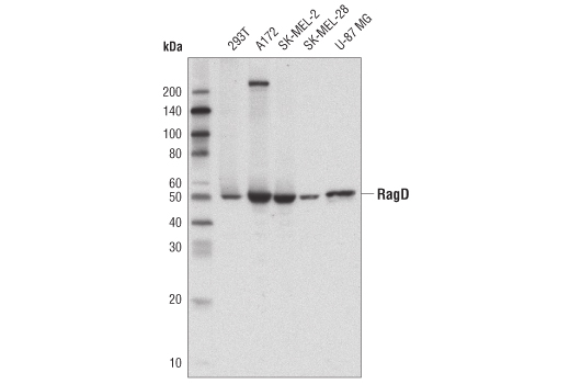
| RagD Antibody | 4470 | 40 µl | 50 kDa | Rabbit |
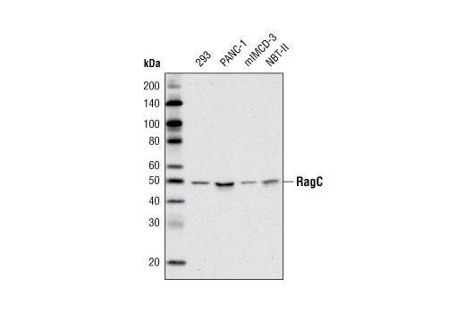
| RagC (D31G9) XP® Rabbit mAb | 5466 | 40 µl | 50 kDa | Rabbit IgG |
| Anti-rabbit IgG, HRP-linked Antibody | 7074 | 100 µl | Goat |
Please visit cellsignal.com for individual component applications, species cross-reactivity, dilutions, protocols, and additional product information.
Description
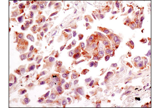
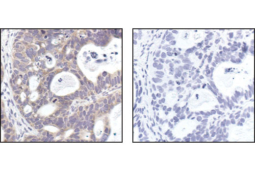
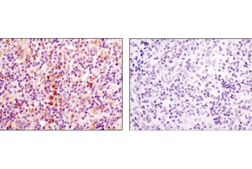
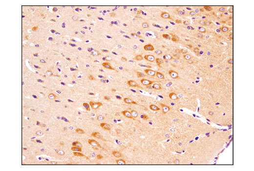
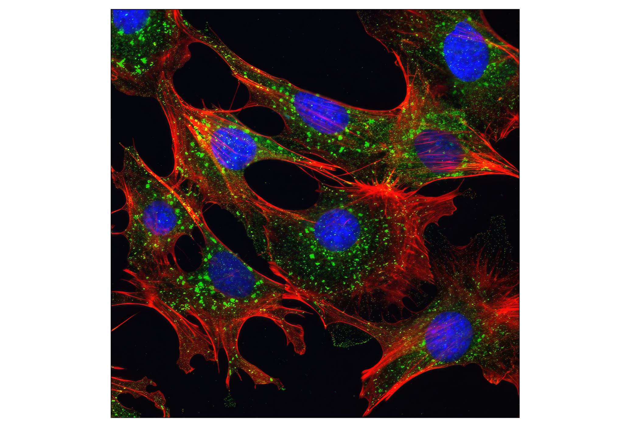
The Rag Protein Antibody Sampler Kit provides an economical means to evaluate the regulation of mTORC1 signaling by such proteins as Raptor, RagA, RagB, RagC, and RagD. The kit contains enough primary and secondary antibodies to perform four Western blot experiments per primary antibody.
Storage
Background
The mTORC1 kinase complex is a critical regulator of cell growth (1,2). Its activity is modulated by energy levels, growth factors, and amino acids via signaling through Akt, MAPK, and AMPK pathways (3,4). Recent studies found that the four related GTPases, RagA, RagB, RagC, and RagD, interact with raptor within the mTORC1 complex (1,2). These interactions are both necessary and sufficient for mTORC1 activation in response to amino acid signals (1,2).
Background References
Trademarks and Patents
Limited Uses
Except as otherwise expressly agreed in a writing signed by a legally authorized representative of CST, the following terms apply to Products provided by CST, its affiliates or its distributors. Any Customer's terms and conditions that are in addition to, or different from, those contained herein, unless separately accepted in writing by a legally authorized representative of CST, are rejected and are of no force or effect.
Products are labeled with For Research Use Only or a similar labeling statement and have not been approved, cleared, or licensed by the FDA or other regulatory foreign or domestic entity, for any purpose. Customer shall not use any Product for any diagnostic or therapeutic purpose, or otherwise in any manner that conflicts with its labeling statement. Products sold or licensed by CST are provided for Customer as the end-user and solely for research and development uses. Any use of Product for diagnostic, prophylactic or therapeutic purposes, or any purchase of Product for resale (alone or as a component) or other commercial purpose, requires a separate license from CST. Customer shall (a) not sell, license, loan, donate or otherwise transfer or make available any Product to any third party, whether alone or in combination with other materials, or use the Products to manufacture any commercial products, (b) not copy, modify, reverse engineer, decompile, disassemble or otherwise attempt to discover the underlying structure or technology of the Products, or use the Products for the purpose of developing any products or services that would compete with CST products or services, (c) not alter or remove from the Products any trademarks, trade names, logos, patent or copyright notices or markings, (d) use the Products solely in accordance with CST Product Terms of Sale and any applicable documentation, and (e) comply with any license, terms of service or similar agreement with respect to any third party products or services used by Customer in connection with the Products.