WB, IP, IHC-P, IF-F, IF-IC, FC-FP
M
Endogenous
22
Rabbit IgG
#Q9EPB4
66824
Product Information
Product Usage Information
| Application | Dilution |
|---|---|
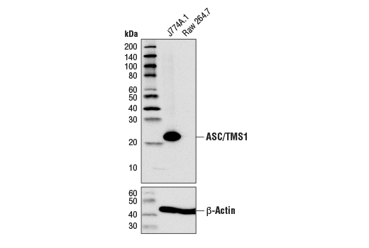
| Western Blotting | 1:1000 |
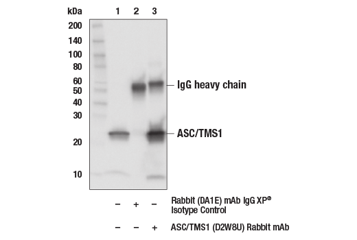
| Immunoprecipitation | 1:100 |
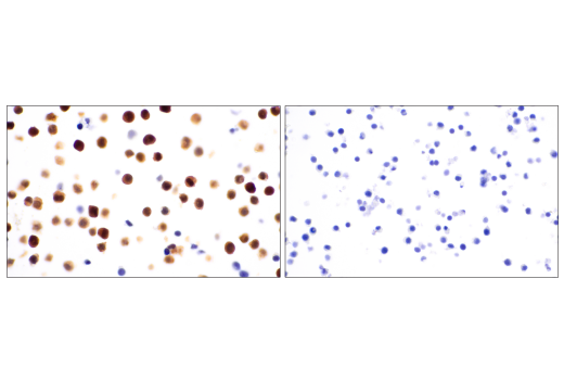
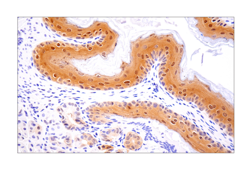
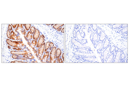
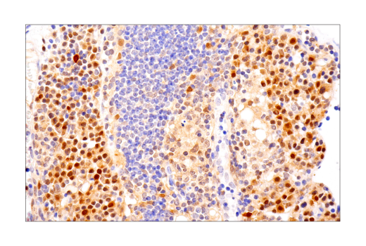
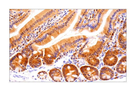
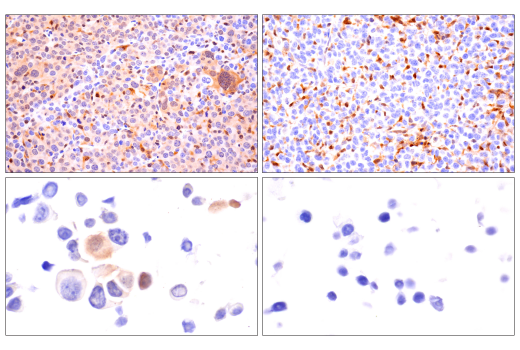
| Immunohistochemistry (Paraffin) | 1:100 - 1:400 |
| Immunofluorescence (Frozen) | 1:400 - 1:1600 |
| Immunofluorescence (Immunocytochemistry) | 1:400 - 1:1600 |
| Flow Cytometry (Fixed/Permeabilized) | 1:400 - 1:1600 |
Storage
For a carrier free (BSA and azide free) version of this product see product #37953.
Specificity / Sensitivity
Species Reactivity:
Mouse
Source / Purification
Monoclonal antibody is produced by immunizing animals with recombinant mouse ASC/TMS1 protein.
Background
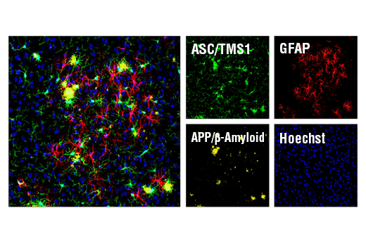
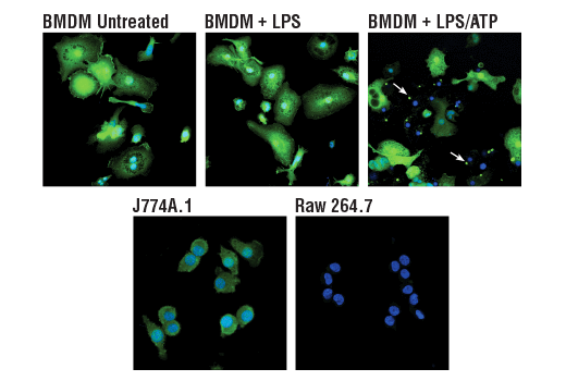
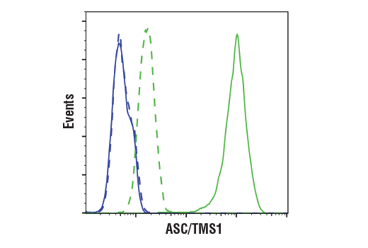
TMS1 (target of methylation-induced silencing)/ASC (apoptosis-associated speck-like protein containing a CARD), also referred to as PYCARD and CARD5, is a 22-kDa pro-apoptotic protein containing an N-terminal pyrin domain (PYD) and a C-terminal caspase recruitment domain (CARD) (1-2). The ASC/TMS1 gene was originally found to be aberrantly methylated and silenced in breast cancer cells (2), and has since been found to be silenced in a number of other cancers, including ovarian cancer (3), glioblastoma (4), melanoma (5), gastric cancer (6), lung cancer (7), and prostate cancer (8). Expression of ASC/TMS1 can be induced by pro-apoptotic/inflammatory stimuli (9). During apoptosis ASC/TMS1 is re-distributed from the cytosol to the mitochondria and associates with mitochondrial Bax to trigger cytochrome c release and subsequent apoptosis (10). ASC/TMS1 has also been found to be a critical component of inflammatory signaling where it associates with and activates caspase-1 in response to pro-inflammatory signals (11).
- Masumoto, J. et al. (1999) J Biol Chem 274, 33835-8.
- Conway, K.E. et al. (2000) Cancer Res 60, 6236-42.
- Terasawa, K. et al. (2004) Clin Cancer Res 10, 2000-6.
- Stone, A.R. et al. (2004) Am J Pathol 165, 1151-61.
- Guan, X. et al. (2003) Int J Cancer 107, 202-8.
- Moriai, R. et al. (2002) Anticancer Res 22, 4163-8.
- Virmani, A. et al. (2003) Int J Cancer 106, 198-204.
- Das, P.M. et al. (2006) Mol Cancer 5, 28.
- Strong, R. et al. (1991) Brain Res 542, 23-8.
- Ohtsuka, T. et al. (2004) Nat Cell Biol 6, 121-8.
- Srinivasula, S.M. et al. (2002) J Biol Chem 277, 21119-22.
Species Reactivity
Species reactivity is determined by testing in at least one approved application (e.g., western blot).
Western Blot Buffer
IMPORTANT: For western blots, incubate membrane with diluted primary antibody in 5% w/v BSA, 1X TBS, 0.1% Tween® 20 at 4°C with gentle shaking, overnight.
Applications Key
WB: Western Blotting IP: Immunoprecipitation IHC-P: Immunohistochemistry (Paraffin) IF-F: Immunofluorescence (Frozen) IF-IC: Immunofluorescence (Immunocytochemistry) FC-FP: Flow Cytometry (Fixed/Permeabilized)
Cross-Reactivity Key
H: human M: mouse R: rat Hm: hamster Mk: monkey Vir: virus Mi: mink C: chicken Dm: D. melanogaster X: Xenopus Z: zebrafish B: bovine Dg: dog Pg: pig Sc: S. cerevisiae Ce: C. elegans Hr: horse GP: Guinea Pig Rab: rabbit All: all species expected
Trademarks and Patents
Limited Uses
Except as otherwise expressly agreed in a writing signed by a legally authorized representative of CST, the following terms apply to Products provided by CST, its affiliates or its distributors. Any Customer's terms and conditions that are in addition to, or different from, those contained herein, unless separately accepted in writing by a legally authorized representative of CST, are rejected and are of no force or effect.
Products are labeled with For Research Use Only or a similar labeling statement and have not been approved, cleared, or licensed by the FDA or other regulatory foreign or domestic entity, for any purpose. Customer shall not use any Product for any diagnostic or therapeutic purpose, or otherwise in any manner that conflicts with its labeling statement. Products sold or licensed by CST are provided for Customer as the end-user and solely for research and development uses. Any use of Product for diagnostic, prophylactic or therapeutic purposes, or any purchase of Product for resale (alone or as a component) or other commercial purpose, requires a separate license from CST. Customer shall (a) not sell, license, loan, donate or otherwise transfer or make available any Product to any third party, whether alone or in combination with other materials, or use the Products to manufacture any commercial products, (b) not copy, modify, reverse engineer, decompile, disassemble or otherwise attempt to discover the underlying structure or technology of the Products, or use the Products for the purpose of developing any products or services that would compete with CST products or services, (c) not alter or remove from the Products any trademarks, trade names, logos, patent or copyright notices or markings, (d) use the Products solely in accordance with CST Product Terms of Sale and any applicable documentation, and (e) comply with any license, terms of service or similar agreement with respect to any third party products or services used by Customer in connection with the Products.