Revision 2
Cell Signaling Technology

Orders: 877-616-CELL (2355) [email protected]

Support: 877-678-TECH (8324)

Web: [email protected] cellsignal.com

3 Trask LaneDanversMassachusetts01923USA
For Research Use Only. Not for Use in Diagnostic Procedures.
Applications:

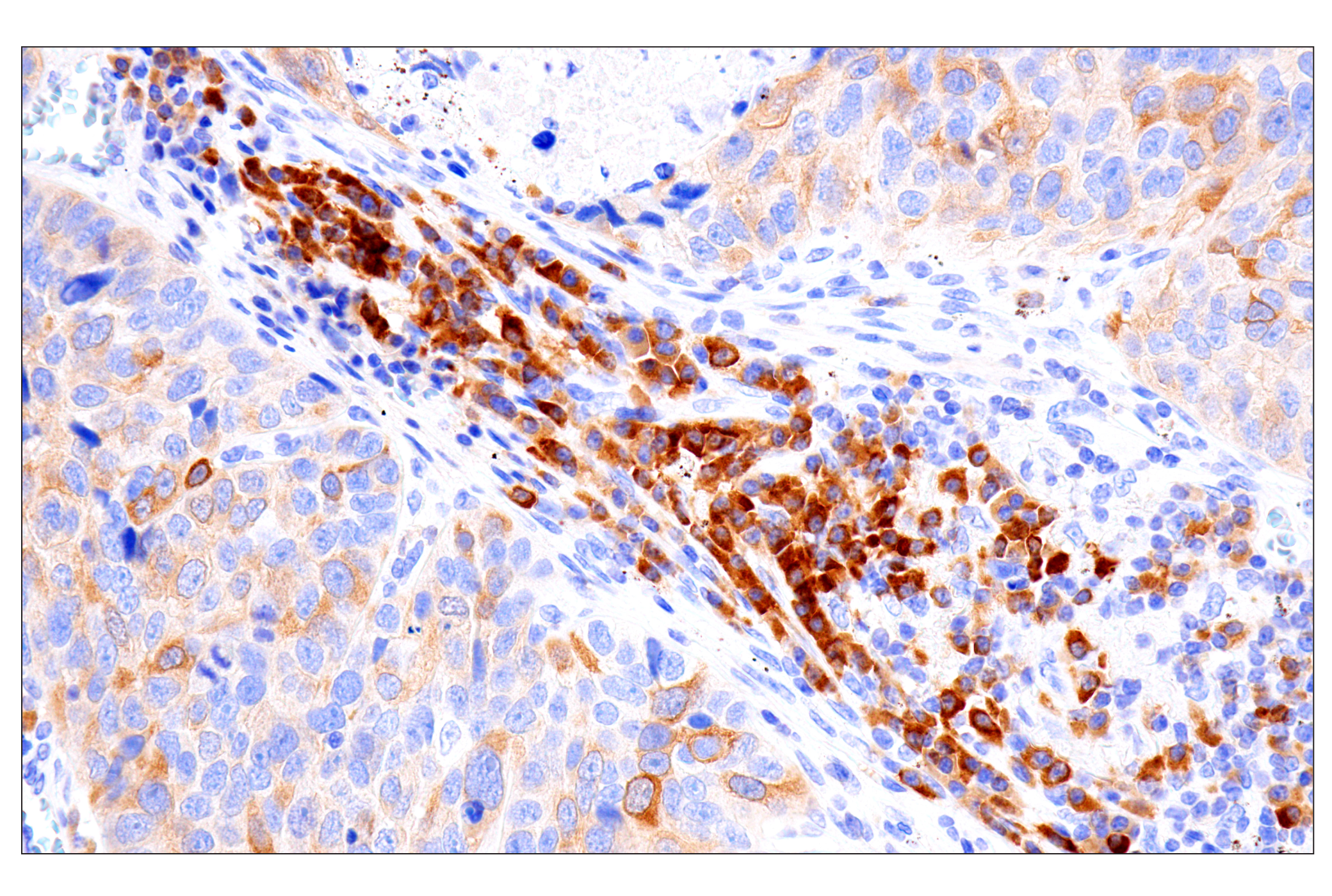
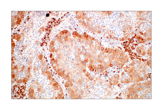
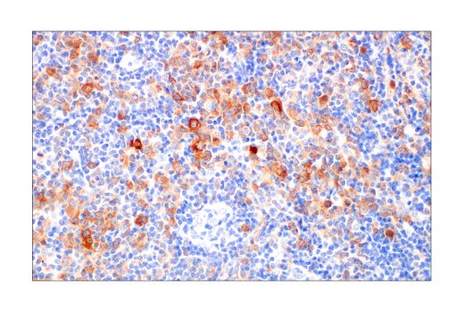
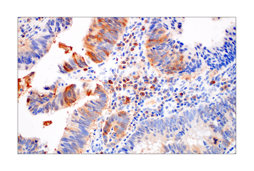
WB, IHC-Bond, IHC-P, FC-FP

REACTIVITY:

H M R

SENSITIVITY:

Endogenous

MW (kDa):

64

Source/Isotype:

Rabbit IgG

UniProt ID:

#P08243

Entrez-Gene Id:

440

Product Information

Product Usage Information

This product is the carrier free version of product #92479. All data were generated using the same antibody clone in the standard formulation which contains BSA and glycerol.

This formulation is ideal for use with technologies requiring specialized or custom antibody labeling, including fluorophores, metals, lanthanides, and oligonucleotides. It is not recommended for ChIP, ChIP-seq, CUT&RUN or CUT&Tag assays. If you require a carrier free formulation for chromatin profiling, please contact us. Optimal dilutions/concentrations should be determined by the end user.

Formulation

Supplied in 1X PBS, BSA and Azide Free.

For standard formulation of this product see product #92479

Storage

Store at -20°C. This product will freeze at -20°C so it is recommended to aliquot into single-use vials to avoid multiple freeze/thaw cycles. A slight precipitate may be present and can be dissolved by gently vortexing. This will not interfere with antibody performance.

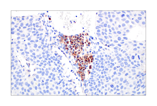
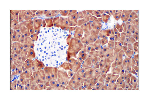
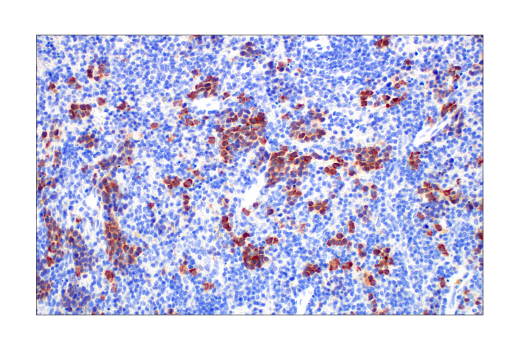
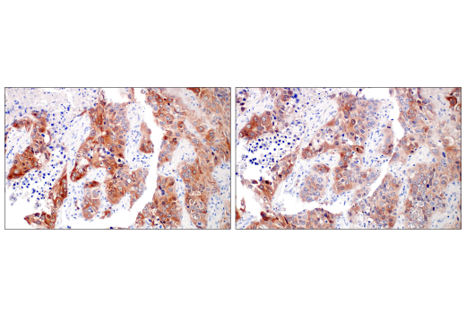
Specificity / Sensitivity

Asparagine Synthetase (E6C2C) XP® Rabbit mAb (BSA and Azide Free) recognizes endogenous levels of total asparagine synthetase protein.

Species Reactivity:

Human, Mouse, Rat

Source / Purification

Monoclonal antibody is produced by immunizing animals with a synthetic peptide corresponding to residues surrounding Lys467 of human asparagine synthetase protein.

Background

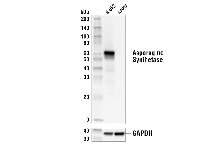
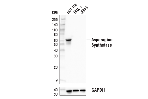
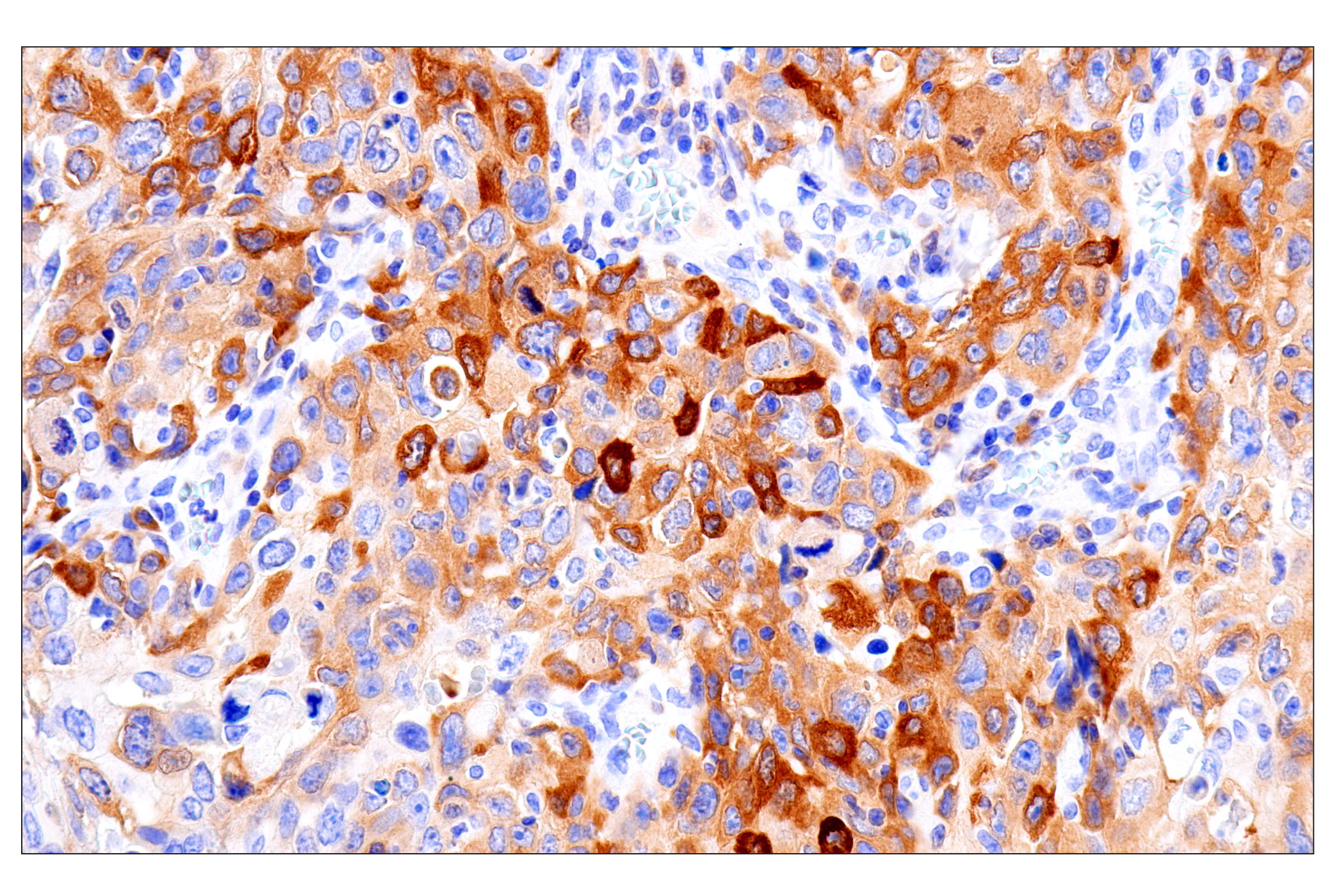
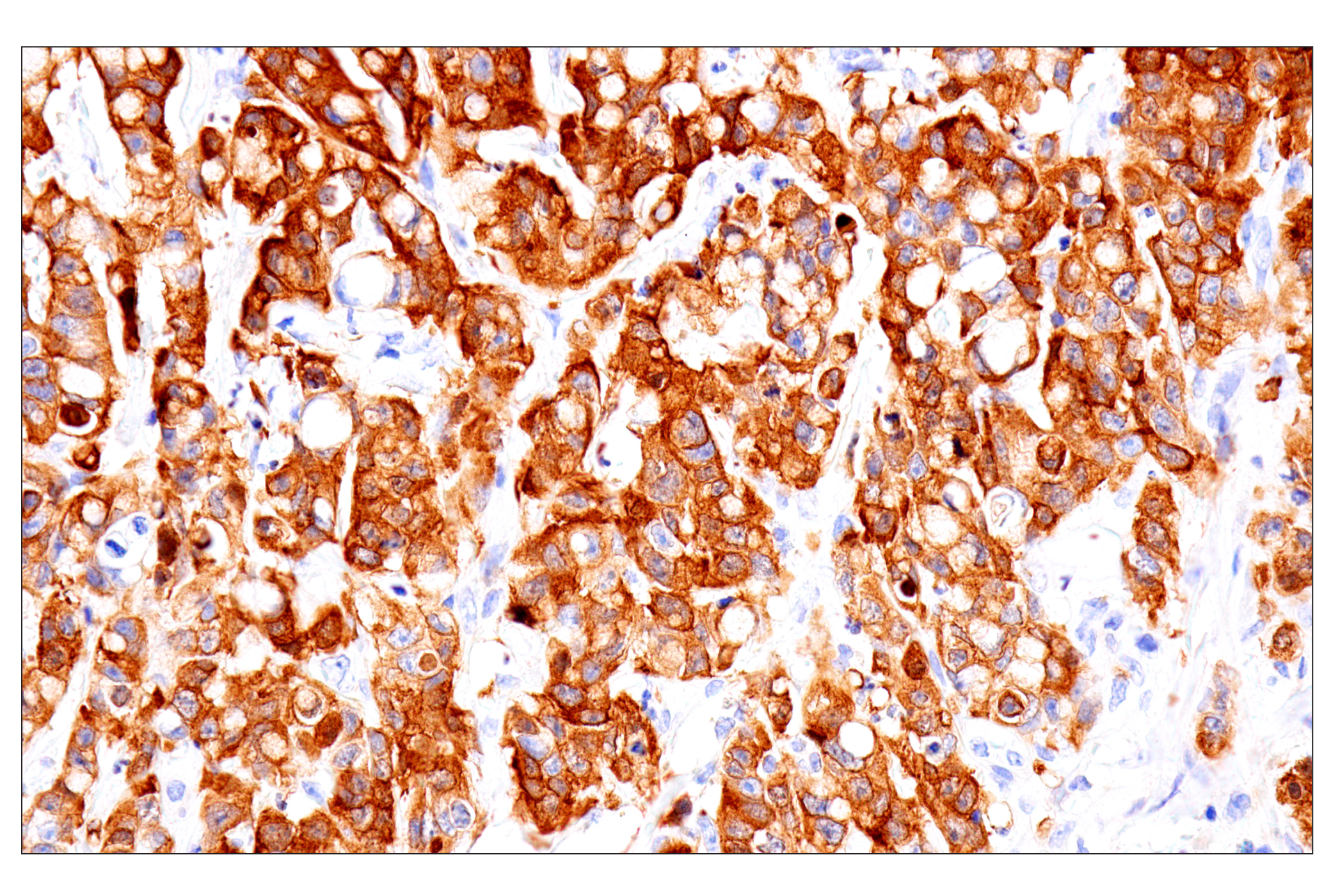
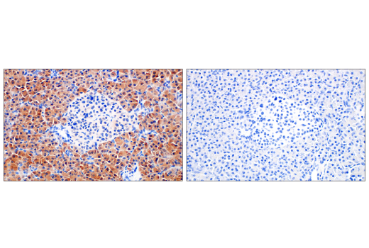
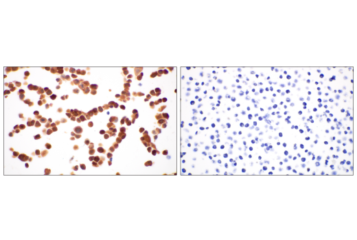
Asparagine synthetase (ASNS) catalyzes the synthesis of asparagine from aspartate and glutamine. Research studies have shown that intracellular asparagine can suppress apoptosis in a large number of human tumors. In addition, ASNS expression levels have been associated with the progression of gliomas and neuroblastomas in patients (1). Furthermore, acute lymphocytic leukemia cells frequently depend upon serum asparagine for their viability, as they lack ASNS (2). Deprivation of asparagine by L-asparaginase has therefore been developed as a therapeutic treatment for acute lymphocytic leukemia (2,3). In subsets of gastric and hepatic cancers, ASNS promoter hypermethylation correlates with low ASNS expression, sensitizing these cancers to the asparaginase treatment (4).

  1. Zhang, J. et al. (2014) Mol Cell 56, 205-18.
  2. Gwinn, D.M. et al. (2018) Cancer Cell 33, 91-107.e6.
  3. Loayza-Puch, F. et al. (2016) Nature 530, 490-4.
  4. Li, H. et al. (2019) Nat Med 25, 850-60.
  5. Nakamura, A. et al. (2018) Proc Natl Acad Sci U S A 115, E7776-E7785.

Species Reactivity

Species reactivity is determined by testing in at least one approved application (e.g., western blot).

Applications Key

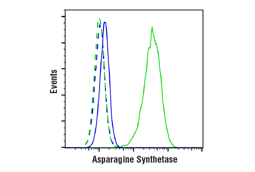
WB: Western Blotting IHC-Bond: IHC Leica Bond IHC-P: Immunohistochemistry (Paraffin) FC-FP: Flow Cytometry (Fixed/Permeabilized)

Cross-Reactivity Key

H: human M: mouse R: rat Hm: hamster Mk: monkey Vir: virus Mi: mink C: chicken Dm: D. melanogaster X: Xenopus Z: zebrafish B: bovine Dg: dog Pg: pig Sc: S. cerevisiae Ce: C. elegans Hr: horse GP: Guinea Pig Rab: rabbit All: all species expected

Trademarks and Patents

Cell Signaling Technology is a trademark of Cell Signaling Technology, Inc.
All other trademarks are the property of their respective owners. Visit cellsignal.com/trademarks for more information.

Limited Uses

Except as otherwise expressly agreed in a writing signed by a legally authorized representative of CST, the following terms apply to Products provided by CST, its affiliates or its distributors. Any Customer's terms and conditions that are in addition to, or different from, those contained herein, unless separately accepted in writing by a legally authorized representative of CST, are rejected and are of no force or effect.

Products are labeled with For Research Use Only or a similar labeling statement and have not been approved, cleared, or licensed by the FDA or other regulatory foreign or domestic entity, for any purpose. Customer shall not use any Product for any diagnostic or therapeutic purpose, or otherwise in any manner that conflicts with its labeling statement. Products sold or licensed by CST are provided for Customer as the end-user and solely for research and development uses. Any use of Product for diagnostic, prophylactic or therapeutic purposes, or any purchase of Product for resale (alone or as a component) or other commercial purpose, requires a separate license from CST. Customer shall (a) not sell, license, loan, donate or otherwise transfer or make available any Product to any third party, whether alone or in combination with other materials, or use the Products to manufacture any commercial products, (b) not copy, modify, reverse engineer, decompile, disassemble or otherwise attempt to discover the underlying structure or technology of the Products, or use the Products for the purpose of developing any products or services that would compete with CST products or services, (c) not alter or remove from the Products any trademarks, trade names, logos, patent or copyright notices or markings, (d) use the Products solely in accordance with CST Product Terms of Sale and any applicable documentation, and (e) comply with any license, terms of service or similar agreement with respect to any third party products or services used by Customer in connection with the Products.

Revision 2
#81128

Asparagine Synthetase (E6C2C) XP® Rabbit mAb (BSA and Azide Free)

Western Blotting Image 1: Asparagine Synthetase (E6C2C) XP® Rabbit mAb (BSA and Azide Free) Expand Image
Western blot analysis of extracts from K-562 and Loucy cells using Asparagine Synthetase (E6C2C) XP® Rabbit mAb (upper) and GAPDH (D16H11) XP® Rabbit mAb #5174 (lower). The absence of detectable asparagine synthetase in Loucy cells is consistent with published data (5), confirming the specificity of the antibody for asparagine synthetase. Data were generated using the standard formulation of this product.
Western Blotting Image 2: Asparagine Synthetase (E6C2C) XP® Rabbit mAb (BSA and Azide Free) Expand Image
Western blot analysis of extracts from HCT 116, TALL-1, and JHH-5 cells using Asparagine Synthetase (E6C2C) XP® Rabbit mAb (upper) and GAPDH (D16H11) XP® Rabbit mAb #5174 (lower). The absence of detectable asparagine synthetase in TALL-1 and JHH-5 cells is consistent with published data (5) and publicly available RNAseq data, respectively, confirming the specificity of the antibody for asparagine synthetase. Data were generated using the standard formulation of this product.
Immunohistochemistry Image 1: Asparagine Synthetase (E6C2C) XP® Rabbit mAb (BSA and Azide Free) Expand Image
Immunohistochemical analysis of paraffin-embedded human oropharyngeal squamous cell carcinoma using Asparagine Synthetase (E6C2C) XP® Rabbit mAb performed on the Leica BOND RX. Data were generated using the standard formulation of this product.
Immunohistochemistry Image 2: Asparagine Synthetase (E6C2C) XP® Rabbit mAb (BSA and Azide Free) Expand Image
Immunohistochemical analysis of paraffin-embedded human colon adenocarcinoma using Asparagine Synthetase (E6C2C) XP® Rabbit mAb performed on the Leica BOND RX. Data were generated using the standard formulation of this product.
Immunohistochemistry Image 3: Asparagine Synthetase (E6C2C) XP® Rabbit mAb (BSA and Azide Free) Expand Image
Immunohistochemical analysis of paraffin-embedded human squamous cell lung carcinoma using Asparagine Synthetase (E6C2C) XP® Rabbit mAb performed on the Leica BOND RX. Data were generated using the standard formulation of this product.
Immunohistochemistry Image 1: Asparagine Synthetase (E6C2C) XP® Rabbit mAb (BSA and Azide Free) Expand Image
Immunohistochemical analysis of paraffin-embedded human non-small cell lung carcinoma using Asparagine Synthetase (E6C2C) XP® Rabbit mAb. Data were generated using the standard formulation of this product.
Immunohistochemistry Image 2: Asparagine Synthetase (E6C2C) XP® Rabbit mAb (BSA and Azide Free) Expand Image
Immunohistochemical analysis of paraffin-embedded human B-cell non-Hodgkin's lymphoma using Asparagine Synthetase (E6C2C) XP® Rabbit mAb. Data were generated using the standard formulation of this product.
Immunohistochemistry Image 3: Asparagine Synthetase (E6C2C) XP® Rabbit mAb (BSA and Azide Free) Expand Image
Immunohistochemical analysis of paraffin-embedded human colon adenocarcinoma using Asparagine Synthetase (E6C2C) XP® Rabbit mAb. Data were generated using the standard formulation of this product.
Immunohistochemistry Image 4: Asparagine Synthetase (E6C2C) XP® Rabbit mAb (BSA and Azide Free) Expand Image
Immunohistochemical analysis of paraffin-embedded JHH-5 xenograft using Asparagine Synthetase (E6C2C) XP® Rabbit mAb. Note the presence of signal in the mouse immune cells and lack of signal in the ASNS negative JHH-5 cells. Data were generated using the standard formulation of this product.
Immunohistochemistry Image 5: Asparagine Synthetase (E6C2C) XP® Rabbit mAb (BSA and Azide Free) Expand Image
Immunohistochemical analysis of paraffin-embedded mouse pancreas using Asparagine Synthetase (E6C2C) XP® Rabbit mAb. Data were generated using the standard formulation of this product.
Immunohistochemistry Image 6: Asparagine Synthetase (E6C2C) XP® Rabbit mAb (BSA and Azide Free) Expand Image
Immunohistochemical analysis of paraffin-embedded mouse spleen using Asparagine Synthetase (E6C2C) XP® Rabbit mAb. Data were generated using the standard formulation of this product.
Immunohistochemistry Image 7: Asparagine Synthetase (E6C2C) XP® Rabbit mAb (BSA and Azide Free) Expand Image
Immunohistochemical analysis of paraffin-embedded human non-small cell lung carcinoma using Asparagine Synthetase (E6C2C) XP® Rabbit mAb (left) or Asparagine Synthetase Rabbit mAb (right). These two antibodies detect independent, unique epitopes on human asparagine synthetase. The similar staining patterns obtained with both antibodies help to confirm the specificity of the staining. Data were generated using the standard formulation of this product.
Immunohistochemistry Image 8: Asparagine Synthetase (E6C2C) XP® Rabbit mAb (BSA and Azide Free) Expand Image
Immunohistochemical analysis of paraffin-embedded human pancreas using Asparagine Synthetase (E6C2C) XP® Rabbit mAb (left) compared to concentration-matched Rabbit (DA1E) mAb IgG XP® Isotype Control #3900 (right). Data were generated using the standard formulation of this product.
Immunohistochemistry Image 9: Asparagine Synthetase (E6C2C) XP® Rabbit mAb (BSA and Azide Free) Expand Image
Immunohistochemical analysis of paraffin-embedded HCT 116 cell pellet (left, positive) or TALL-1 cell pellet (right, negative) using Asparagine Synthetase (E6C2C) XP® Rabbit mAb. Data were generated using the standard formulation of this product.
Flow Cytometry Image 1: Asparagine Synthetase (E6C2C) XP® Rabbit mAb (BSA and Azide Free) Expand Image
Flow cytometric analysis of Loucy cells (blue) and K-562 cells (green) using Asparagine Synthetase (E6C2C) XP® Rabbit mAb (solid lines) or a concentration-matched Rabbit (DA1E) mAb IgG XP® Isotype Control #3900 (dashed lines). Anti-rabbit IgG (H+L), F(ab')2 Fragment (Alexa Fluor® 488 Conjugate) #4412 was used as a secondary antibody. Data were generated using the standard formulation of this product.