| Product Includes | Product # | Quantity | Mol. Wt | Isotype/Source |
|---|---|---|---|---|
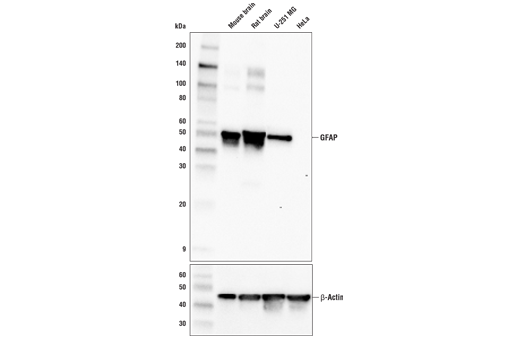
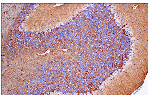
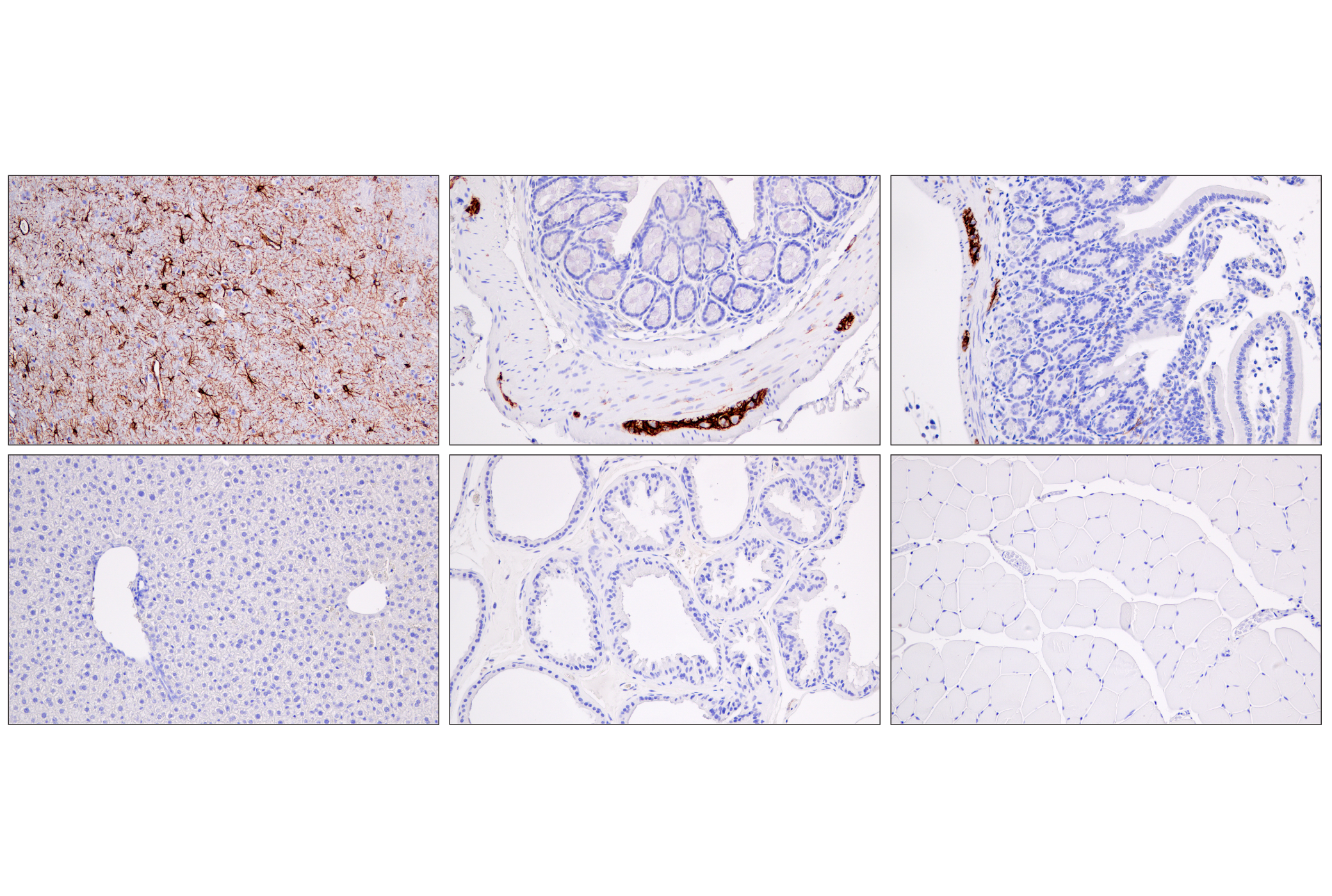
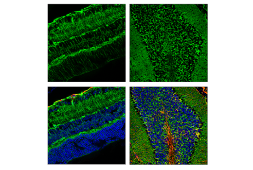
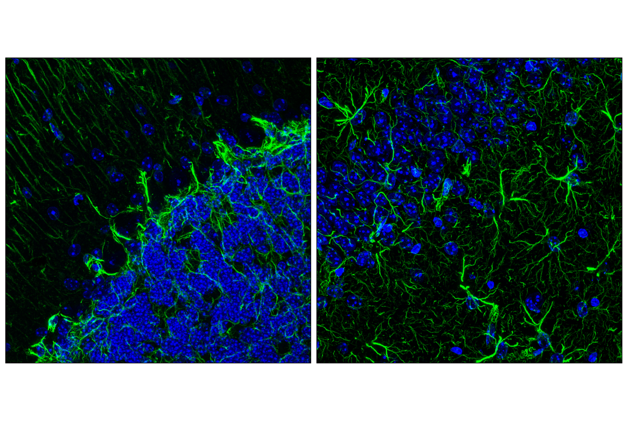
| GFAP (E4L7M) XP® Rabbit mAb | 80788 | 20 µl | 50 kDa | Rabbit IgG |
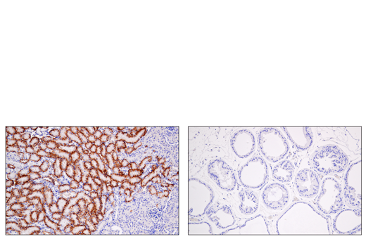
| EAAT1 (D44E2) XP® Rabbit mAb | 5684 | 20 µl | 58 kDa | Rabbit IgG |
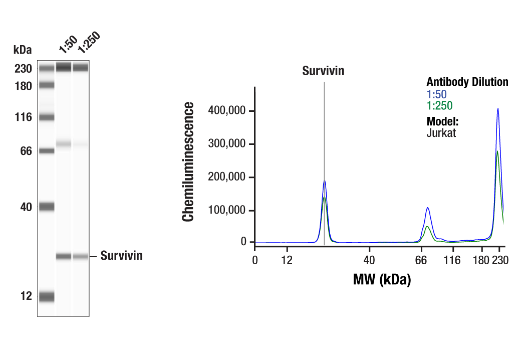
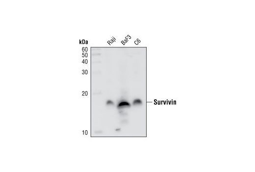
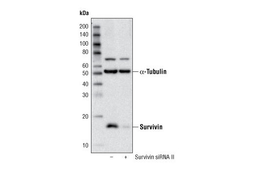
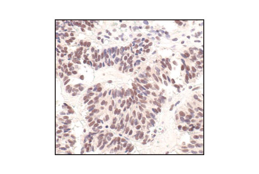
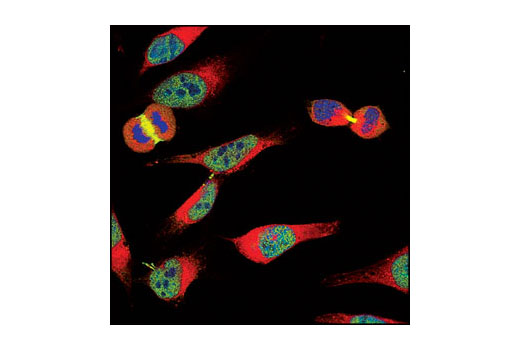
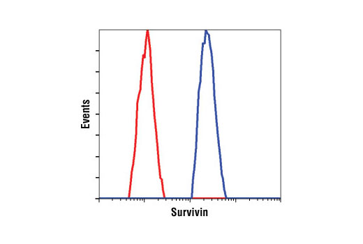
| Survivin (71G4B7) Rabbit mAb | 2808 | 20 µl | 16 kDa | Rabbit IgG |
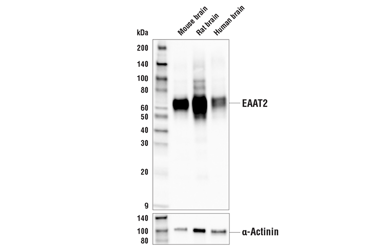
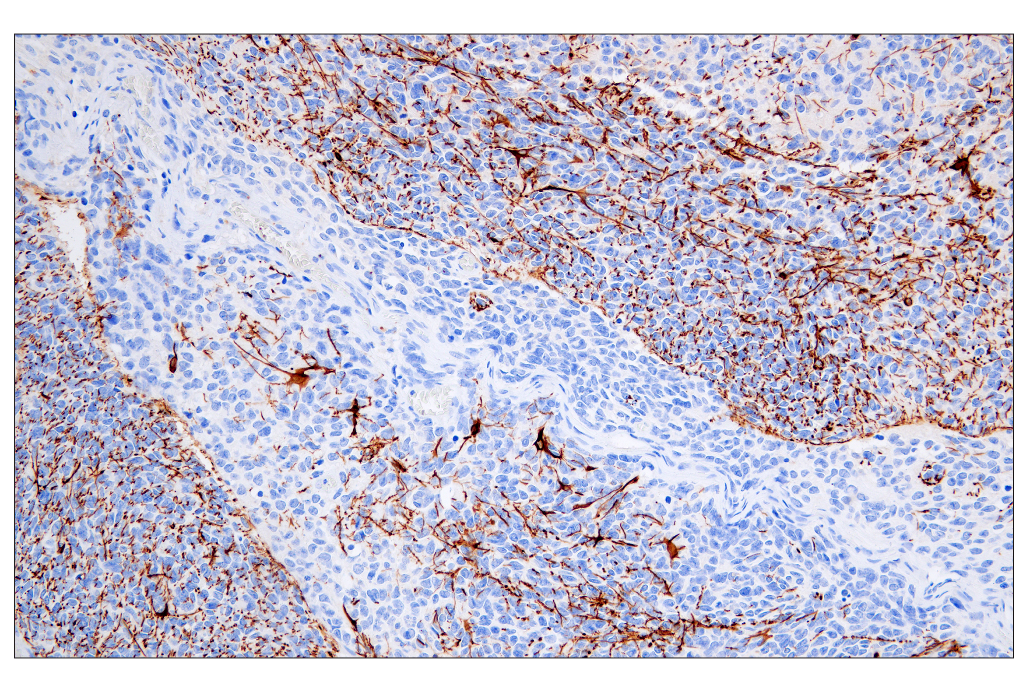
| EAAT2 (E3P5K) Rabbit mAb | 20848 | 20 µl | 65 kDa | Rabbit IgG |
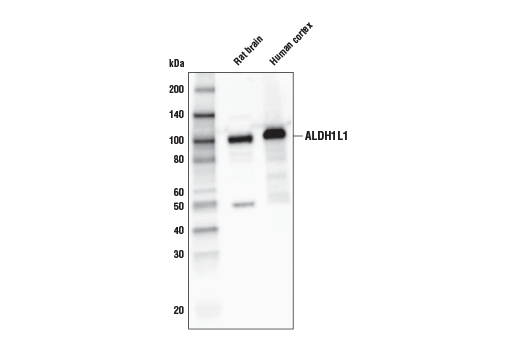
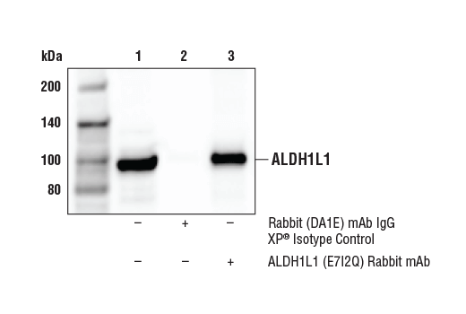
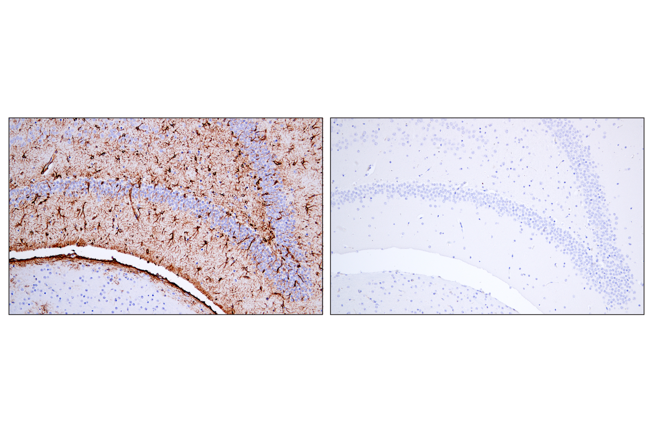
| ALDH1L1 (E7I2Q) Rabbit mAb | 85828 | 20 µl | 98 kDa | Rabbit IgG |
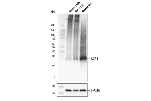
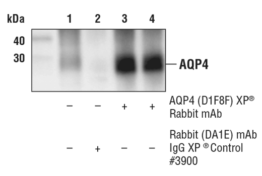
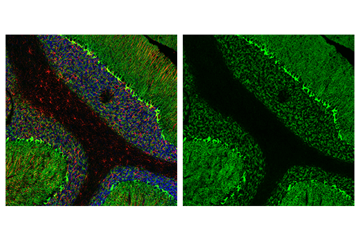
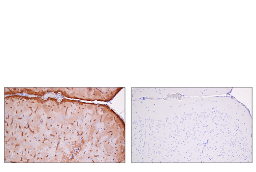
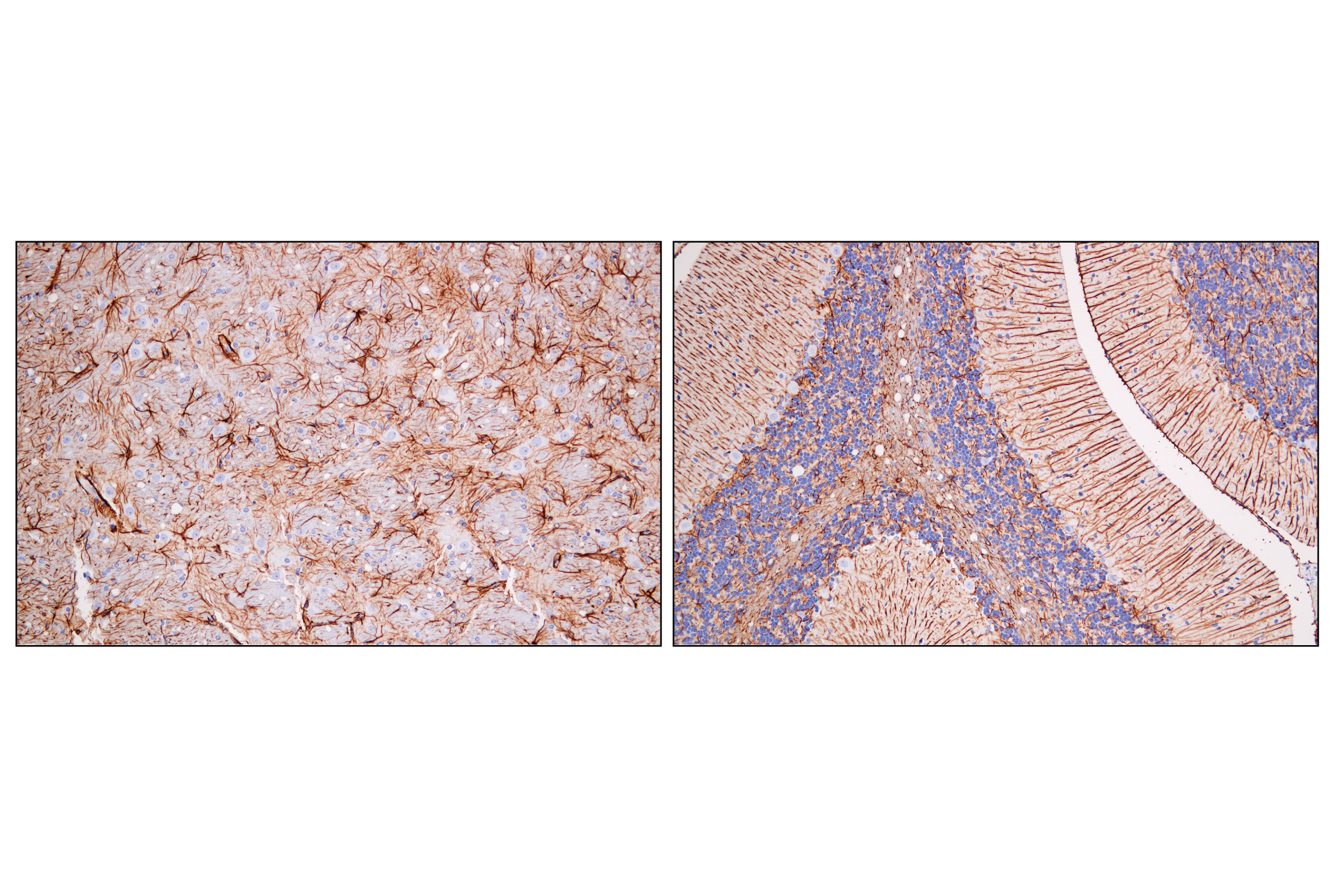
| AQP4 (D1F8E) XP® Rabbit mAb | 59678 | 20 µl | 28 kDa | Rabbit IgG |
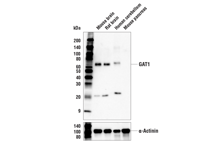
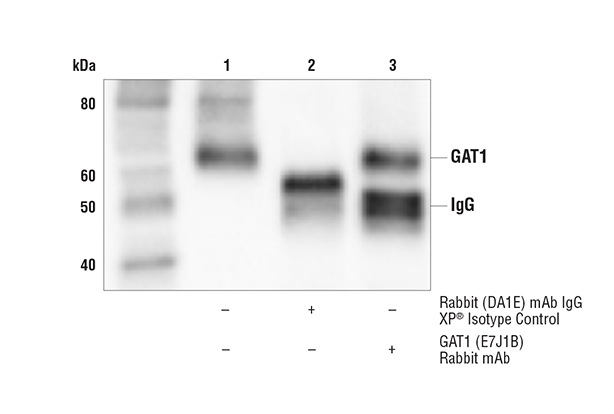
| GAT1 (E7J1B) Rabbit mAb | 37342 | 20 µl | 65 kDa | Rabbit IgG |
| Anti-rabbit IgG, HRP-linked Antibody | 7074 | 100 µl | Goat |
Please visit cellsignal.com for individual component applications, species cross-reactivity, dilutions, protocols, and additional product information.
Description
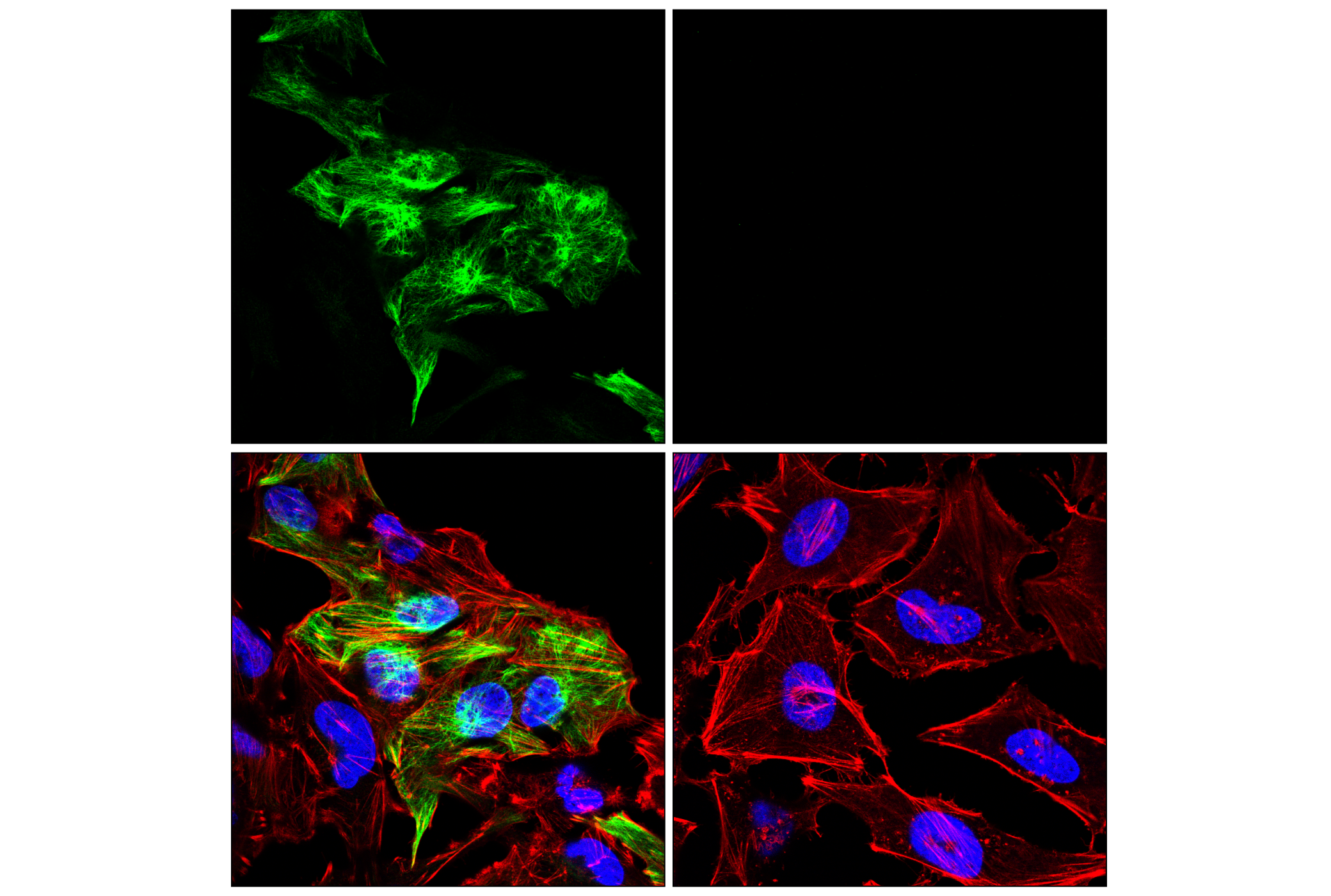
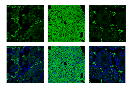
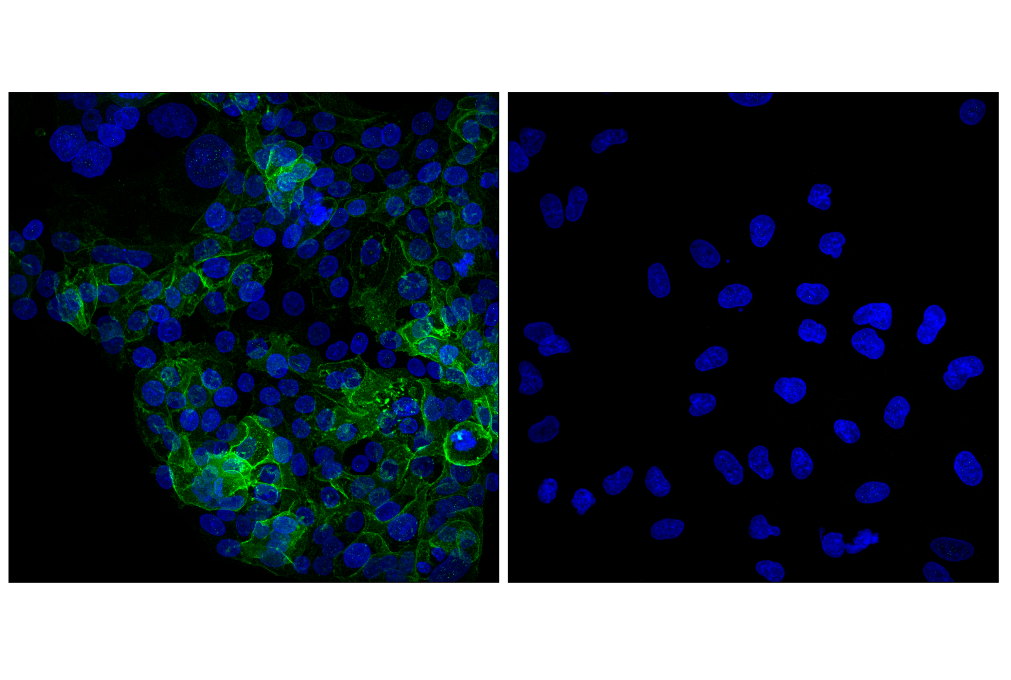
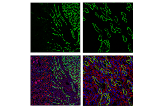
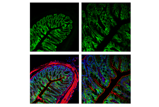
The Astrocyte Markers Antibody Sampler Kit provides an economical means of detecting astrocyte markers by Immunofluorescence or Western Blot. The kit includes enough antibodies to perform at least two western blot or twenty IF tests with each primary antibody.
Storage
Background
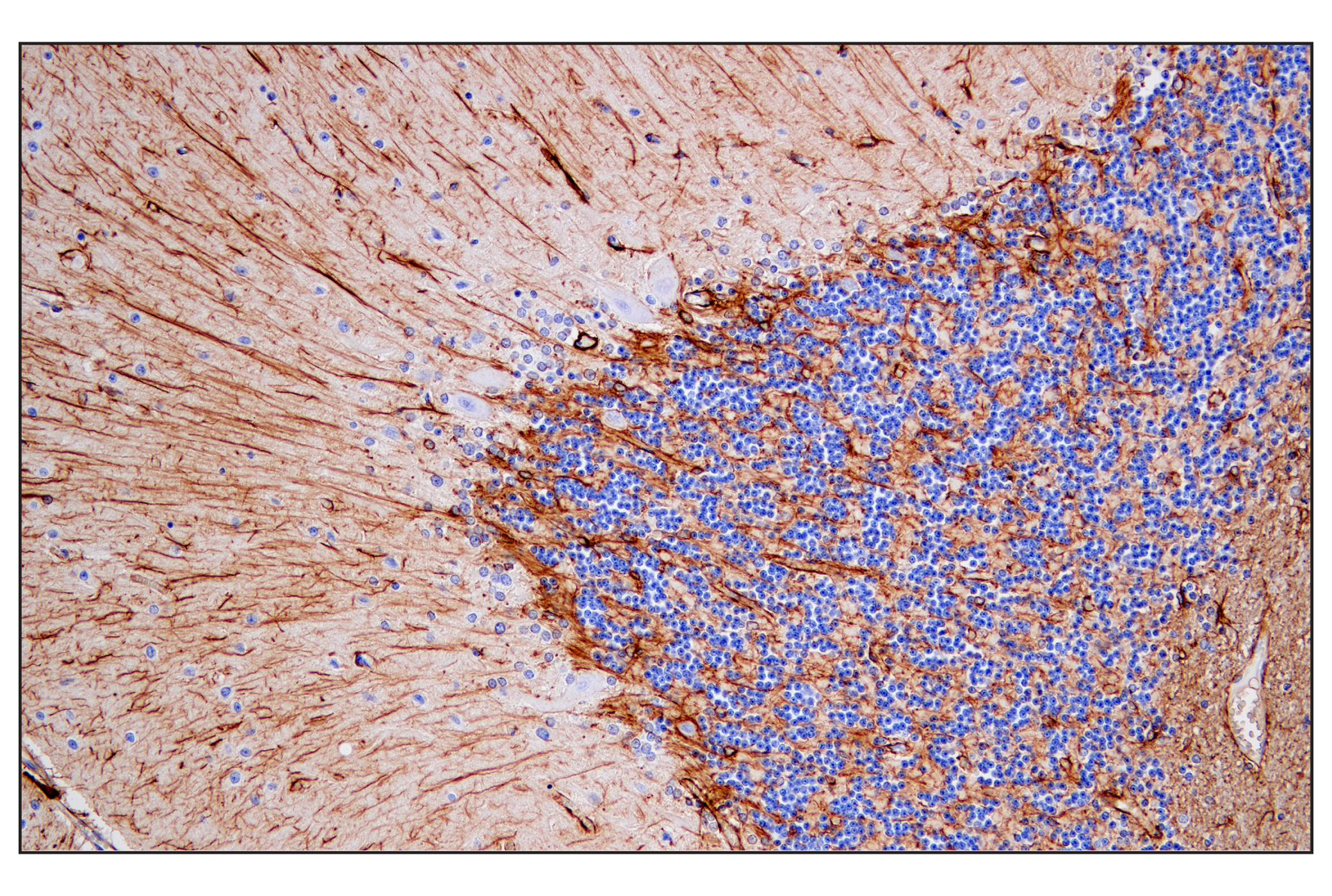
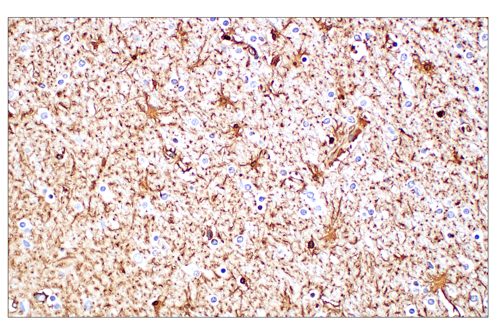
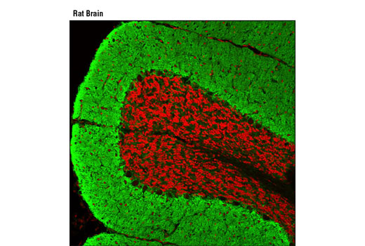
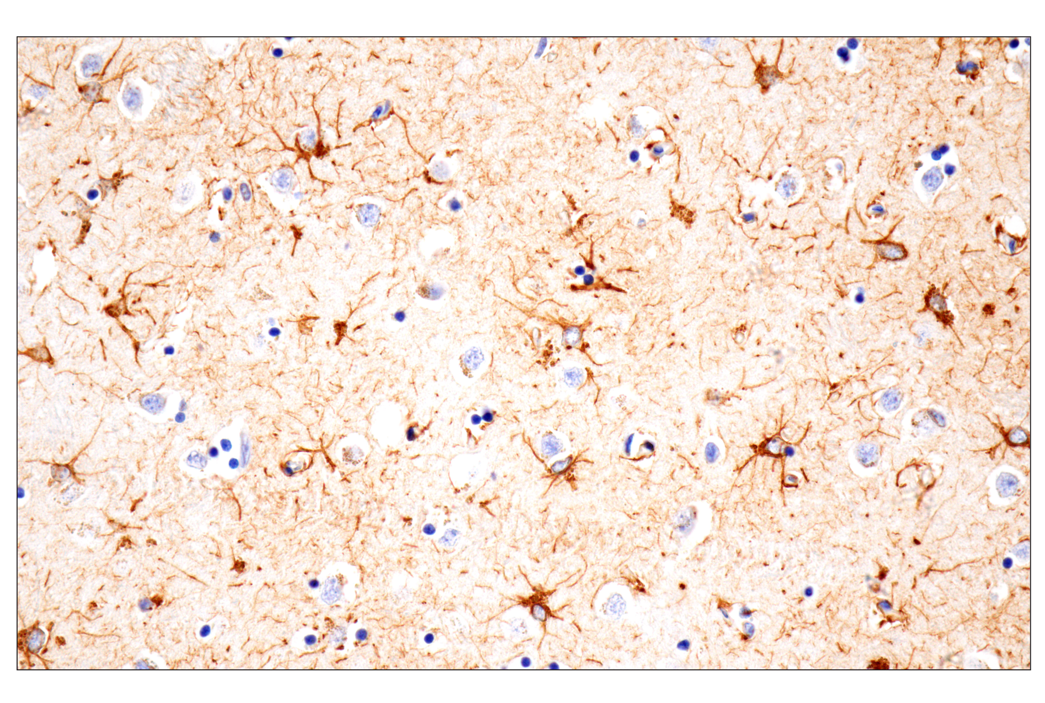
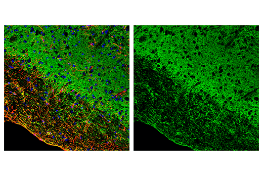
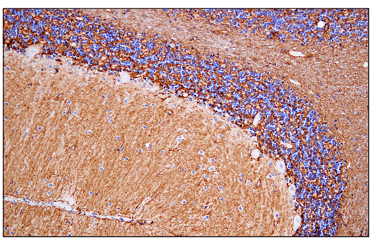
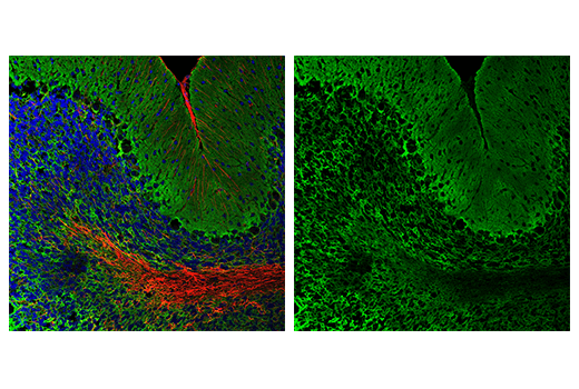
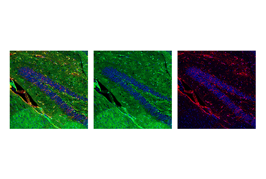
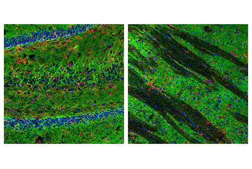
Astrocytes are a population of cells with distinctive morphological and functional characteristics that differ within specific areas of the brain. Postnatally, astrocyte progenitors migrate to reach their brain area and related properties. They have a regulatory role in brain functions that are implicated in neurogenesis and synaptogenesis, controlling blood-brain barrier permeability and maintaining extracellular homeostasis. Mature astrocytes also express some genes enriched in cell progenitors, suggesting they can retain proliferative potential (1). Astrocytes in the human brain are characterized by a heavy expression of the glial fibrillary acidic protein (GFAP), comprised of interlaminar that are located in layers I and II, protoplasmic in layers III and IV, varicose projections in layers V and VI, and fibrous astroglia in white matter (2,3).
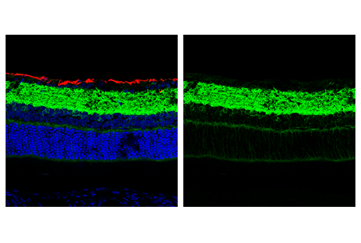
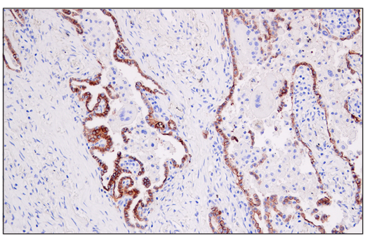
Aquaporins (AQP) are integral membrane proteins that serve as channels in the transfer of water and small solutes across the membrane. AQP4 is present in the brain and it is enriched in astrocytes to regulate water homeostasis, preventing cerebral edema caused by solute imbalance (4,5). Excitatory amino acid transporters are members of sodium-dependent, high-affinity transporters that mediate the uptake of L-glutamate and D-aspartate (6). GABA transmitters are Na+/Cl--dependent transporters that regulate neurotransmitter transport, including GAT1 (SLC6), GAT2, GAT3, and BGT1 (7). GAT1 expresses in the brain, preferentially in glial cells, but is also found in neurons, regulating the uptake and release of neurotransmitters in terminal clefts (8-10). EAAT2, also known as GLT-1, is primarily expressed in astrocytes accounting for up to 90% of the total glutamate transport in the brain. EAAT1 upregulates increased concentrations of glutamate in astrocytes and has a neuroprotective potential following ischemia since reactive astrocytes and activated microglia express EAAT1 but not EAAT2 (11-13).
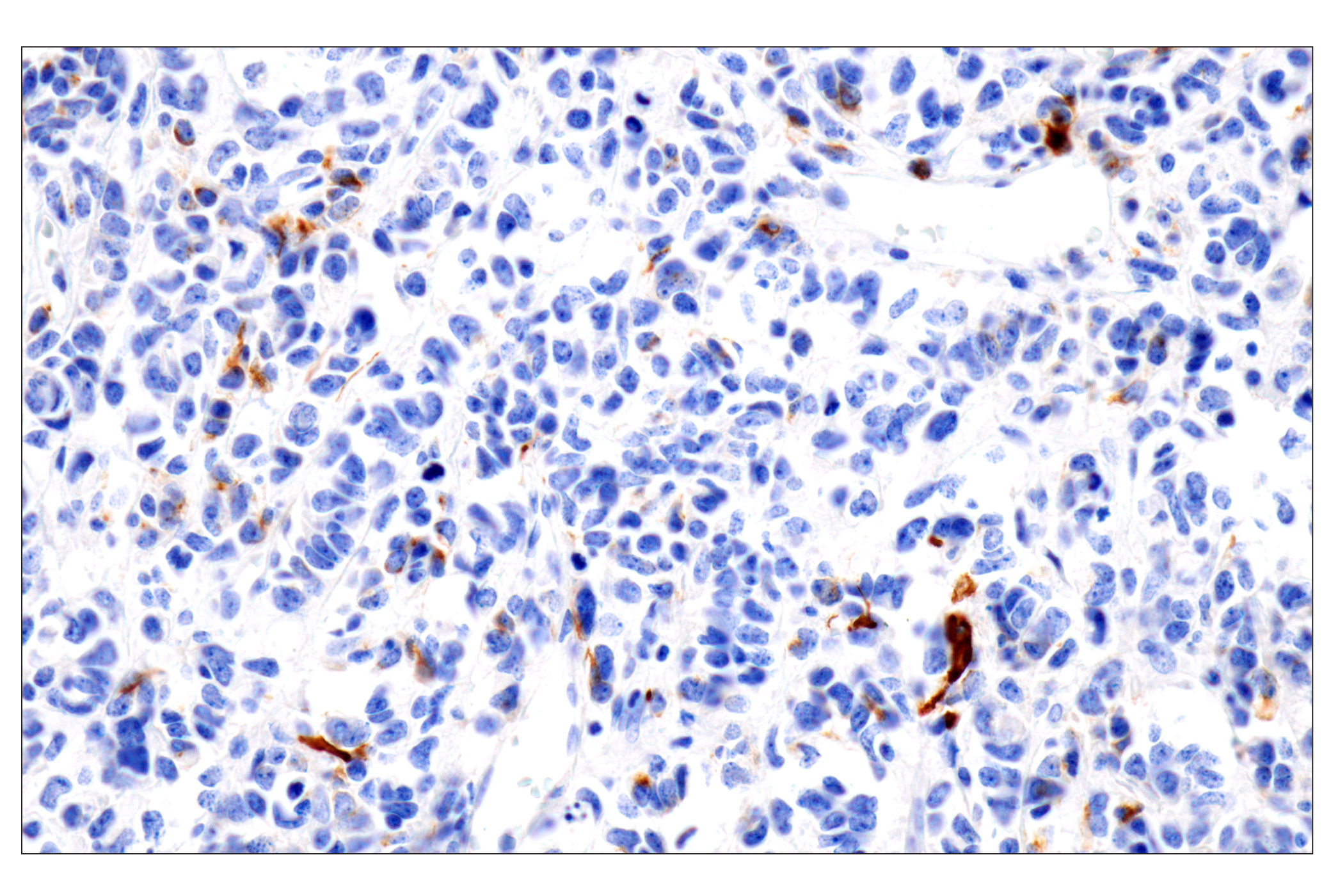
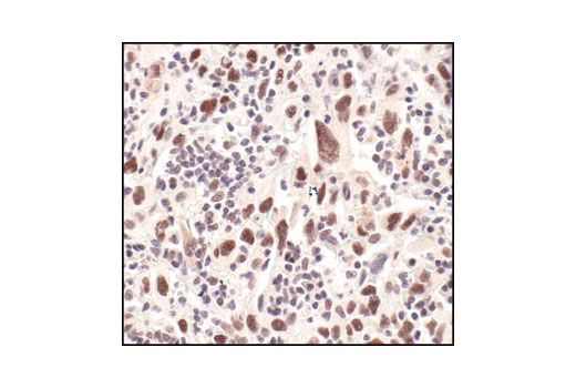
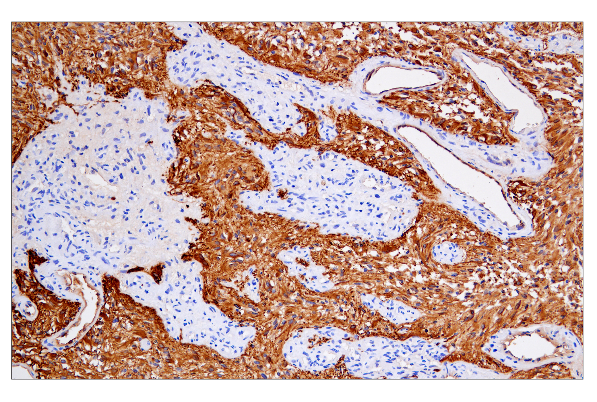
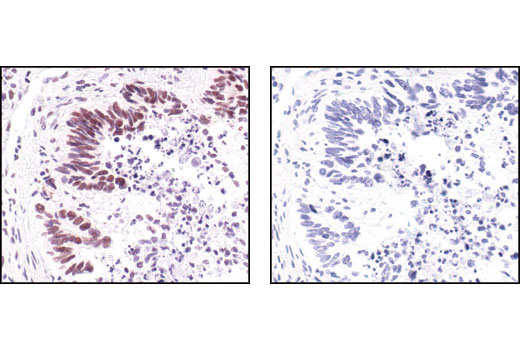
Aldehyde dehydrogenase 1 family member L1 (ALDH1L1) is a member of the aldehyde dehydrogenase super family (14,15). ALDH1L1 has also been shown to be a useful astrocyte marker throughout the grey and white matter of the brain, labeling both the cell body and processes of astrocytes. ALDH1L1 does not label neurons or oligodendrocytes (16). It has been shown that expression of ALDH1L1 in astrocytes is stably expressed through normal tissue and during astrogliosis (17). Survivin is a 16 kDa anti-apoptotic protein highly expressed during fetal development and cancer malignancy. It binds and inhibits caspase-3, controlling the checkpoint in the G2/M-phase of the cell cycle by inhibiting apoptosis and promoting cell division (18). Survivin expression is associated with malignant phenotypes and prognosis of glioma (19).
- Siracusa, R. et al. (2019) Front Pharmacol 10, 1114.
- Vasile, F. et al. (2017) Brain Struct Funct 222, 2017-2029.
- Eng, L.F. et al. (2000) Neurochem Res 25, 1439-51.
- Takata, K. et al. (2004) Prog Histochem Cytochem 39, 1-83.
- Kobayashi, H. et al. (2004) J Pharmacol Sci 96, 264-70.
- Amara, S.G. and Fontana, A.C. (2002) Neurochem Int 41, 313-8.
- Kristensen, A.S. et al. (2011) Pharmacol Rev 63, 585-640.
- Borden, L.A. (1996) Neurochem Int 29, 335-56.
- Moldavan, M. et al. (2017) J Neurophysiol 118, 3092-3106.
- Lorenz-Guertin, J.M. and Jacob, T.C. (2018) Dev Neurobiol 78, 238-270.
- Hediger, M.A. (1999) Am J Physiol 277, F487-92.
- Gegelashvili, G. et al. (1996) Neuroreport 8, 261-5.
- Beschorner, R. et al. (2007) Histopathology 50, 897-910.
- Krupenko, S.A. (2009) Chem Biol Interact 178, 84-93.
- Krupenko, S.A. and Oleinik, N.V. (2002) Cell Growth Differ 13, 227-36.
- Cahoy, J.D. et al. (2008) J Neurosci 28, 264-78.
- Michalovicz, L.T. et al. (2019) J Neurochem 150, 420-440.
- Reed, J.C. and Reed, S.I. (1999) Nat Cell Biol 1, E199-200.
- Tong, X. et al. (2019) Oncol Lett 18, 359-367.
Background References
Trademarks and Patents
Limited Uses
Except as otherwise expressly agreed in a writing signed by a legally authorized representative of CST, the following terms apply to Products provided by CST, its affiliates or its distributors. Any Customer's terms and conditions that are in addition to, or different from, those contained herein, unless separately accepted in writing by a legally authorized representative of CST, are rejected and are of no force or effect.
Products are labeled with For Research Use Only or a similar labeling statement and have not been approved, cleared, or licensed by the FDA or other regulatory foreign or domestic entity, for any purpose. Customer shall not use any Product for any diagnostic or therapeutic purpose, or otherwise in any manner that conflicts with its labeling statement. Products sold or licensed by CST are provided for Customer as the end-user and solely for research and development uses. Any use of Product for diagnostic, prophylactic or therapeutic purposes, or any purchase of Product for resale (alone or as a component) or other commercial purpose, requires a separate license from CST. Customer shall (a) not sell, license, loan, donate or otherwise transfer or make available any Product to any third party, whether alone or in combination with other materials, or use the Products to manufacture any commercial products, (b) not copy, modify, reverse engineer, decompile, disassemble or otherwise attempt to discover the underlying structure or technology of the Products, or use the Products for the purpose of developing any products or services that would compete with CST products or services, (c) not alter or remove from the Products any trademarks, trade names, logos, patent or copyright notices or markings, (d) use the Products solely in accordance with CST Product Terms of Sale and any applicable documentation, and (e) comply with any license, terms of service or similar agreement with respect to any third party products or services used by Customer in connection with the Products.