Revision 1
Cell Signaling Technology

Orders: 877-616-CELL (2355) [email protected]

Support: 877-678-TECH (8324)

Web: [email protected] cellsignal.com

3 Trask LaneDanversMassachusetts01923USA
For Research Use Only. Not for Use in Diagnostic Procedures.
:

:

:

:

UniProt ID:

#P05412

Entrez-Gene Id:

3725

Product Information

Storage

Supplied in 10 mM sodium HEPES (pH 7.5), 150 mM NaCl, 100 µg/ml BSA, 50% glycerol and less than 0.02% sodium azide. Store at –20°C. Do not aliquot the antibody.

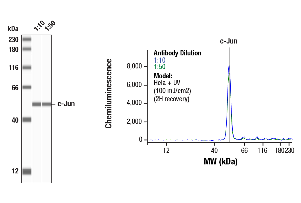
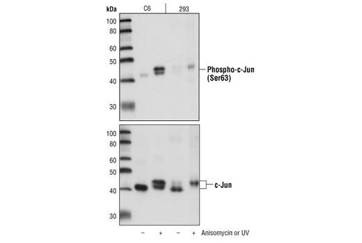
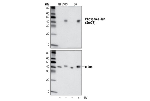
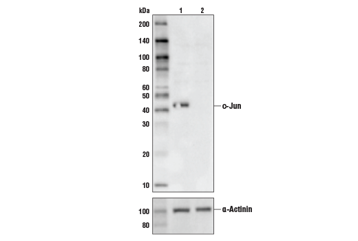
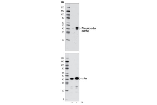
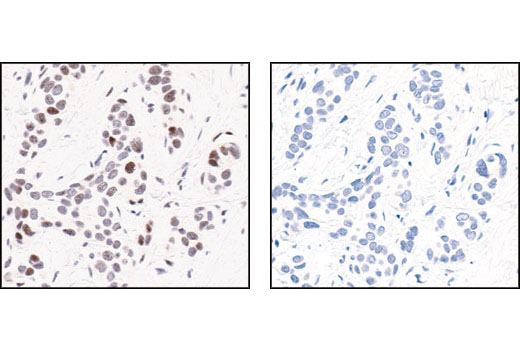
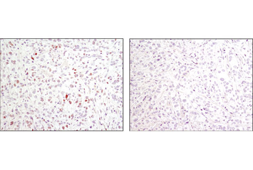
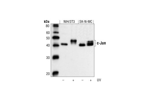
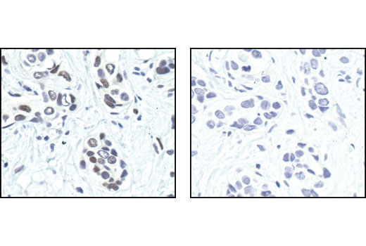
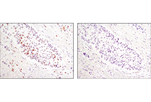
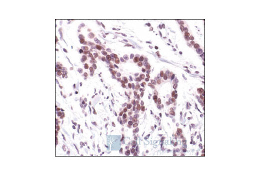
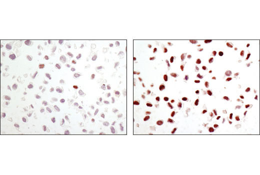
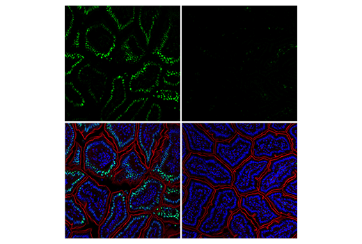
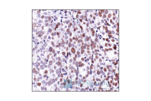
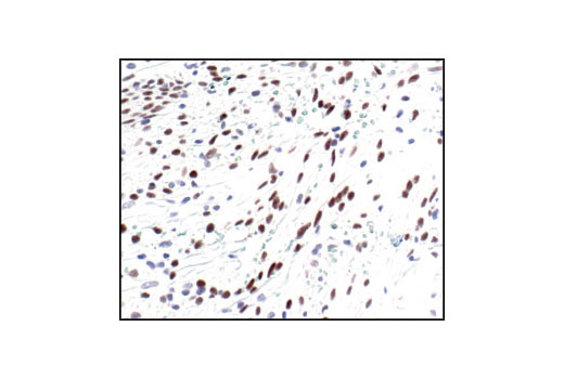
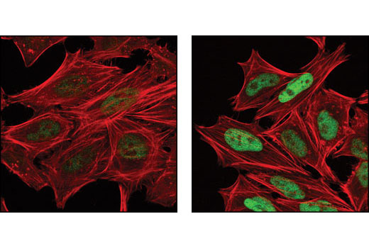
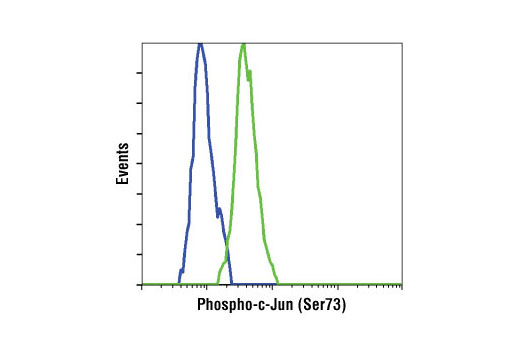
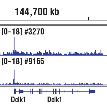
Specificity / Sensitivity

Phospho-c-Jun (Ser63) (54B3) Rabbit mAb detects endogenous levels of c-Jun only when phosphorylated at Ser63. Phospho-c-Jun (Ser73) Antibody detects endogenous levels of c-Jun only when phosphorylated at Ser73. This antibody also recognizes phosphorylation of JunD at Ser100. c-Jun (60A8) Rabbit mAb detects endogenous levels of total c-Jun protein.

Source / Purification

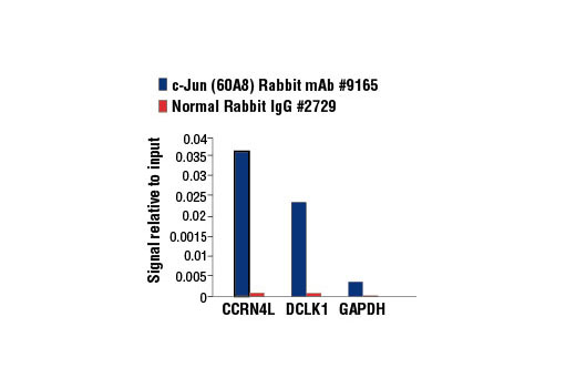
Phospho-specific polyclonal antibodies are produced by immunizing rabbits with a synthetic phosphopeptide corresponding to residues surrounding Ser63 or Ser73 of human c-Jun, and purified by protein A and peptide affinity chromatography. Monoclonal antibody is produced by immunizing animals with a synthetic peptide corresponding to the amino-terminal of human c-Jun.

Product Description

The PhosphoPlus (R) c-Jun (Ser63) and c-Jun (Ser73) Antibody Kit provides reagents and controls for rapid analysis of c-Jun phosphorylation status.
MW (kDa) 43, 50

Background

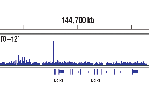
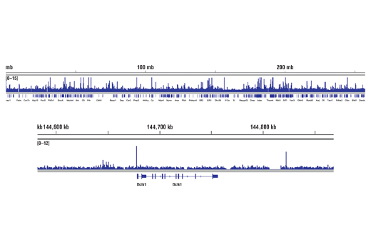
c-Jun is a member of the Jun family containing c-Jun, JunB, and JunD, and is a component of the transcription factor activator protein-1 (AP-1). AP-1 is composed of dimers of Fos, Jun, and ATF family members and binds to and activates transcription at TRE/AP-1 elements (reviewed in 1). Extracellular signals, including growth factors, chemokines, and stress, activate AP-1-dependent transcription. The transcriptional activity of c-Jun is regulated by phosphorylation at Ser63 and Ser73 through SAPK/JNK (reviewed in 2). Knockout studies in mice have shown that c-Jun is essential for embryogenesis (3), and subsequent studies have demonstrated roles for c-Jun in various tissues and developmental processes, including axon regeneration (4), liver regeneration (5), and T cell development (6). AP-1 regulated genes exert diverse biological functions, including cell proliferation, differentiation, and apoptosis, as well as transformation, invasion and metastasis, depending on cell type and context (7-9). Other target genes regulate survival, as well as hypoxia and angiogenesis (8,10). Research studies have implicated c-Jun as a promising therapeutic target for cancer, vascular remodeling, acute inflammation, and rheumatoid arthritis (11,12).

  1. Jochum, W. et al. (2001) Oncogene 20, 2401-12.
  2. Davis, R.J. (2000) Cell 103, 239-52.
  3. Hilberg, F. et al. (1993) Nature 365, 179-81.
  4. Raivich, G. et al. (2004) Neuron 43, 57-67.
  5. Behrens, A. et al. (2002) EMBO J 21, 1782-90.
  6. Riera-Sans, L. and Behrens, A. (2007) J Immunol 178, 5690-700.
  7. Leppä, S. and Bohmann, D. (1999) Oncogene 18, 6158-62.
  8. Shaulian, E. and Karin, M. (2002) Nat Cell Biol 4, E131-6.
  9. Weiss, C. and Bohmann, D. (2004) Cell Cycle 3, 111-3.
  10. Karamouzis, M.V. et al. (2007) Mol Cancer Res 5, 109-20.
  11. Kim, S. and Iwao, H. (2003) J Pharmacol Sci 91, 177-81.
  12. Dass, C.R. and Choong, P.F. (2008) Pharmazie 63, 411-4.

Species Reactivity

Species reactivity is determined by testing in at least one approved application (e.g., western blot).

Cross-Reactivity Key

H: human M: mouse R: rat Hm: hamster Mk: monkey Vir: virus Mi: mink C: chicken Dm: D. melanogaster X: Xenopus Z: zebrafish B: bovine Dg: dog Pg: pig Sc: S. cerevisiae Ce: C. elegans Hr: horse GP: Guinea Pig Rab: rabbit All: all species expected

Trademarks and Patents

Cell Signaling Technology is a trademark of Cell Signaling Technology, Inc.
PhosphoPlus is a trademark of Cell Signaling Technology, Inc.
All other trademarks are the property of their respective owners. Visit cellsignal.com/trademarks for more information.

Limited Uses

Except as otherwise expressly agreed in a writing signed by a legally authorized representative of CST, the following terms apply to Products provided by CST, its affiliates or its distributors. Any Customer's terms and conditions that are in addition to, or different from, those contained herein, unless separately accepted in writing by a legally authorized representative of CST, are rejected and are of no force or effect.

Products are labeled with For Research Use Only or a similar labeling statement and have not been approved, cleared, or licensed by the FDA or other regulatory foreign or domestic entity, for any purpose. Customer shall not use any Product for any diagnostic or therapeutic purpose, or otherwise in any manner that conflicts with its labeling statement. Products sold or licensed by CST are provided for Customer as the end-user and solely for research and development uses. Any use of Product for diagnostic, prophylactic or therapeutic purposes, or any purchase of Product for resale (alone or as a component) or other commercial purpose, requires a separate license from CST. Customer shall (a) not sell, license, loan, donate or otherwise transfer or make available any Product to any third party, whether alone or in combination with other materials, or use the Products to manufacture any commercial products, (b) not copy, modify, reverse engineer, decompile, disassemble or otherwise attempt to discover the underlying structure or technology of the Products, or use the Products for the purpose of developing any products or services that would compete with CST products or services, (c) not alter or remove from the Products any trademarks, trade names, logos, patent or copyright notices or markings, (d) use the Products solely in accordance with CST Product Terms of Sale and any applicable documentation, and (e) comply with any license, terms of service or similar agreement with respect to any third party products or services used by Customer in connection with the Products.

Revision 1
#9260

PhosphoPlus® c-Jun (Ser63) and c-Jun (Ser73) Antibody Kit

PhosphoPlus® c-Jun (Ser63) and c-Jun (Ser73) Antibody Kit: Image 1 Expand Image

Simple Western™ analysis of lysates (0.1 mg/mL) from HeLa cells treated with UV (100 mJ/cm2; 2H recovery) using c-Jun (60A8) Rabbit mAb #9165. The virtual lane view (left) shows the target band (as indicated) at 1:10 and 1:50 dilutions of primary antibody. The corresponding electropherogram view (right) plots chemiluminescence by molecular weight along the capillary at 1:10 (blue line) and 1:50 (green line) dilutions of primary antibody. This experiment was performed under reducing conditions on the Jess™ ​​​​​​​ Simple Western instrument from ProteinSimple, a BioTechne brand, using the 12-230 kDa separation module.

PhosphoPlus® c-Jun (Ser63) and c-Jun (Ser73) Antibody Kit: Image 2 Expand Image
Western blot analysis of extracts from untreated or anisomycin-treated C6 cells, or untreated or UV-treated 293 cells, using Phospho-c-Jun (Ser63) (54B3) Rabbit mAb (upper) or c-Jun (60A8) Rabbit mAb #9165 (lower).
PhosphoPlus® c-Jun (Ser63) and c-Jun (Ser73) Antibody Kit: Image 3 Expand Image
Western blot analysis of extracts from NIH/3T3 or C6 cells, untreated or UV-treated, using Phospho-c-Jun (Ser73) (D47G9) XP® Rabbit mAb (upper) or c-Jun (60A8) Rabbit mAb #9165 (lower).
PhosphoPlus® c-Jun (Ser63) and c-Jun (Ser73) Antibody Kit: Image 4 Expand Image
CUT&RUN was performed with PC-12 cells starved overnight and treated with Human β-Nerve Growth Factor (hβ-NGF) #5221 (50 ng/ml) for 2h and Phospho-c-Jun (Ser73) (D47G9) XP® Rabbit mAb, using CUT&RUN Assay Kit #86652. DNA Library was prepared using DNA Library Prep Kit for Illumina® (ChIP-seq, CUT&RUN) #56795. The figure shows binding across Lmna, a known target gene of Phospho-c-Jun (see additional figure containing CUT&RUN-qPCR data).
PhosphoPlus® c-Jun (Ser63) and c-Jun (Ser73) Antibody Kit: Image 5 Expand Image
After the primary antibody is bound to the target protein, a complex with HRP-linked secondary antibody is formed. The LumiGLO® is added and emits light during enzyme catalyzed decomposition.
PhosphoPlus® c-Jun (Ser63) and c-Jun (Ser73) Antibody Kit: Image 6 Expand Image
After the primary antibody is bound to the target protein, a complex with HRP-linked secondary antibody is formed. The LumiGLO® is added and emits light during enzyme catalyzed decomposition.
PhosphoPlus® c-Jun (Ser63) and c-Jun (Ser73) Antibody Kit: Image 7 Expand Image
Western blot analysis of extracts from control HeLa cells (lane 1) or c-Jun knockout HeLa cells (lane 2) using c-Jun (60A8) Rabbit mAb #9165. The absence of signal in the c-Jun knockout HeLa cells confirms specificity of the antibody for c-Jun.
PhosphoPlus® c-Jun (Ser63) and c-Jun (Ser73) Antibody Kit: Image 8 Expand Image
Western blot analysis of c-Jun Control Cell Extracts using Phospho-c-Jun (Ser73) (D47G9) XP® Rabbit mAb #3270 (upper) and c-Jun (60A8) Rabbit mAb #9165 (lower).
PhosphoPlus® c-Jun (Ser63) and c-Jun (Ser73) Antibody Kit: Image 9 Expand Image
Immunohistochemical analysis of paraffin-embedded human breast carcinoma, using Phospho-c-Jun (Ser63) (54B3) Rabbit mAb in the presence of control peptide (left) or Phospho-c-Jun (Ser63) II Blocking Peptide (#1020) (right).
PhosphoPlus® c-Jun (Ser63) and c-Jun (Ser73) Antibody Kit: Image 10 Expand Image
Immunohistochemical analysis of paraffin-embedded human breast carcinoma, control (left) or lambda phosphatase-treated (right), using Phospho-c-Jun (Ser73) (D47G9) XP® Rabbit mAb.
PhosphoPlus® c-Jun (Ser63) and c-Jun (Ser73) Antibody Kit: Image 11 Expand Image
CUT&RUN was performed with PC-12 cells starved overnight and treated with Human β-Nerve Growth Factor (hβ-NGF) #5221 (50 ng/ml) for 2h and Phospho-c-Jun (Ser73) (D47G9) XP® Rabbit mAb, using CUT&RUN Assay Kit #86652. DNA Library was prepared using DNA Library Prep Kit for Illumina® (ChIP-seq, CUT&RUN) #56795. The figures show binding across chromosome 2 (upper), including Lmna (lower), a known target gene of Phospho-c-Jun (see additional figure containing CUT&RUN-qPCR data).
PhosphoPlus® c-Jun (Ser63) and c-Jun (Ser73) Antibody Kit: Image 12 Expand Image
Western blot analysis of extracts from NIH/3T3 and SK-N-MC cells, untreated or UV-treated, using c-Jun (60A8) Rabbit mAb.
PhosphoPlus® c-Jun (Ser63) and c-Jun (Ser73) Antibody Kit: Image 13 Expand Image
Immunohistochemical analysis of paraffin-embedded human breast carcinoma untreated (left) or lambda phosphatase-treated (right), using Phospho-c-Jun (Ser63) (54B3) Rabbit mAb.
PhosphoPlus® c-Jun (Ser63) and c-Jun (Ser73) Antibody Kit: Image 14 Expand Image
Immunohistochemical analysis of parafin-embedded human colon carcinoma using Phospho-c-Jun (Ser73) (D47G9) XP® Rabbit mAb in the presence of control peptide (left) or Phospho-c-Jun (Ser73) Blocking Peptide (right).
PhosphoPlus® c-Jun (Ser63) and c-Jun (Ser73) Antibody Kit: Image 15 Expand Image
CUT&RUN was performed with PC-12 cells starved overnight and treated with Human β-Nerve Growth Factor (hβ-NGF) #5221 (50 ng/ml) for 2h and either Phospho-c-Jun (Ser73) (D47G9) XP® Rabbit mAb or Rabbit (DA1E) mAb IgG XP® Isotype Control (CUT&RUN) #66362, using CUT&RUN Assay Kit #86652. The enriched DNA was quantified by real-time PCR using rat Lmna promoter primer, rat Cic intron 1 primer and rat Phf17 promoter primer. The amount of immunoprecipitated DNA in each sample is represented as signal relative to the total amount of input chromatin, which is equivalent to one.
PhosphoPlus® c-Jun (Ser63) and c-Jun (Ser73) Antibody Kit: Image 16 Expand Image
Immunohistochemical analysis of paraffin-embedded human astrocytoma, using c-Jun (60A8) Rabbit mAb.
PhosphoPlus® c-Jun (Ser63) and c-Jun (Ser73) Antibody Kit: Image 17 Expand Image
Immunohistochemical analysis of paraffin-embedded human lung carcinoma, showing nuclear localization, using Phospho-c-Jun (Ser63) (54B3) Rabbit mAb.
PhosphoPlus® c-Jun (Ser63) and c-Jun (Ser73) Antibody Kit: Image 18 Expand Image
Immunohistochemical analysis of paraffin-embedded human lung carcinoma using Phospho-c-Jun (Ser73) (D47G9) XP® Rabbit mAb.
PhosphoPlus® c-Jun (Ser63) and c-Jun (Ser73) Antibody Kit: Image 19 Expand Image
Immunohistochemical analysis of paraffin-embedded human colon carcinoma, using c-Jun (60A8) Rabbit mAb.
PhosphoPlus® c-Jun (Ser63) and c-Jun (Ser73) Antibody Kit: Image 20 Expand Image
Immunohistochemical analysis of paraffin-embedded NIH/3T3 cell pellets, control (left) or anisomycin-treated (right), using Phospho-c-Jun (Ser63) (54B3) Rabbit mAb.
PhosphoPlus® c-Jun (Ser63) and c-Jun (Ser73) Antibody Kit: Image 21 Expand Image
Confocal immunofluorescent analysis of mouse small intestine, untreated (left) or treated with λ-phosphatase (right), using Phospho-c-Jun (Ser73) (D47G9) XP® Rabbit mAb #3270 (green). Actin filaments have been labeled with DY-554 phalloidin (red). Samples were mounted in ProLong® Gold Antifade Reagent with DAPI #8961 (blue).
PhosphoPlus® c-Jun (Ser63) and c-Jun (Ser73) Antibody Kit: Image 22 Expand Image
Immunohistochemical analysis of paraffin-embedded human ovarian carcinoma, using c-Jun (60A8) Rabbit mAb.
PhosphoPlus® c-Jun (Ser63) and c-Jun (Ser73) Antibody Kit: Image 23 Expand Image
Immunohistochemical analysis of paraffin-embedded human skin (normal adjacent to hemangioma), using Phospho-c-Jun (Ser63) (54B3) Rabbit mAb.
PhosphoPlus® c-Jun (Ser63) and c-Jun (Ser73) Antibody Kit: Image 24 Expand Image
Confocal immunofluorescent analysis of HeLa cells, untreated (left) or anisomycin-treated (right), using Phospho-c-Jun (Ser73) (D47G9) XP® Rabbit mAb (green). Actin filaments have been labeled with DY-554 phalloidin (red).
PhosphoPlus® c-Jun (Ser63) and c-Jun (Ser73) Antibody Kit: Image 25 Expand Image
Flow cytometric analysis of HeLa cells, untreated (blue) or UV treated (green), using Phospho-c-Jun (Ser73) (D47G9) XP® Rabbit mAb.
PhosphoPlus® c-Jun (Ser63) and c-Jun (Ser73) Antibody Kit: Image 26 Expand Image
Chromatin immunoprecipitations were performed with cross-linked chromatin from PC-12 cells starved overnight and treated with Human β-Nerve Growth Factor (hβ-NGF) #5221 (50 ng/ml) for 2h and either Phospho-c-Jun (Ser73) (D47G9) XP® Rabbit mAb or c-Jun (60A8) Rabbit mAb #9165, using SimpleChIP® Plus Enzymatic Chromatin IP Kit (Magnetic Beads) #9005. DNA Libraries were prepared using SimpleChIP® ChIP-seq DNA Library Prep Kit for Illumina® #56795. The figure shows binding across Dclk1, a known target gene of both Phospho-c-Jun and c-Jun (see additional figures containing ChIP-qPCR data). For additional ChIP-seq tracks, please download the product datasheet.
PhosphoPlus® c-Jun (Ser63) and c-Jun (Ser73) Antibody Kit: Image 27 Expand Image
Confocal immunofluorescent analysis of HeLa cells, using c-Jun (60A8) Rabbit mAb (green). Actin filaments have been labeled with Alexa Fluor® 555 phalloidin (red).
PhosphoPlus® c-Jun (Ser63) and c-Jun (Ser73) Antibody Kit: Image 28 Expand Image
Chromatin immunoprecipitations were performed with cross-linked chromatin from PC-12 cells starved overnight and treated with Human β-Nerve Growth Factor (hβ-NGF) #5221 (50 ng/ml) for 2h and either Phospho-c-Jun (Ser73) (D47G9) XP® Rabbit mAb or c-Jun (60A8) Rabbit mAb #9165, using SimpleChIP® Plus Enzymatic Chromatin IP Kit (Magnetic Beads) #9005. DNA Libraries were prepared using DNA Library Prep Kit for Illumina® (ChIP-seq, CUT&RUN) #56795. The figure shows binding across chromosome 2 (upper), including Dclk1 (lower), a known target gene of both Phospho-c-Jun and c-Jun (see additional figures containing ChIP-qPCR data).
PhosphoPlus® c-Jun (Ser63) and c-Jun (Ser73) Antibody Kit: Image 29 Expand Image
Flow cytometric analysis of Jurkat cells using c-Jun (60A8) Rabbit mAb (solid line) compared to concentration-matched Rabbit (DA1E) mAb IgG XP® Isotype Control #3900 (dashed line). Anti-rabbit IgG (H+L), F(ab')2 Fragment (Alexa Fluor® 488 Conjugate) #4412 was used as a secondary antibody.
PhosphoPlus® c-Jun (Ser63) and c-Jun (Ser73) Antibody Kit: Image 30 Expand Image
Chromatin immunoprecipitations were performed with cross-linked chromatin from cells starved overnight and treated with Human β-Nerve Growth Factor (hβ-NGF) #5221 (50 ng/ml) for 2h and either Phospho-c-Jun (Ser73) (D47G9) XP® Rabbit mAb or Normal Rabbit IgG #2729 using SimpleChIP® Enzymatic Chromatin IP Kit (Magnetic Beads) #9003. The enriched DNA was quantified by real-time PCR SimpleChIP® using Rat CCRN4L Promoter Primers #7983, rat DCLK1 promoter primers, and SimpleChIP® Rat GAPDH Promoter Primers #7964. The amount of immunoprecipitated DNA in each sample is represented as signal relative to the total amount of input chromatin, which is equivalent to one.
PhosphoPlus® c-Jun (Ser63) and c-Jun (Ser73) Antibody Kit: Image 31 Expand Image
Chromatin immunoprecipitations were performed with cross-linked chromatin from PC-12 cells starved overnight and treated with β-NGF #5221 (50ng/ml) for 2h and c-Jun (60A8) Rabbit mAb, using SimpleChIP® Plus Enzymatic Chromatin IP Kit (Magnetic Beads) #9005. DNA Library Prep Kit for Illumina® (ChIP-seq, CUT&RUN) #56795. The figure shows binding across Dclk1, a known target gene of c-Jun (see additional figure containing ChIP-qPCR data).
PhosphoPlus® c-Jun (Ser63) and c-Jun (Ser73) Antibody Kit: Image 32 Expand Image
Chromatin immunoprecipitations were performed with cross-linked chromatin from PC-12 cells starved overnight and treated with β-NGF #5221 (50ng/ml) for 2h and c-Jun (60A8) Rabbit mAb, using SimpleChIP® Plus Enzymatic Chromatin IP Kit (Magnetic Beads) #9005. DNA Library Prep Kit for Illumina® (ChIP-seq, CUT&RUN) #56795. The figure shows binding across chromosome 2 (upper), including Dclk1 (lower), a known target gene of c-Jun (see additional figure containing ChIP-qPCR data).
PhosphoPlus® c-Jun (Ser63) and c-Jun (Ser73) Antibody Kit: Image 33 Expand Image
Chromatin immunoprecipitations were performed with cross-linked chromatin from PC-12 cells starved overnight and treated with β-NGF #5221 (50ng/ml) for 2h and either of c-Jun (60A8) Rabbit mAb or Normal Rabbit IgG #2729 using SimpleChIP® Enzymatic Chromatin IP Kit (Magnetic Beads) #9003. The enriched DNA was quantified by real-time PCR using SimpleChIP® Rat CCRN4L Promoter Primers #7983, rat DCLK1 promoter primers, and SimpleChIP® Rat GAPDH Promoter Primers #7964. The amount of immunoprecipitated DNA in each sample is represented as signal relative to the total amount of input chromatin, which is equivalent to one.