WB, IP, IF-F, IF-IC
H M R
Endogenous
80
Rabbit IgG
#P17655
824
Product Information
Product Usage Information
| Application | Dilution |
|---|---|
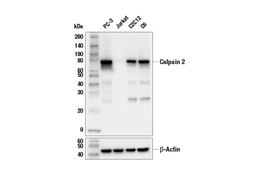
| Western Blotting | 1:1000 |
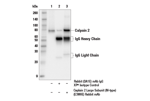
| Immunoprecipitation | 1:50 |
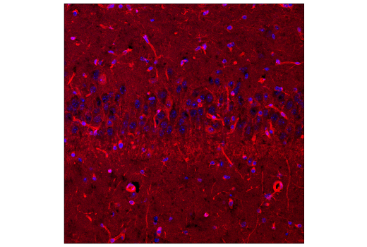
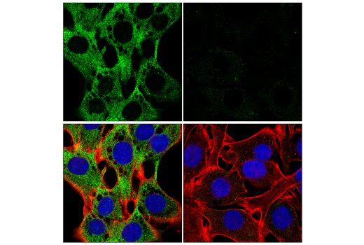
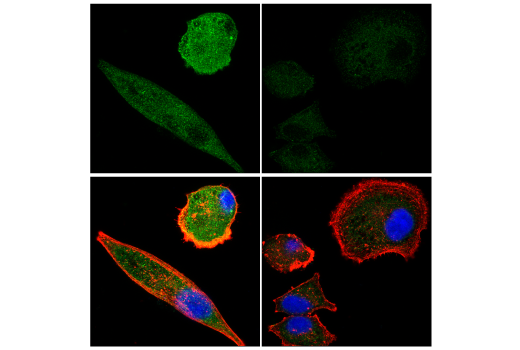
| Immunofluorescence (Frozen) | 1:50 - 1:100 |
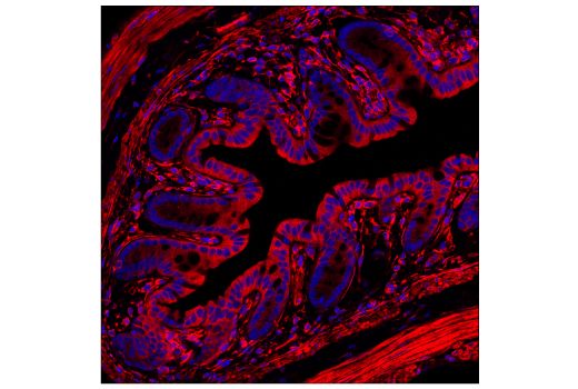
| Immunofluorescence (Immunocytochemistry) | 1:200 - 1:400 |
Storage
Specificity / Sensitivity
Species Reactivity:
Human, Mouse, Rat
Source / Purification
Monoclonal antibody is produced by immunizing animals with a synthetic peptide corresponding to residues near the amino terminus of human calpain 2 (large subunit) protein.
Background
Calpain is a calcium-dependent thiol proteinase that is functionally active as a heterodimer composed of a small regulatory subunit and one of at least two large catalytic subunits (calpain 1 or calpain 2). In vitro, calpain 1 (mu-calpain) requires micromolar levels of calcium, while calpain 2 (M-calpain) requires millimolar levels of calcium for activation. The regulation of calpain in vivo is the subject of many current studies, which suggest that proteolytic activity is regulated post-transcriptionally by mechanisms such as calcium requirements, subcellular localization of the heterodimer, phosphorylation via the EGFR-Erk signaling cascade, endogenous inhibitors (calpastatin), and autoproteolytic cleavage (1). Calpastatin negatively regulates autoproteolytic cleavage of calpain 1 between Gly27 and Leu28 (2). Calpain influences cell migration by modifying rather than degrading its substrates responsible for cell adhesion and cytoskeletal arrangement. Control of calpain activity has caught the attention of drug development since limiting its activity could mute invasiveness of tumors or chronic inflammation (1).
Species Reactivity
Species reactivity is determined by testing in at least one approved application (e.g., western blot).
Western Blot Buffer
IMPORTANT: For western blots, incubate membrane with diluted primary antibody in 5% w/v nonfat dry milk, 1X TBS, 0.1% Tween® 20 at 4°C with gentle shaking, overnight.
Applications Key
WB: Western Blotting IP: Immunoprecipitation IF-F: Immunofluorescence (Frozen) IF-IC: Immunofluorescence (Immunocytochemistry)
Cross-Reactivity Key
H: human M: mouse R: rat Hm: hamster Mk: monkey Vir: virus Mi: mink C: chicken Dm: D. melanogaster X: Xenopus Z: zebrafish B: bovine Dg: dog Pg: pig Sc: S. cerevisiae Ce: C. elegans Hr: horse GP: Guinea Pig Rab: rabbit All: all species expected
Trademarks and Patents
Limited Uses
Except as otherwise expressly agreed in a writing signed by a legally authorized representative of CST, the following terms apply to Products provided by CST, its affiliates or its distributors. Any Customer's terms and conditions that are in addition to, or different from, those contained herein, unless separately accepted in writing by a legally authorized representative of CST, are rejected and are of no force or effect.
Products are labeled with For Research Use Only or a similar labeling statement and have not been approved, cleared, or licensed by the FDA or other regulatory foreign or domestic entity, for any purpose. Customer shall not use any Product for any diagnostic or therapeutic purpose, or otherwise in any manner that conflicts with its labeling statement. Products sold or licensed by CST are provided for Customer as the end-user and solely for research and development uses. Any use of Product for diagnostic, prophylactic or therapeutic purposes, or any purchase of Product for resale (alone or as a component) or other commercial purpose, requires a separate license from CST. Customer shall (a) not sell, license, loan, donate or otherwise transfer or make available any Product to any third party, whether alone or in combination with other materials, or use the Products to manufacture any commercial products, (b) not copy, modify, reverse engineer, decompile, disassemble or otherwise attempt to discover the underlying structure or technology of the Products, or use the Products for the purpose of developing any products or services that would compete with CST products or services, (c) not alter or remove from the Products any trademarks, trade names, logos, patent or copyright notices or markings, (d) use the Products solely in accordance with CST Product Terms of Sale and any applicable documentation, and (e) comply with any license, terms of service or similar agreement with respect to any third party products or services used by Customer in connection with the Products.