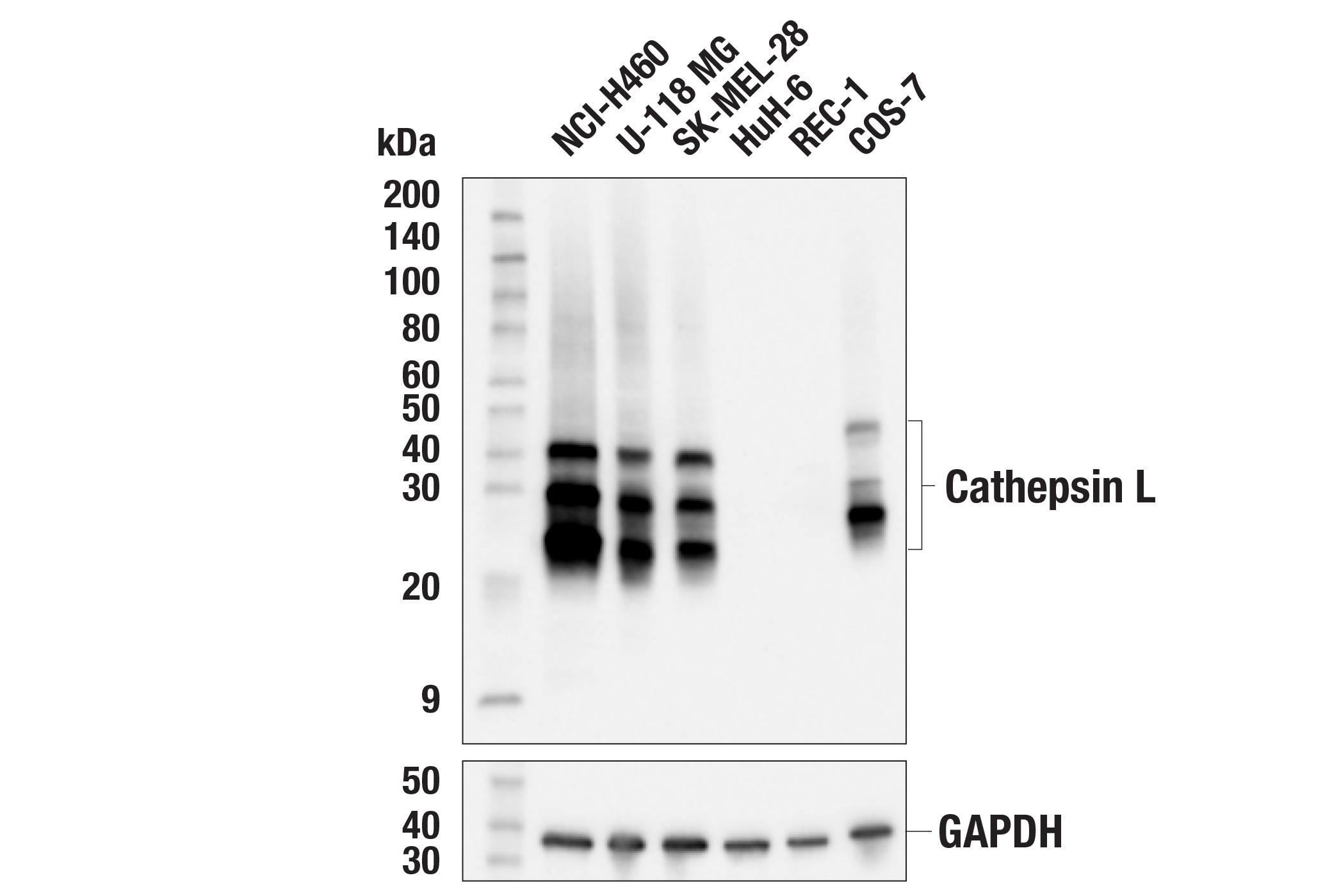
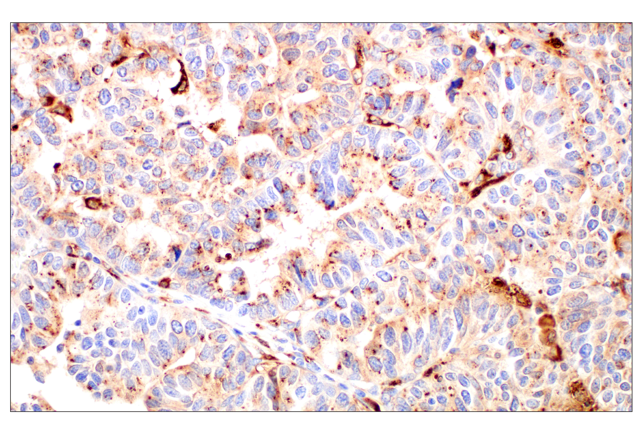
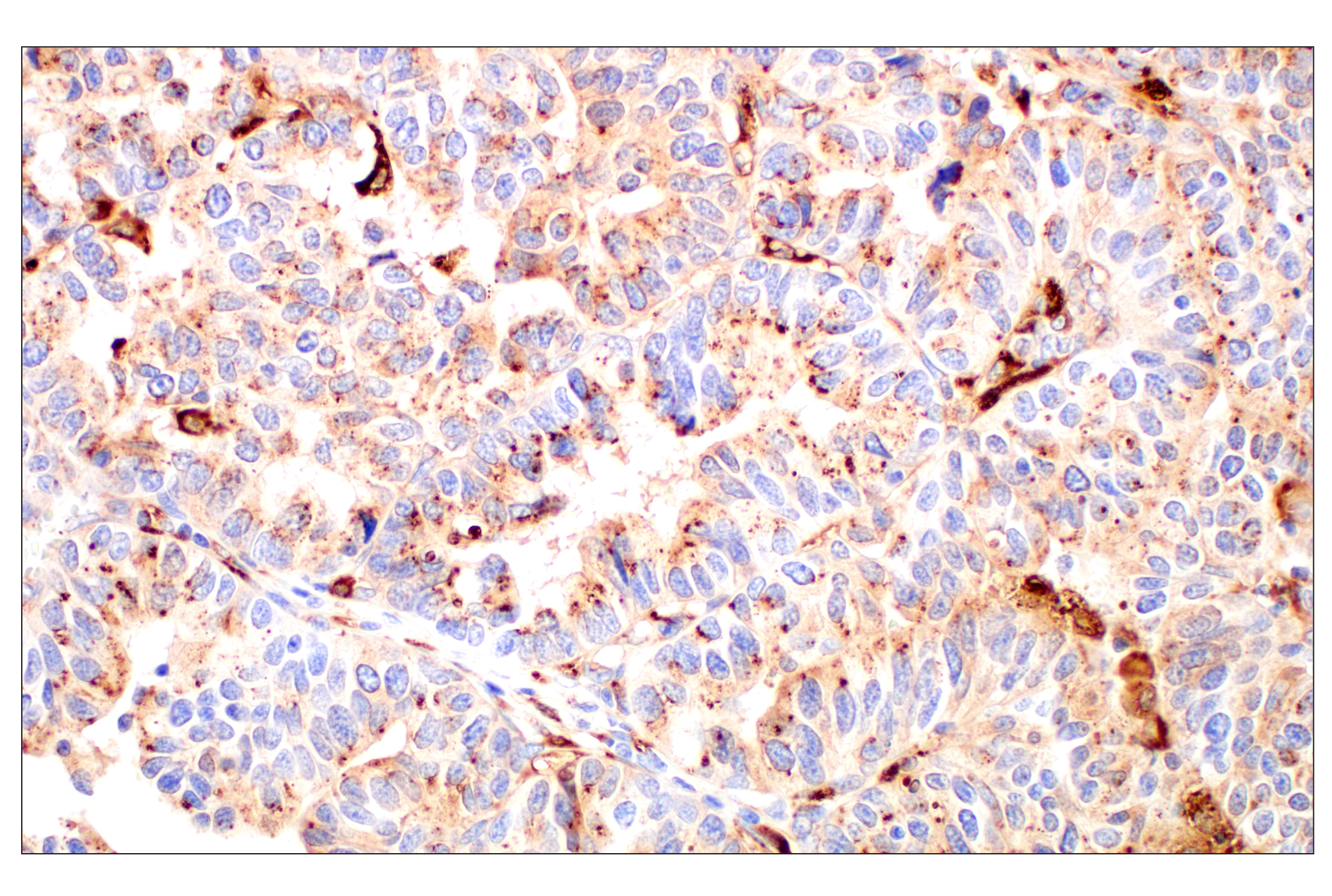
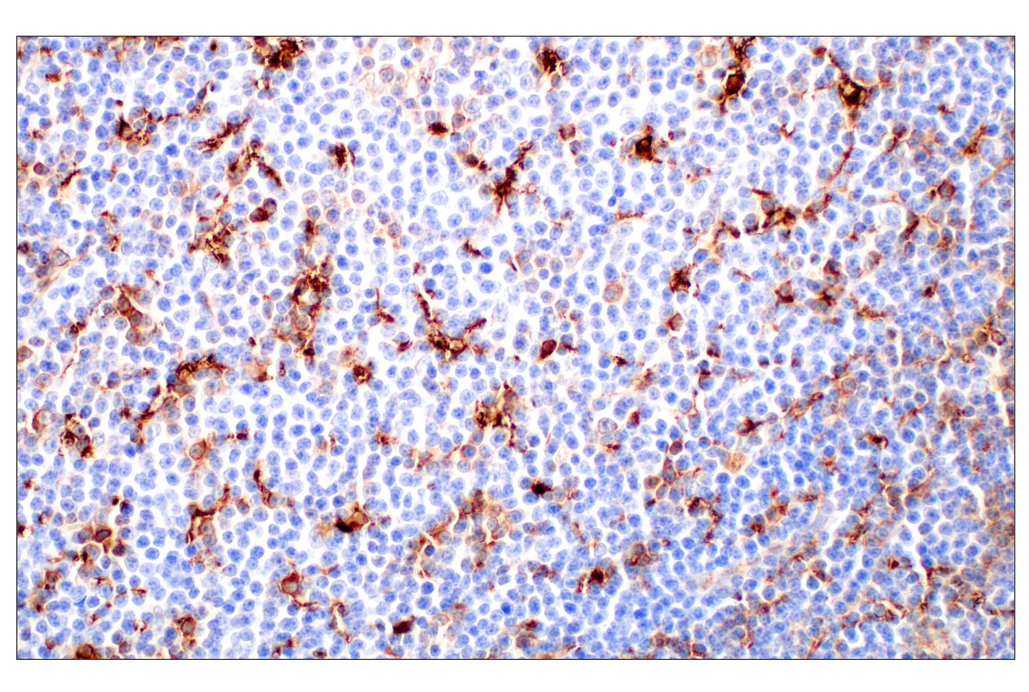
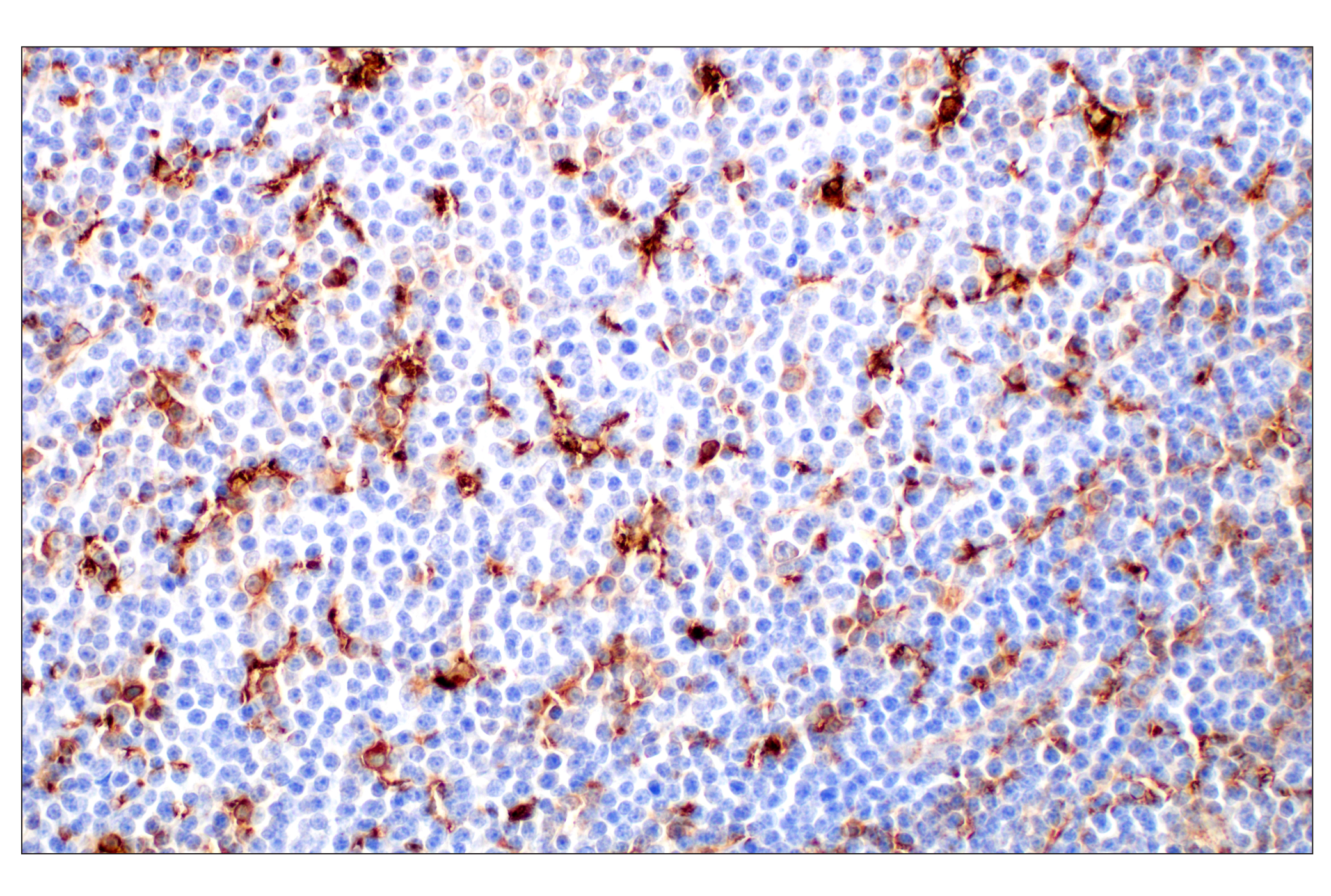
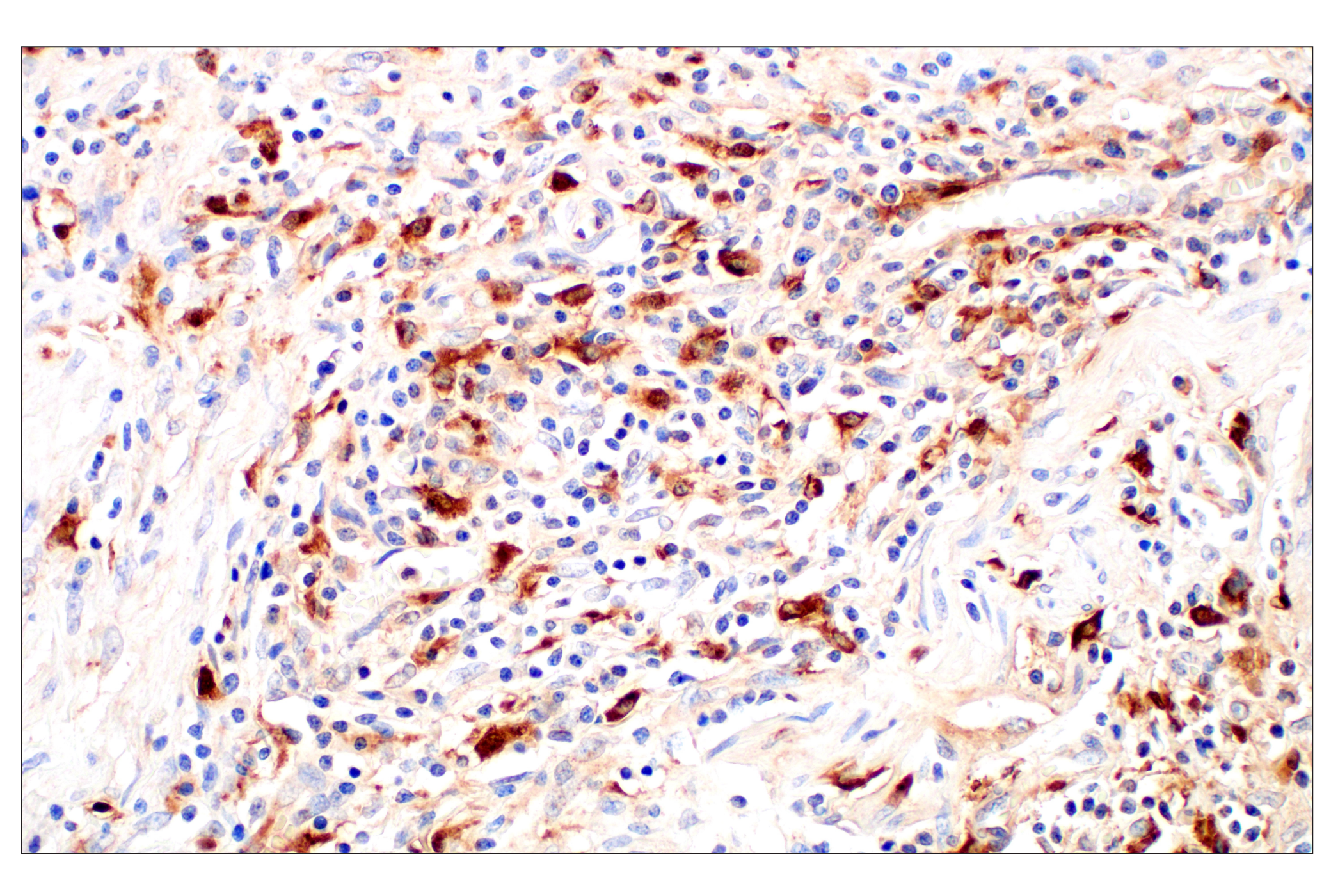
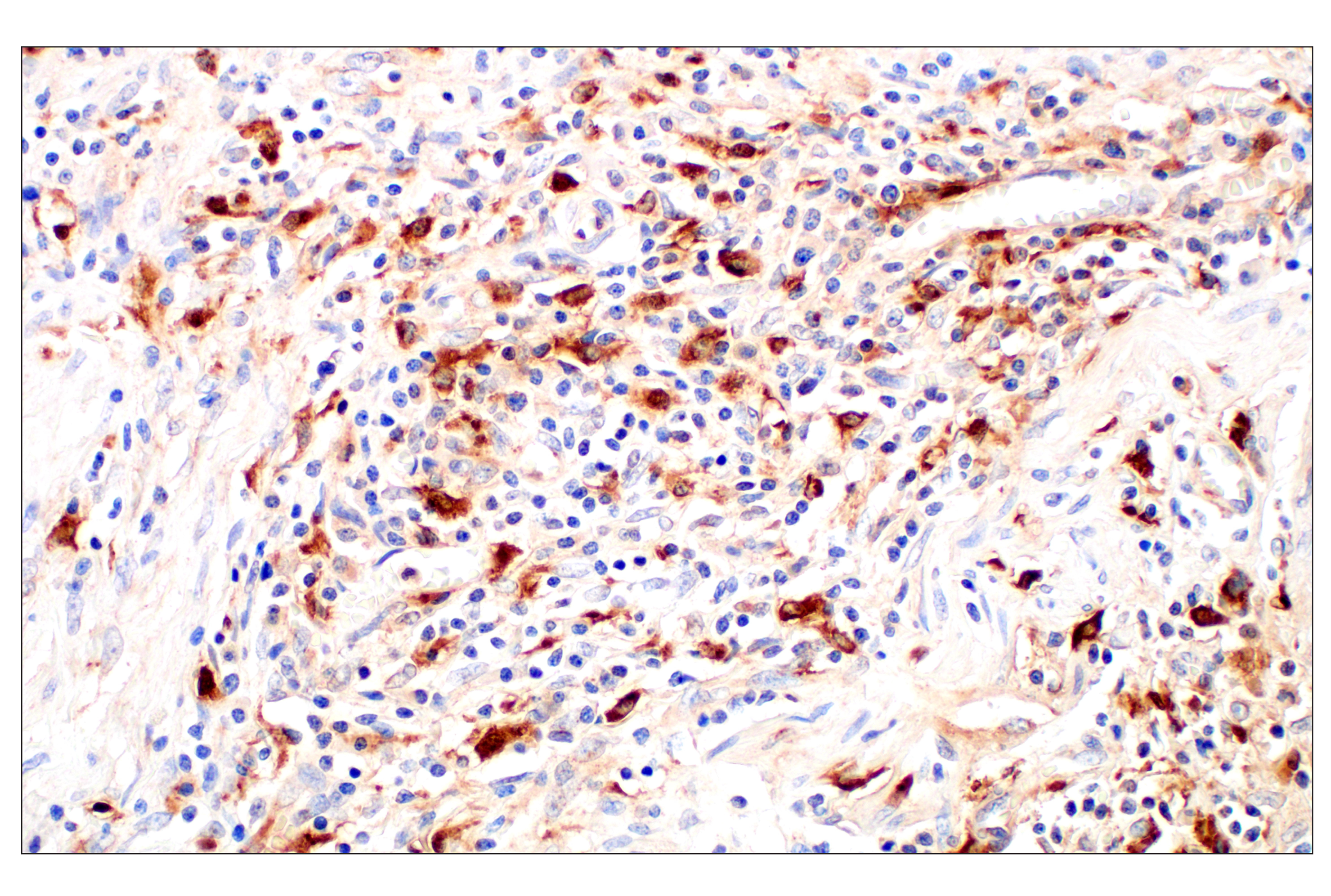
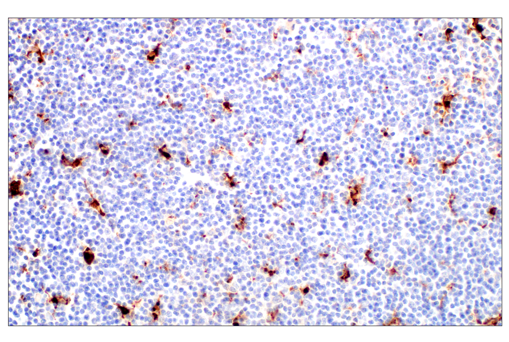
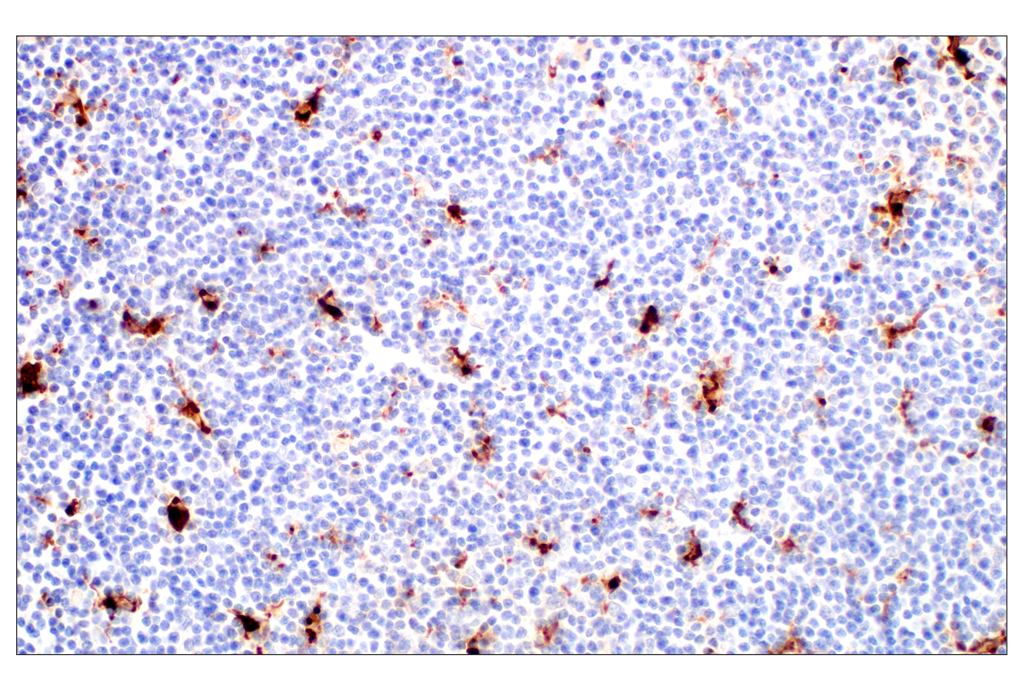
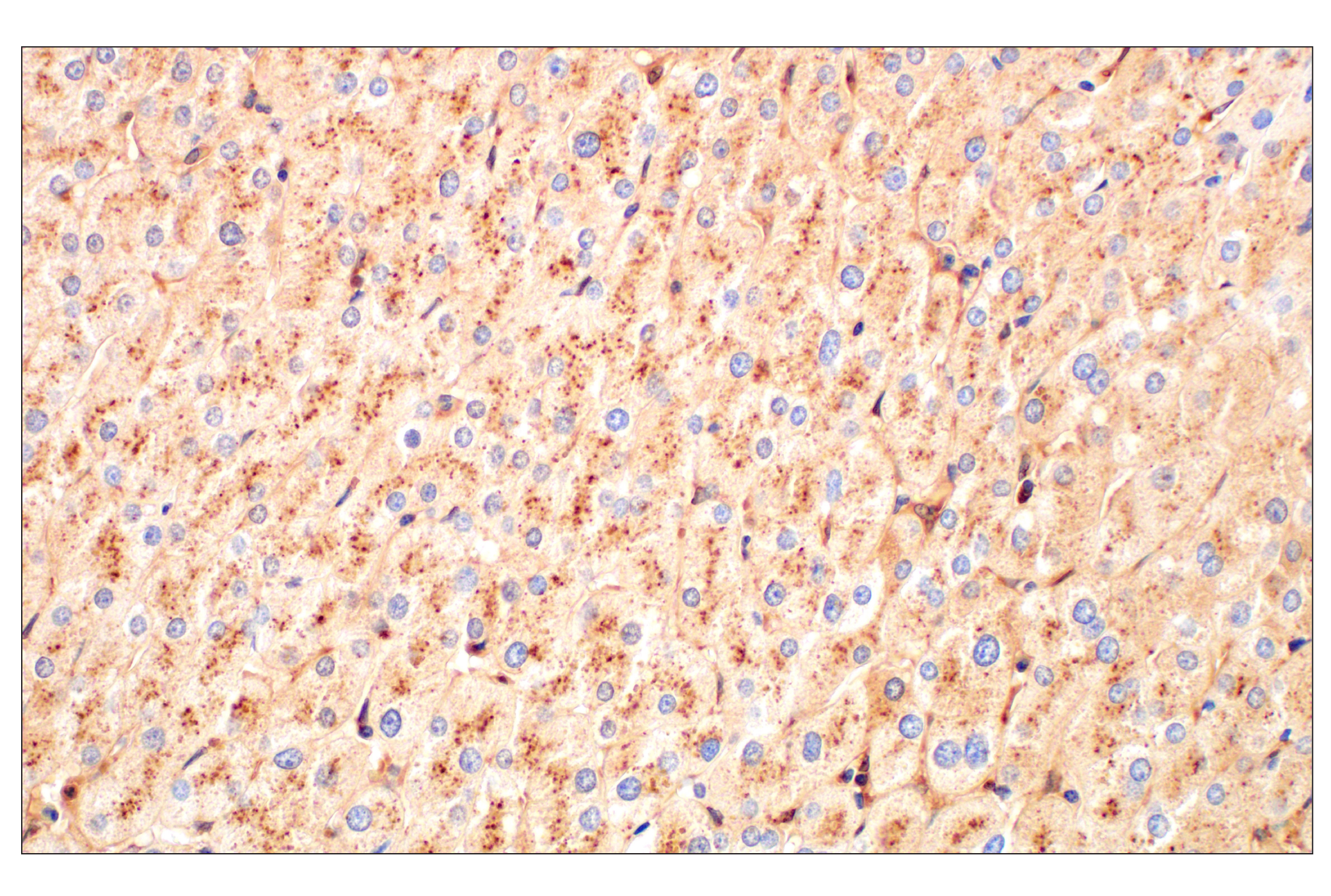
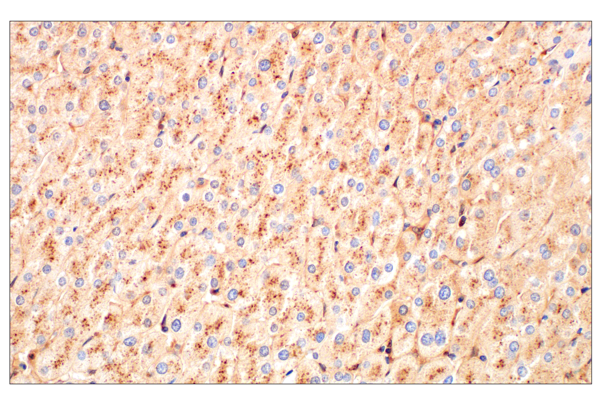
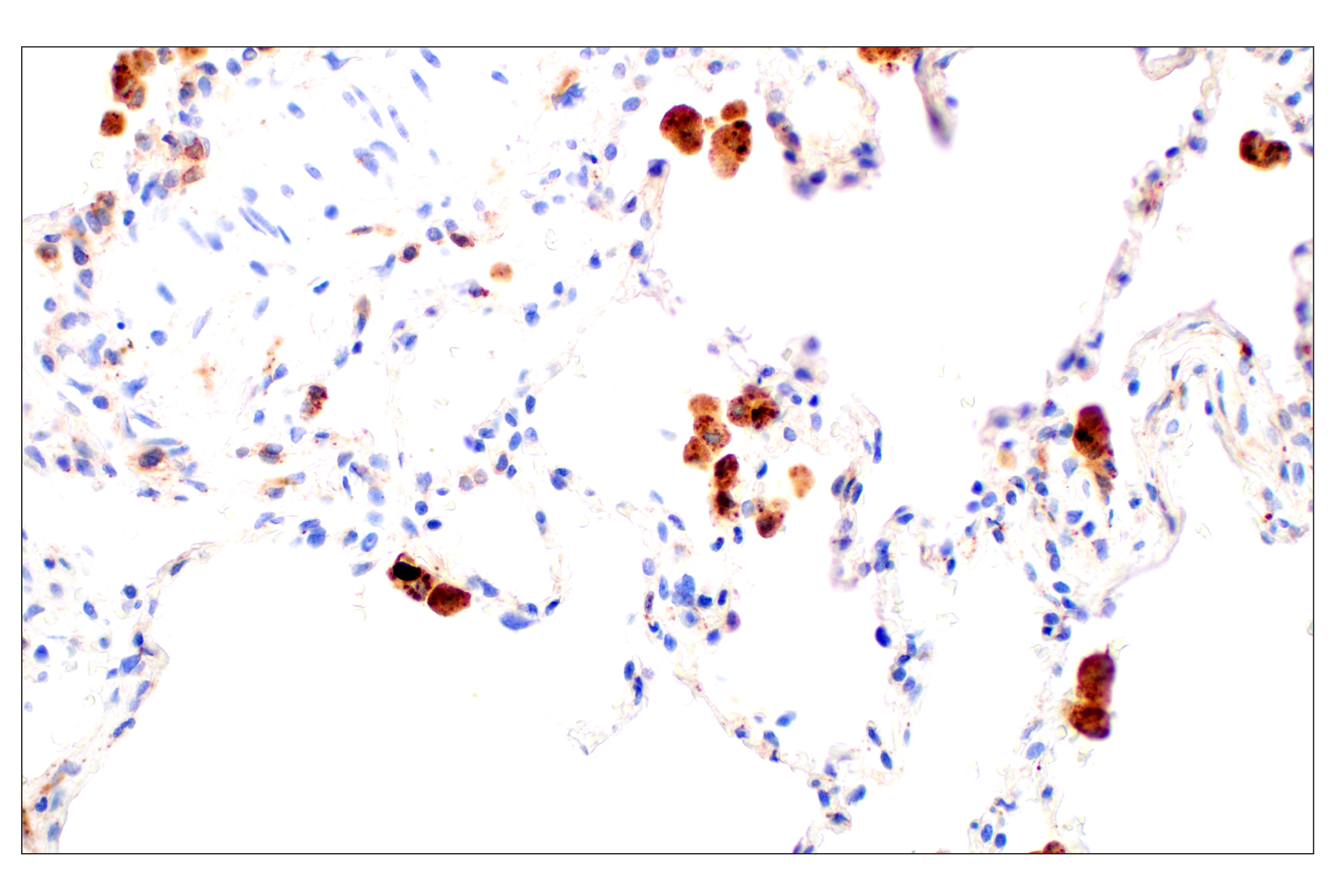
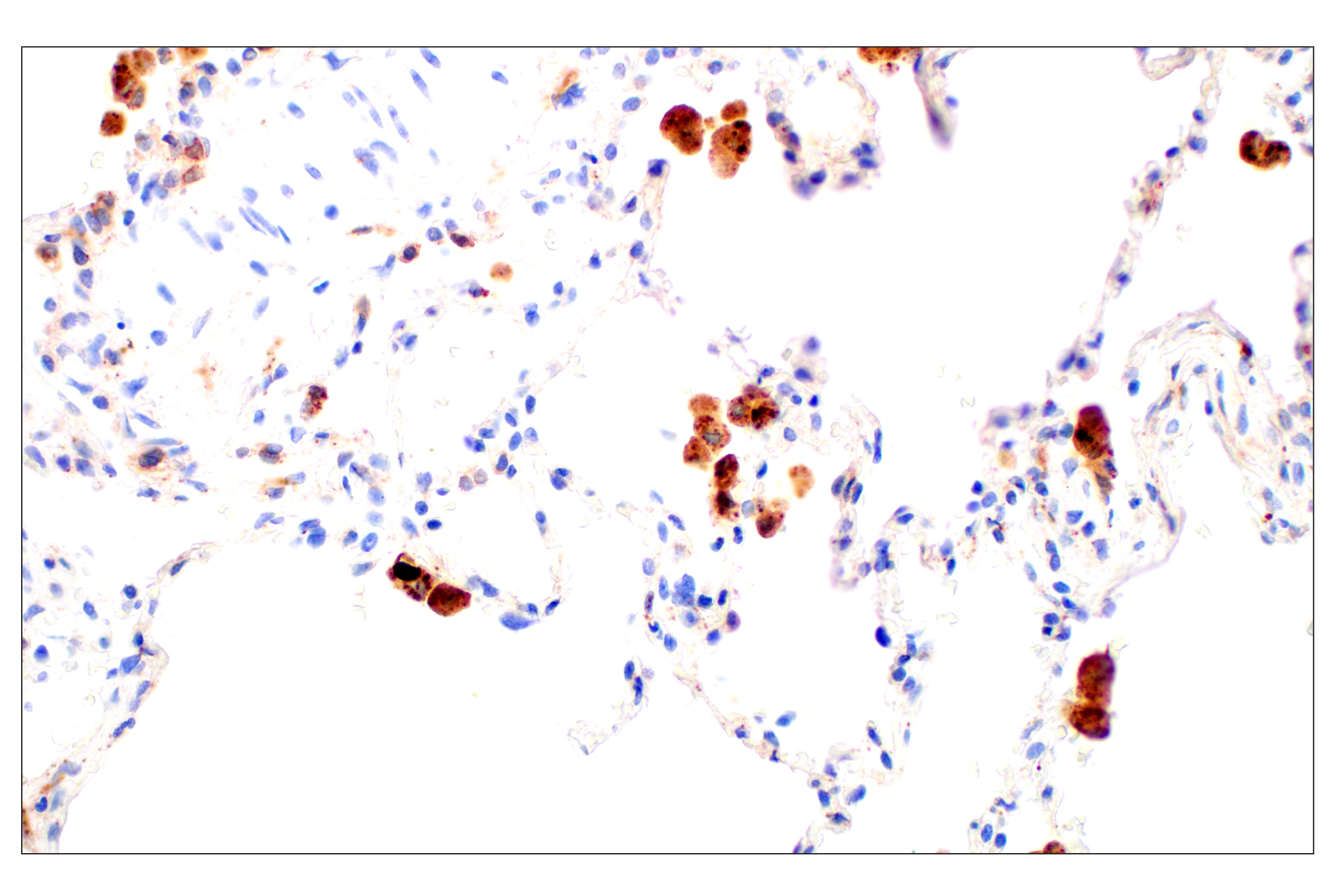
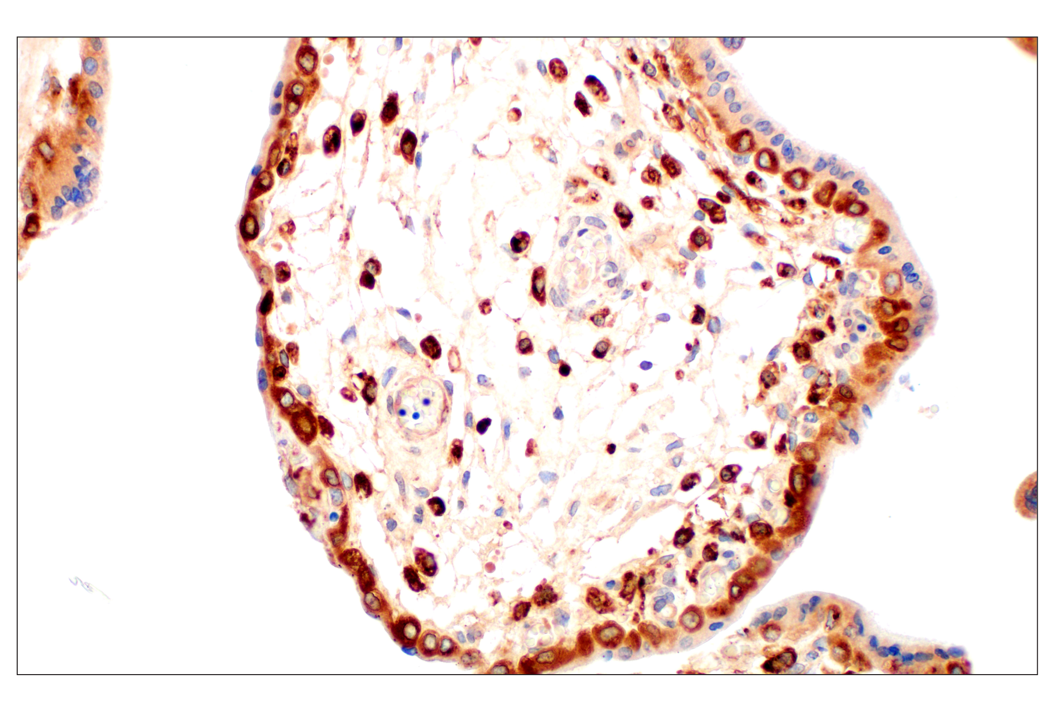
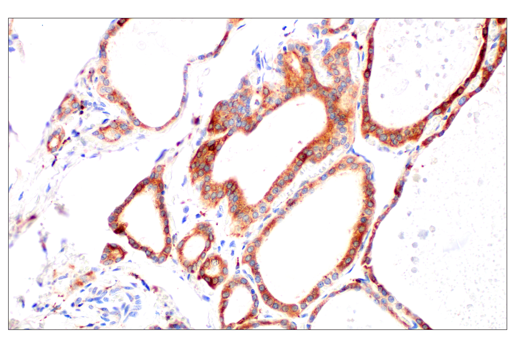
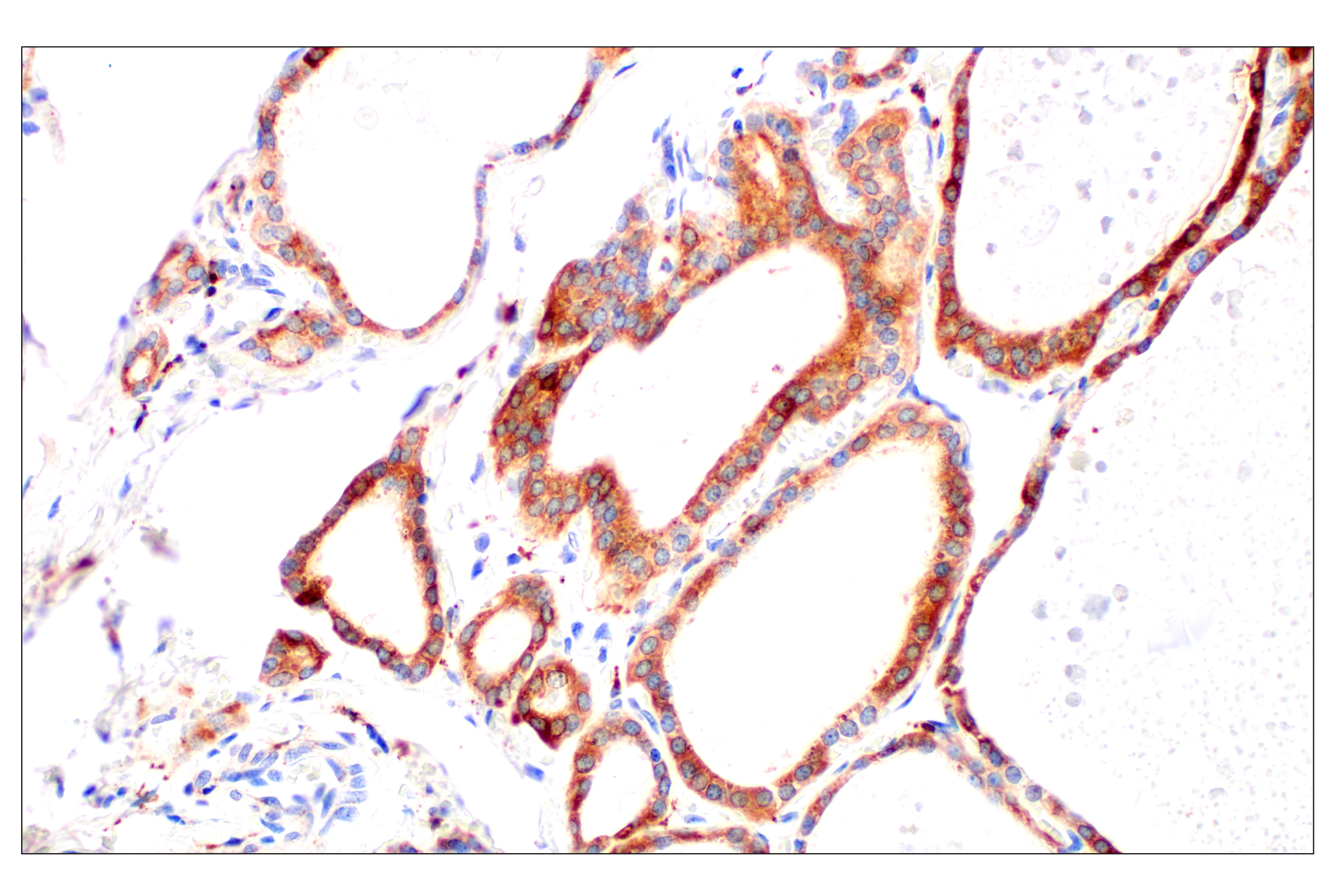
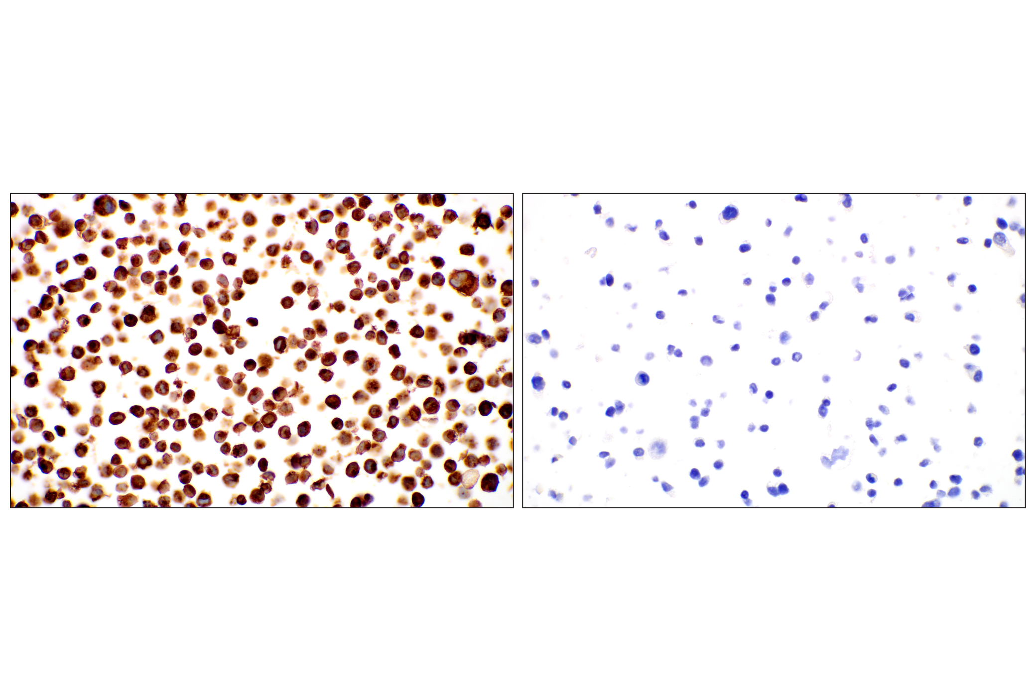
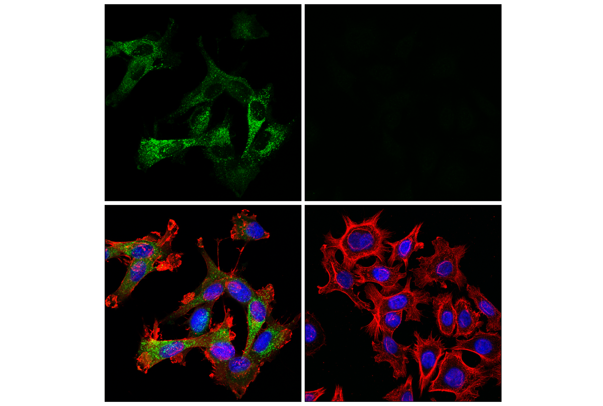
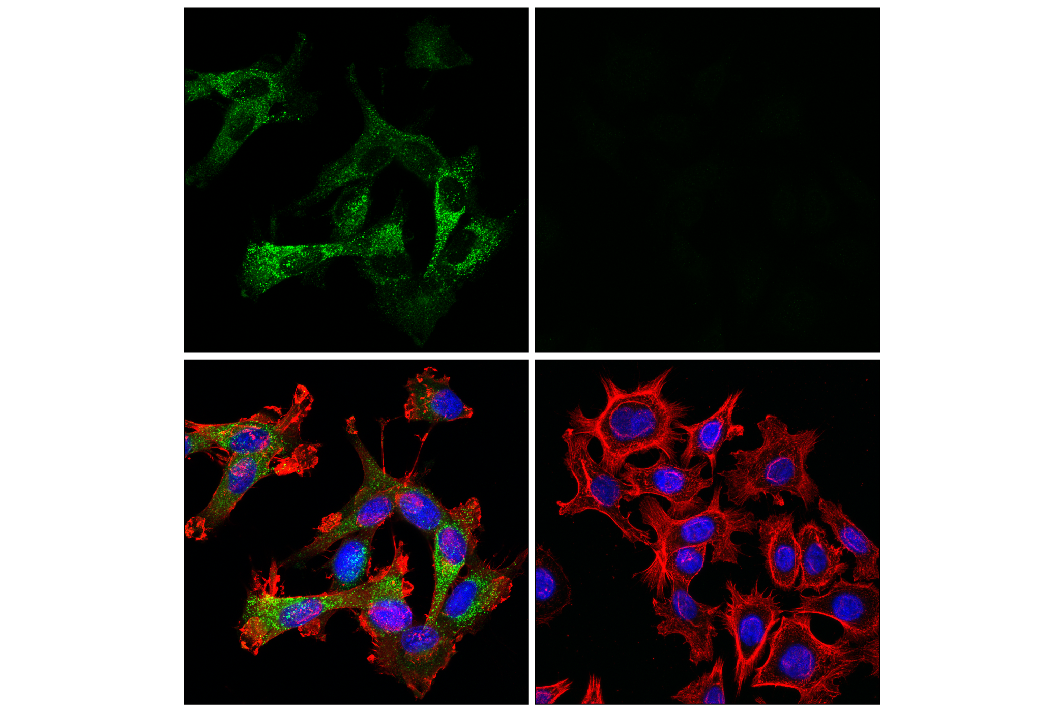
Revision 4

#55914Store at -20C

Cell Signaling Technology

Orders: 877-616-CELL (2355) [email protected]

Support: 877-678-TECH (8324)

Web: [email protected] cellsignal.com

3 Trask LaneDanversMassachusetts01923USA
For Research Use Only. Not for Use in Diagnostic Procedures.
Applications:

WB, IHC-P, IF-IC

REACTIVITY:

H Mk

SENSITIVITY:

Endogenous

MW (kDa):

25-42

Source/Isotype:

Rabbit IgG

UniProt ID:

#P07711

Entrez-Gene Id:

1514

Product Information

Product Usage Information

Application Dilution
Western Blotting 1:1000
Immunohistochemistry (Paraffin) 1:500 - 1:2000
Immunofluorescence (Immunocytochemistry) 1:50 - 1:200

Storage

Supplied in 10 mM sodium HEPES (pH 7.5), 150 mM NaCl, 100 µg/mL BSA, 50% glycerol, and less than 0.02% sodium azide. Store at –20°C. Do not aliquot the antibody.

For a carrier free (BSA and azide free) version of this product see product #74355.

Specificity / Sensitivity

Cathepsin L (E3R3P) Rabbit mAb recognizes endogenous levels of total cathepsin L protein. This antibody detects full-length cathepsin L pro-enzyme in addition to the processed (active) form of cathepsin L.

Species Reactivity:

Human, Monkey

Source / Purification

Monoclonal antibody is produced by immunizing animals with recombinant protein corresponding to a fragment of human cathepsin L protein spanning residues Ala114 to Thr288.

Background

Cathepsin L belongs to the C1 family of peptidases, a large and diverse family of cysteine (thiol) proteases that play fundamental roles in protein processing and degradation. The catalytic activity of C1 peptidases maps to a nucleophilic cysteine thiol, usually found within a catalytic dyad or triad (1). Cathepsin L, initially synthesized as procathepsin L before undergoing proteolytic cleavage to its active form, is a ubiquitously expressed thiol protease, expressed primarily in lysosomes but which is also secreted by some cell types. Its diverse functions include general protein catabolism and autophagy (2), antigen processing (3), promoting angiogenesis (4), and extracellular matrix degradation (5,6). An isoform of cathepsin L has also been reported in the nucleus, where it was shown to be involved in the regulation of transcription factor activity (7). Targeted deletion of the Ctsl gene in mice embryos was non-lethal, but resulted in periodic hair loss and skin defects, including epidermal hyperplasia, acanthosis, and hyperkeratosis (8). More recently, research studies have reported that cathepsin L is involved in viral protein processing during viral replication, and that cathepsin L inhibitors may have utility in combating infection by SARS-CoV-2 and related coronaviruses (9).

  1. Rawlings, N.D. et al. (2018) Nucleic Acids Res 46, D624-D632.
  2. Kaminskyy, V. and Zhivotovsky, B. (2012) Biochim Biophys Acta 1824, 44-50.
  3. Nakagawa, T. et al. (1998) Science 280, 450-3.
  4. Pan, T. et al. (2020) Gastric Cancer 23, 974-987.
  5. Dykes, S.S. et al. (2019) Oncotarget 10, 5560-5568.
  6. Felbor, U. et al. (2000) EMBO J 19, 1187-94.
  7. Burton, L.J. et al. (2017) Mol Cell Biol 37, e00297-16. doi: 10.1128/MCB.00297-16.
  8. Roth, W. et al. (2000) FASEB J 14, 2075-86.
  9. Zhao, M.M. et al. (2021) Signal Transduct Target Ther 6, 134.

Species Reactivity

Species reactivity is determined by testing in at least one approved application (e.g., western blot).

Western Blot Buffer

IMPORTANT: For western blots, incubate membrane with diluted primary antibody in 5% w/v BSA, 1X TBS, 0.1% Tween® 20 at 4°C with gentle shaking, overnight.

Applications Key

WB: Western Blotting IHC-P: Immunohistochemistry (Paraffin) IF-IC: Immunofluorescence (Immunocytochemistry)

Cross-Reactivity Key

H: human M: mouse R: rat Hm: hamster Mk: monkey Vir: virus Mi: mink C: chicken Dm: D. melanogaster X: Xenopus Z: zebrafish B: bovine Dg: dog Pg: pig Sc: S. cerevisiae Ce: C. elegans Hr: horse GP: Guinea Pig Rab: rabbit All: all species expected

Trademarks and Patents

Cell Signaling Technology is a trademark of Cell Signaling Technology, Inc.
KARPAS cell line source: Dr. Abraham Karpas at the University of Cambridge.
All other trademarks are the property of their respective owners. Visit cellsignal.com/trademarks for more information.

Limited Uses

Except as otherwise expressly agreed in a writing signed by a legally authorized representative of CST, the following terms apply to Products provided by CST, its affiliates or its distributors. Any Customer's terms and conditions that are in addition to, or different from, those contained herein, unless separately accepted in writing by a legally authorized representative of CST, are rejected and are of no force or effect.

Products are labeled with For Research Use Only or a similar labeling statement and have not been approved, cleared, or licensed by the FDA or other regulatory foreign or domestic entity, for any purpose. Customer shall not use any Product for any diagnostic or therapeutic purpose, or otherwise in any manner that conflicts with its labeling statement. Products sold or licensed by CST are provided for Customer as the end-user and solely for research and development uses. Any use of Product for diagnostic, prophylactic or therapeutic purposes, or any purchase of Product for resale (alone or as a component) or other commercial purpose, requires a separate license from CST. Customer shall (a) not sell, license, loan, donate or otherwise transfer or make available any Product to any third party, whether alone or in combination with other materials, or use the Products to manufacture any commercial products, (b) not copy, modify, reverse engineer, decompile, disassemble or otherwise attempt to discover the underlying structure or technology of the Products, or use the Products for the purpose of developing any products or services that would compete with CST products or services, (c) not alter or remove from the Products any trademarks, trade names, logos, patent or copyright notices or markings, (d) use the Products solely in accordance with CST Product Terms of Sale and any applicable documentation, and (e) comply with any license, terms of service or similar agreement with respect to any third party products or services used by Customer in connection with the Products.

Revision 4
#55914

Cathepsin L (E3R3P) Rabbit mAb

Western Blotting Image 1: Cathepsin L (E3R3P) Rabbit mAb Expand Image
Western blot analysis of extracts from various cell lines using Cathepsin L (E3R3P) Rabbit mAb (upper) or GAPDH (D16H11) XP® Rabbit mAb #5174 (lower). Negative expression of cathepsin L protein in HuH-6 and REC-1 cells is consistent with the predicted expression pattern.
Immunohistochemistry Image 1: Cathepsin L (E3R3P) Rabbit mAb Expand Image
Immunohistochemical analysis of paraffin-embedded human urothelial carcinoma using Cathepsin L (E3R3P) Rabbit mAb.
Immunohistochemistry Image 2: Cathepsin L (E3R3P) Rabbit mAb Expand Image
Immunohistochemical analysis of paraffin-embedded human urothelial carcinoma using Cathepsin L (E3R3P) Rabbit mAb.
Immunohistochemistry Image 3: Cathepsin L (E3R3P) Rabbit mAb Expand Image
Immunohistochemical analysis of paraffin-embedded human B-cell non-Hodgkin lymphoma using Cathepsin L (E3R3P) Rabbit mAb.
Immunohistochemistry Image 4: Cathepsin L (E3R3P) Rabbit mAb Expand Image
Immunohistochemical analysis of paraffin-embedded human B-cell non-Hodgkin lymphoma using Cathepsin L (E3R3P) Rabbit mAb.
Immunohistochemistry Image 5: Cathepsin L (E3R3P) Rabbit mAb Expand Image
Immunohistochemical analysis of paraffin-embedded human colon adenocarcinoma using Cathepsin L (E3R3P) Rabbit mAb.
Immunohistochemistry Image 6: Cathepsin L (E3R3P) Rabbit mAb Expand Image
Immunohistochemical analysis of paraffin-embedded human colon adenocarcinoma using Cathepsin L (E3R3P) Rabbit mAb.
Immunohistochemistry Image 7: Cathepsin L (E3R3P) Rabbit mAb Expand Image
Immunohistochemical analysis of paraffin-embedded human follicular hyperplasia of the tonsil using Cathepsin L (E3R3P) Rabbit mAb.
Immunohistochemistry Image 8: Cathepsin L (E3R3P) Rabbit mAb Expand Image
Immunohistochemical analysis of paraffin-embedded human follicular hyperplasia of the tonsil using Cathepsin L (E3R3P) Rabbit mAb.
Immunohistochemistry Image 9: Cathepsin L (E3R3P) Rabbit mAb Expand Image
Immunohistochemical analysis of paraffin-embedded normal human liver using Cathepsin L (E3R3P) Rabbit mAb.
Immunohistochemistry Image 10: Cathepsin L (E3R3P) Rabbit mAb Expand Image
Immunohistochemical analysis of paraffin-embedded normal human liver using Cathepsin L (E3R3P) Rabbit mAb.
Immunohistochemistry Image 11: Cathepsin L (E3R3P) Rabbit mAb Expand Image
Immunohistochemical analysis of paraffin-embedded normal human lung using Cathepsin L (E3R3P) Rabbit mAb.
Immunohistochemistry Image 12: Cathepsin L (E3R3P) Rabbit mAb Expand Image
Immunohistochemical analysis of paraffin-embedded normal human lung using Cathepsin L (E3R3P) Rabbit mAb.
Immunohistochemistry Image 13: Cathepsin L (E3R3P) Rabbit mAb Expand Image
Immunohistochemical analysis of paraffin-embedded normal human placenta using Cathepsin L (E3R3P) Rabbit mAb.
Immunohistochemistry Image 14: Cathepsin L (E3R3P) Rabbit mAb Expand Image
Immunohistochemical analysis of paraffin-embedded normal human placenta using Cathepsin L (E3R3P) Rabbit mAb.
Immunohistochemistry Image 15: Cathepsin L (E3R3P) Rabbit mAb Expand Image
Immunohistochemical analysis of paraffin-embedded normal human thyroid using Cathepsin L (E3R3P) Rabbit mAb.
Immunohistochemistry Image 16: Cathepsin L (E3R3P) Rabbit mAb Expand Image
Immunohistochemical analysis of paraffin-embedded normal human thyroid using Cathepsin L (E3R3P) Rabbit mAb.
Immunohistochemistry Image 17: Cathepsin L (E3R3P) Rabbit mAb Expand Image
Immunohistochemical analysis of paraffin-embedded KARPAS 299 cell pellet (left, positive) or AGS cell pellet (right, negative) using Cathepsin L (E3R3P) Rabbit mAb.
Immunohistochemistry Image 18: Cathepsin L (E3R3P) Rabbit mAb Expand Image
Immunohistochemical analysis of paraffin-embedded Karpas-299 cell pellet (left, positive) or AGS cell pellet (right, negative) using Cathepsin L (E3R3P) Rabbit mAb.
Immunofluorescence Image 1: Cathepsin L (E3R3P) Rabbit mAb Expand Image
Confocal immunofluorescent analysis of U-118 MG cells (left, positive) or AGS cells (right, negative) using Cathepsin L (E3R3P) Rabbit mAb (green), β-Actin (8H10D10) Mouse mAb #3700 (red), and DAPI #4083 (blue).
Immunofluorescence Image 2: Cathepsin L (E3R3P) Rabbit mAb Expand Image
Confocal immunofluorescent analysis of U-118 MG cells (left, positive) or AGS cells (right, negative) using Cathepsin L (E3R3P) Rabbit mAb (green), β-Actin (8H10D10) Mouse mAb #3700 (red), and DAPI #4083 (blue).