WB, IP
H M R
Endogenous
22
Rabbit IgG
#O00299
1192
Product Information
Product Usage Information
| Application | Dilution |
|---|---|
| Western Blotting | 1:1000 |
| Immunoprecipitation | 1:50 |
Storage
Specificity / Sensitivity
Species Reactivity:
Human, Mouse, Rat
Source / Purification
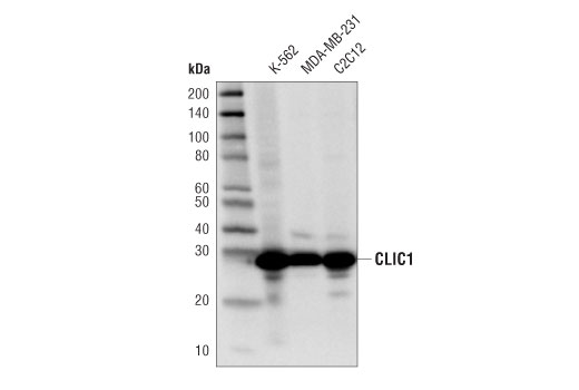
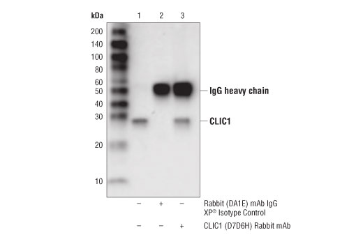
Monoclonal antibody is produced by immunizing animals with a synthetic peptide corresponding to residues surrounding Glu234 of human CLIC1 protein.
Background
Chloride intracellular channel (CLIC) proteins belong to a family of highly conserved transport proteins found as both soluble and membrane-bound forms (1). Although CLIC proteins have putative, selective chloride ion channel activity, they are structural homologs to members of the glutathione-S-transferase protein superfamily and are likewise regulated by redox status (2). CLIC proteins are distinct from other ion channels in that they are found as both soluble and integral membrane forms, and their form determines their function (3-6). Chloride intracellular channel proteins are ubiquitously expressed in numerous tissue types and are involved in diverse biological functions (1,2).
CLIC1 is a member of the CLIC protein family. It is ubiquitously expressed in many tissues and organs (7). CLIC1 is overexpressed in multiple tumor types and has been implicated in the proliferation, migration, and invasion of these tumors (8-11). In the central nervous system, CLIC1 protein expression is elevated upon amyloid β-peptide treatment in neonatal rat microglia. Inhibition of CLIC1 prevents neuronal apoptosis in neurons co-cultured with amyloid β-peptide treated microglia (12). Further studies indicate that CLIC1 translocates from the cytosol to the plasma membrane of microglia upon exposure to amyloid β-peptide, and contributes to the subsequent neurotoxicity through generation of superoxide anions (13). These discoveries implicate CLIC1 as a possible therapeutic target for Alzheimer’s disease.
- Littler, D.R. et al. (2010) FEBS Lett 584, 2093-101.
- Oakley, A.J. (2005) Curr Opin Struct Biol 15, 716-23.
- Littler, D.R. et al. (2005) FEBS J 272, 4996-5007.
- Singh, H. and Ashley, R.H. (2006) Biophys J 90, 1628-38.
- Suh, K.S. et al. (2004) J Biol Chem 279, 4632-41.
- Fernández-Salas, E. et al. (2002) Mol Cell Biol 22, 3610-20.
- Ulmasov, B. et al. (2007) BMC Cell Biol 8, 8.
- Wei, X. et al. (2015) J Gastroenterol Hepatol 30, 208-16.
- Gurski, L.A. et al. (2015) Mol Cancer Res 13, 273-80.
- Ding, Q. et al. (2015) Tumour Biol 36, 193-8.
- Tian, Y. et al. (2014) Cancer Biother Radiopharm 29, 339-44.
- Novarino, G. et al. (2004) J Neurosci 24, 5322-30.
- Milton, R.H. et al. (2008) J Neurosci 28, 11488-99.
Species Reactivity
Species reactivity is determined by testing in at least one approved application (e.g., western blot).
Western Blot Buffer
IMPORTANT: For western blots, incubate membrane with diluted primary antibody in 5% w/v BSA, 1X TBS, 0.1% Tween® 20 at 4°C with gentle shaking, overnight.
Applications Key
WB: Western Blotting IP: Immunoprecipitation
Cross-Reactivity Key
H: human M: mouse R: rat Hm: hamster Mk: monkey Vir: virus Mi: mink C: chicken Dm: D. melanogaster X: Xenopus Z: zebrafish B: bovine Dg: dog Pg: pig Sc: S. cerevisiae Ce: C. elegans Hr: horse GP: Guinea Pig Rab: rabbit All: all species expected
Trademarks and Patents
Limited Uses
Except as otherwise expressly agreed in a writing signed by a legally authorized representative of CST, the following terms apply to Products provided by CST, its affiliates or its distributors. Any Customer's terms and conditions that are in addition to, or different from, those contained herein, unless separately accepted in writing by a legally authorized representative of CST, are rejected and are of no force or effect.
Products are labeled with For Research Use Only or a similar labeling statement and have not been approved, cleared, or licensed by the FDA or other regulatory foreign or domestic entity, for any purpose. Customer shall not use any Product for any diagnostic or therapeutic purpose, or otherwise in any manner that conflicts with its labeling statement. Products sold or licensed by CST are provided for Customer as the end-user and solely for research and development uses. Any use of Product for diagnostic, prophylactic or therapeutic purposes, or any purchase of Product for resale (alone or as a component) or other commercial purpose, requires a separate license from CST. Customer shall (a) not sell, license, loan, donate or otherwise transfer or make available any Product to any third party, whether alone or in combination with other materials, or use the Products to manufacture any commercial products, (b) not copy, modify, reverse engineer, decompile, disassemble or otherwise attempt to discover the underlying structure or technology of the Products, or use the Products for the purpose of developing any products or services that would compete with CST products or services, (c) not alter or remove from the Products any trademarks, trade names, logos, patent or copyright notices or markings, (d) use the Products solely in accordance with CST Product Terms of Sale and any applicable documentation, and (e) comply with any license, terms of service or similar agreement with respect to any third party products or services used by Customer in connection with the Products.