| Product Includes | Product # | Quantity | Mol. Wt | Isotype/Source |
|---|---|---|---|---|
| ACE2 Antibody | 4355 | 20 µl | 120-135 kDa | Rabbit |
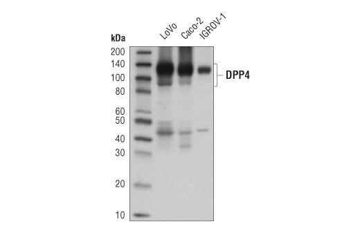
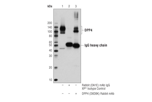
| DPP4/CD26 (D6D8K) Rabbit mAb | 67138 | 20 µl | 90, 120 kDa | Rabbit IgG |
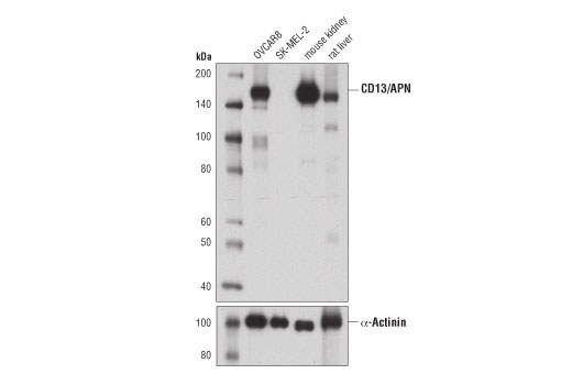
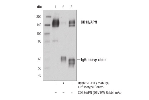
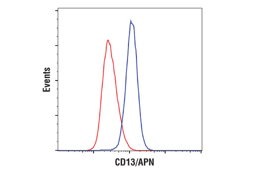
| CD13/APN (D6V1W) Rabbit mAb | 32720 | 20 µl | 160 kDa | Rabbit IgG |
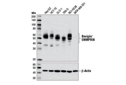
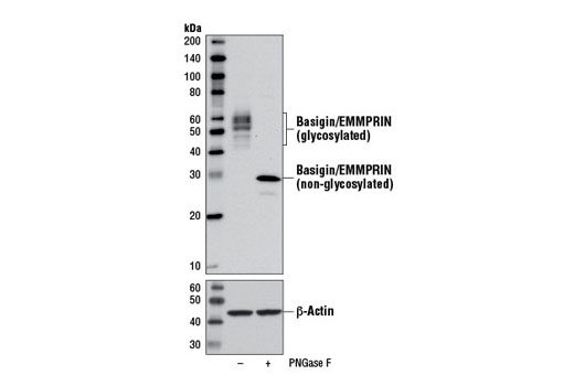
| Basigin/EMMPRIN (E1S1V) Rabbit mAb | 13287 | 20 µl | 38-58 kDa | Rabbit IgG |
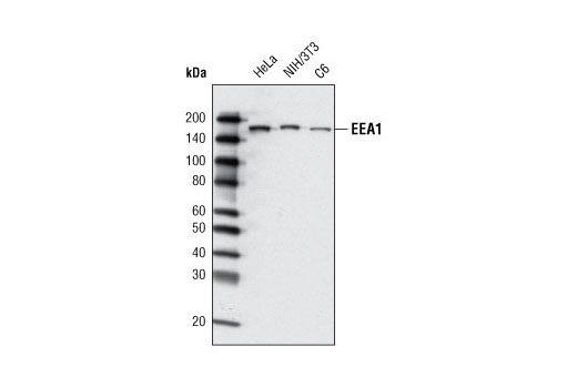
| EEA1 (C45B10) Rabbit mAb | 3288 | 20 µl | 170 kDa | Rabbit IgG |
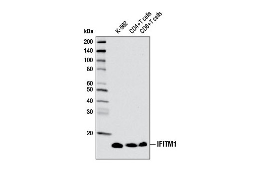
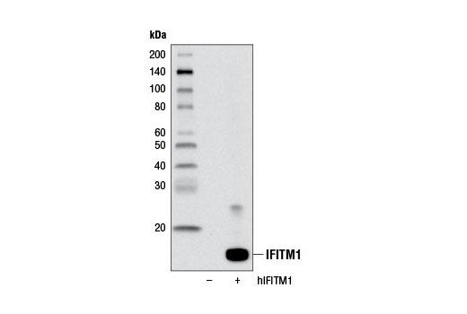
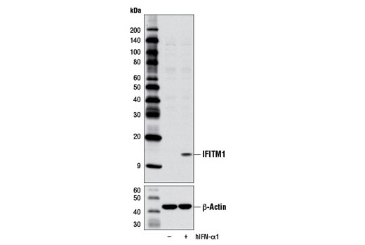
| IFITM1 Antibody | 13126 | 20 µl | 14 kDa | Rabbit |
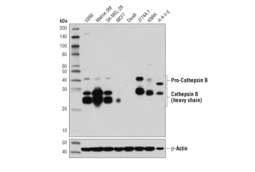
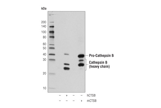
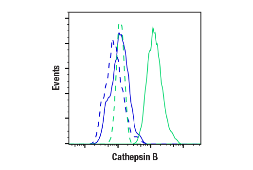
| Cathepsin B (D1C7Y) XP® Rabbit mAb | 31718 | 20 µl | 44, 27, 24 kDa | Rabbit IgG |
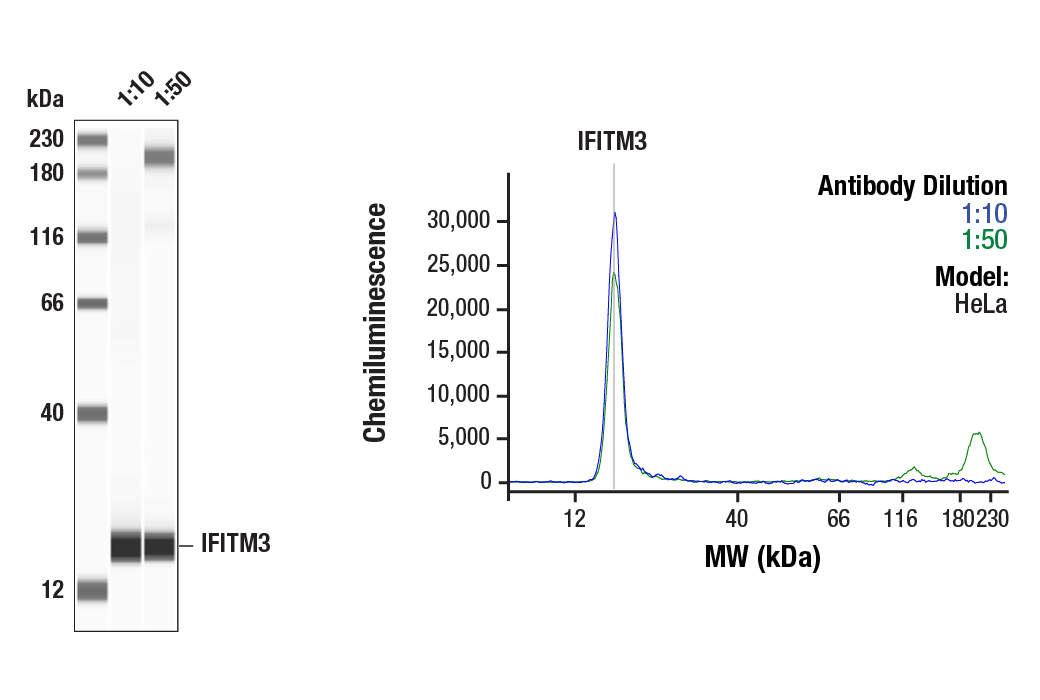
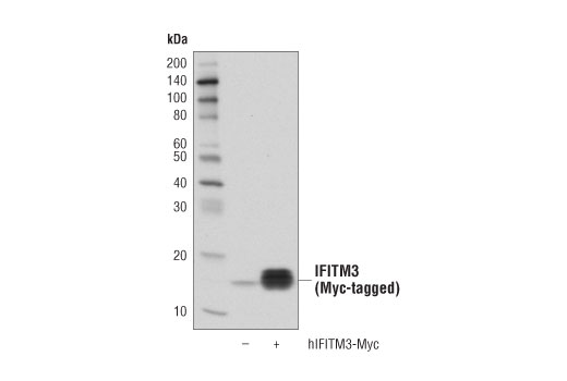
| IFITM3 (D8E8G) XP® Rabbit mAb | 59212 | 20 µl | 15 kDa | Rabbit IgG |
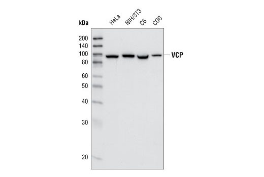
| VCP (7F3) Rabbit mAb | 2649 | 20 µl | 89 kDa | Rabbit IgG |
| Anti-rabbit IgG, HRP-linked Antibody | 7074 | 100 µl | Goat |
Please visit cellsignal.com for individual component applications, species cross-reactivity, dilutions, protocols, and additional product information.
Description
The Coronavirus Host Cell Attachment and Entry Antibody Sampler Kit provides an economical means of detecting key host cell proteins involved in the attachment and cellular entry of coronaviruses. The kit includes enough antibodies to perform two western blot experiments with each primary antibody.
Storage
Background
Coronaviruses are a group of viruses that contain single-stranded, positive-sense RNA genomes. Several members of this group, which include severe acute respiratory syndrome coronaviruses (SARS-CoV and SARS-CoV-2) and Middle East respiratory syndrome coronavirus (MERS-CoV), are highly pathogenic and have caused significant disease outbreaks in human hosts. In order for human coronaviruses to transcribe and replicate their genomes within host cells, they must first attach and gain entry into host cells using a variety of cell surface receptors and components of the endocytic machinery.
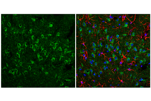
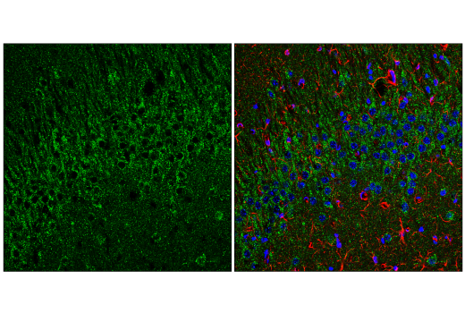
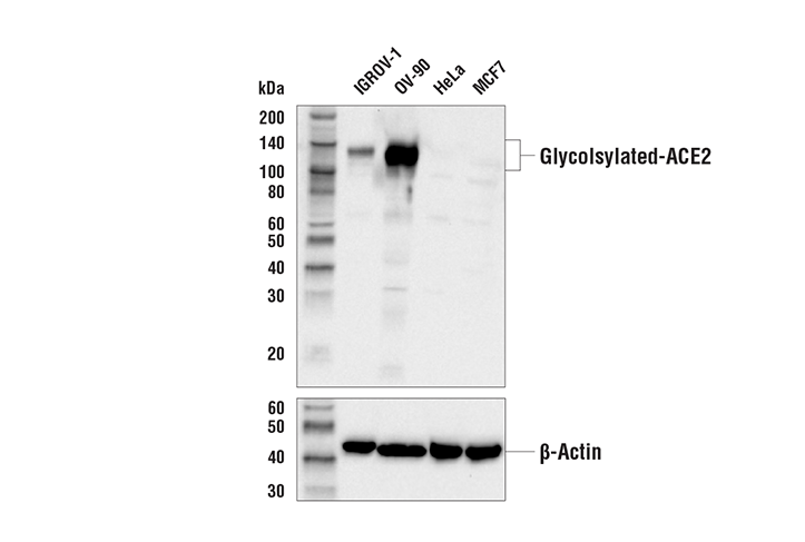
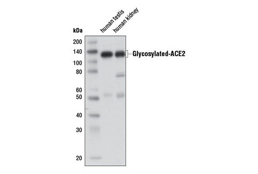
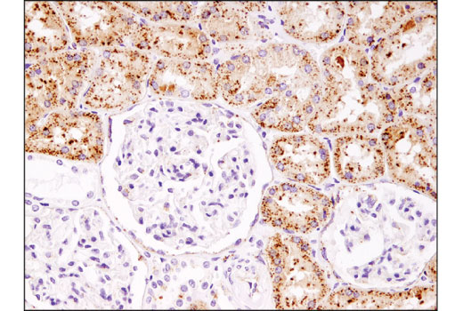
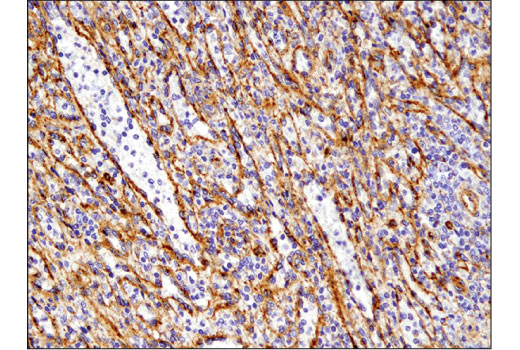
ACE2 is a carboxypeptidase that catalyses the conversion of angiotensin I to angiotensin 1-9, or of angiotensin II to the vasodilator angiotensin 1-7 (1). Research studies have identified ACE2 as the receptor for SARS and SARS-CoV-2 coronaviruses (2-4).
DPP4 (CD26) is a type II transmembrane glycoprotein expressed ubiquitously in most tissues and different cell types (5,6). In addition to its peptidase activity, DPP4 interacts with multiple important cell surface ligands, such as adenosine deaminase, fibronectin, and IGF2 receptor to influence processes like T cell activation, cell migration, and proliferation (7). Research studies have shown that DPP4 serves as a cellular receptor for the MERS-CoV spike protein (8).
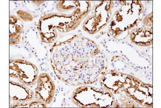
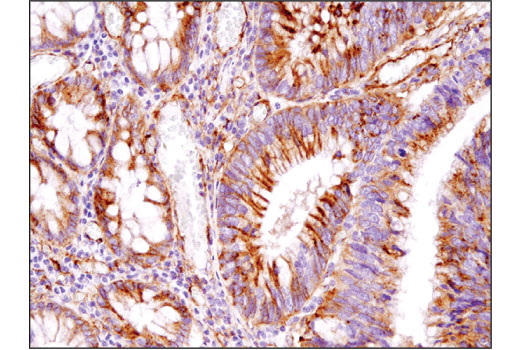
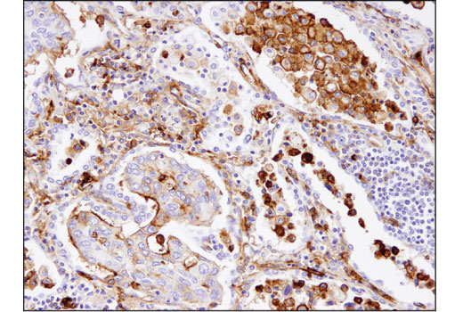
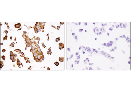
Aminopeptidase N (APN, CD13) is a widely expressed, membrane-bound proteolytic enzyme that breaks down peptides during digestion, cleaves cell surface antigens during antigen presentation, and acts as a receptor for human viruses, including several coronaviruses. This multifunctional protein is implicated in the regulation of many biological processes, including angiogenesis, cell proliferation, cell migration, inflammation, and immune response (9,10).
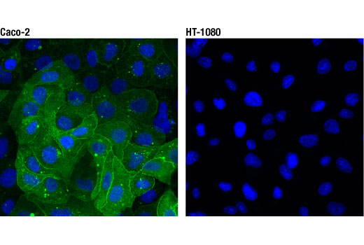
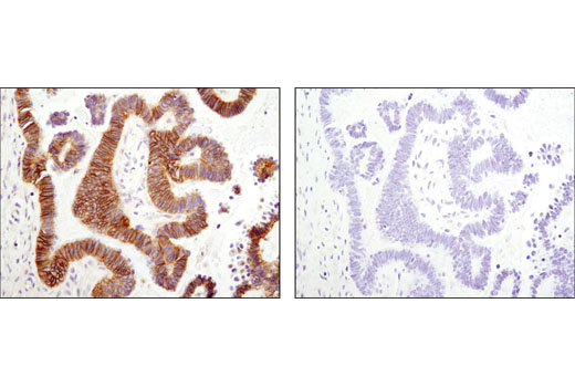
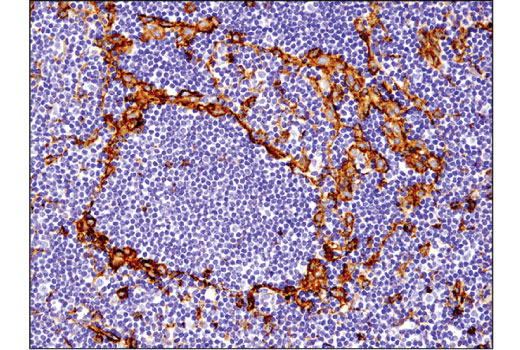
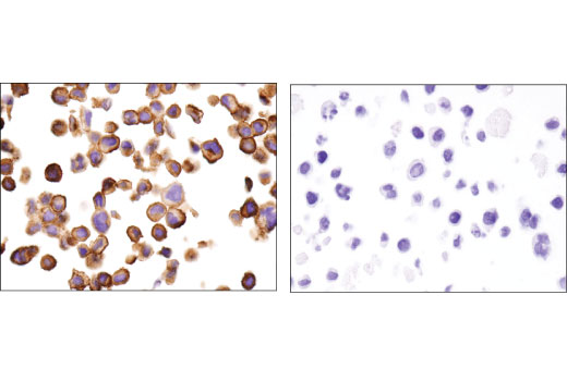
Basigin (EMMPRIN, CD147) is a type I integral membrane receptor protein belonging to the immunoglobulin superfamily (11). Multiple functions have been ascribed to Basigin; foremost among these is stimulating the secretion of extracellular matrix metalloproteinases by adjacent fibroblasts, a function which has been implicated in promoting tumor progression (12-14). Research studies have suggested that Basigin serves as a novel host cell surface receptor for SARS-CoV-2 (15).
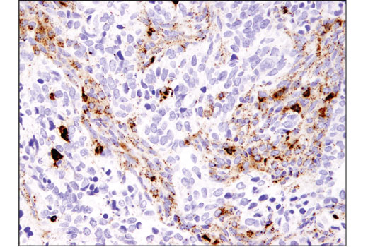
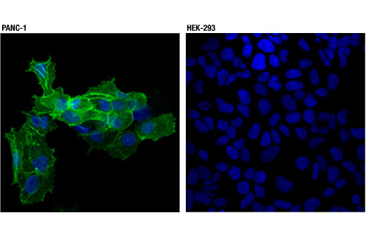
EEA1 is an early endosomal marker and a Rab5 effector protein essential for early endosomal membrane fusion and trafficking (16,17). Research studies have shown that efficient coronavirus host cell entry and replication relies upon early endosomes containing EEA1 (18).
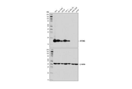
Interferon-induced transmembrane protein (IFITM) family members are composed of short amino- and carboxy-termini, two transmembrane domains, and a cytoplasmic domain (19). The primary function of IFITM family proteins appears to be viral restriction, as IFITM proteins inhibit cytosolic entry of coronaviruses by preventing fusion of viral and host membranes (20,21).
Valosin-containing protein (VCP) is a highly conserved and abundant 97 kDa protein that belongs to the AAA family of proteins. These protein complexes participate in many cellular functions, including vesicle transport and fusion, fragmentation and reassembly of the golgi stacks during mitosis, nuclear envelope formation and spindle disassembly following mitosis, cell cycle regulation, DNA damage repair, apoptosis, B and T cell activation, NF-κB-mediated transcriptional regulation, endoplasmic reticulum (ER)-associated degradation, and protein degradation (22). Research studies have shown that VCP facilitates the release of some coronaviruses from the early endosomal compartment (23).
Cathepsin B, part of the papain family of proteases, is a widely expressed lysosomal cysteine endopeptidase (24,25). Research studies have suggested that cathepsin B facilitates host cell entry of SARS-CoV by promoting fusion of viral and endosomal membranes (26).
- Schmidt, B.L. et al. (2000) J Clin Microbiol 38, 1279-82.
- Li, W. et al. (2005) EMBO J 24, 1634-43.
- Hoffmann, M. et al. (2020) Cell 181, 271-280.e8.
- Lan, J. et al. (2020) Nature 581, 215-220.
- Mentzel, S. et al. (1996) J Histochem Cytochem 44, 445-61.
- Röhrborn, D. et al. (2015) Front Immunol 6, 386.
- Zhong, J. et al. (2015) J Diabetes Res 2015, 606031.
- Wang, N. et al. (2013) Cell Res 23, 986-93.
- Luan, Y. and Xu, W. (2007) Curr Med Chem 14, 639-47.
- Mina-Osorio, P. (2008) Trends Mol Med 14, 361-71.
- Biswas, C. et al. (1995) Cancer Res 55, 434-9.
- Liao, C.G. et al. (2011) Mol Cell Biol 31, 2591-604.
- Sweeny, L. et al. (2012) Exp Cell Res 318, 1788-98.
- Lescaille, G. et al. (2012) BMC Cancer 12, 115.
- Wang, K. et al. (2020) Signal Transduct Target Ther 5, 283.
- Mu, F.T. et al. (1995) J Biol Chem 270, 13503-11.
- Christoforidis, S. et al. (1999) Nature 397, 621-5.
- Burkard, C. et al. (2014) PLoS Pathog 10, e1004502.
- Diamond, M.S. and Farzan, M. (2013) Nat Rev Immunol 13, 46-57.
- Brass, A.L. et al. (2009) Cell 139, 1243-54.
- Feeley, E.M. et al. (2011) PLoS Pathog 7, e1002337.
- Wang, Q. et al. J Struct Biol 146, 44-57.
- Wong, H.H. et al. (2015) J Virol 89, 11116-28.
- Chan, S.J. et al. (1986) Proc Natl Acad Sci U S A 83, 7721-5.
- Fong, D. et al. (1986) Proc Natl Acad Sci U S A 83, 2909-13.
- Simmons, G. et al. (2005) Proc Natl Acad Sci U S A 102, 11876-81.
Background References
Trademarks and Patents
Limited Uses
Except as otherwise expressly agreed in a writing signed by a legally authorized representative of CST, the following terms apply to Products provided by CST, its affiliates or its distributors. Any Customer's terms and conditions that are in addition to, or different from, those contained herein, unless separately accepted in writing by a legally authorized representative of CST, are rejected and are of no force or effect.
Products are labeled with For Research Use Only or a similar labeling statement and have not been approved, cleared, or licensed by the FDA or other regulatory foreign or domestic entity, for any purpose. Customer shall not use any Product for any diagnostic or therapeutic purpose, or otherwise in any manner that conflicts with its labeling statement. Products sold or licensed by CST are provided for Customer as the end-user and solely for research and development uses. Any use of Product for diagnostic, prophylactic or therapeutic purposes, or any purchase of Product for resale (alone or as a component) or other commercial purpose, requires a separate license from CST. Customer shall (a) not sell, license, loan, donate or otherwise transfer or make available any Product to any third party, whether alone or in combination with other materials, or use the Products to manufacture any commercial products, (b) not copy, modify, reverse engineer, decompile, disassemble or otherwise attempt to discover the underlying structure or technology of the Products, or use the Products for the purpose of developing any products or services that would compete with CST products or services, (c) not alter or remove from the Products any trademarks, trade names, logos, patent or copyright notices or markings, (d) use the Products solely in accordance with CST Product Terms of Sale and any applicable documentation, and (e) comply with any license, terms of service or similar agreement with respect to any third party products or services used by Customer in connection with the Products.