Revision 1
#9384
Store at -20C
Cytokeratin Antibody Sampler Kit
1 Kit
(6 x 20 microliters)
877-616-CELL (2355)
877-678-TECH (8324)
3 Trask Lane | Danvers | Massachusetts | 01923 | USA
For Research Use Only. Not for Use in Diagnostic Procedures.
| Product Includes | Product # | Quantity | Mol. Wt | Isotype/Source |
|---|---|---|---|---|
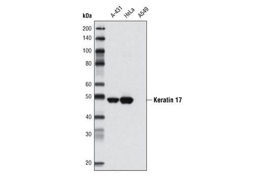
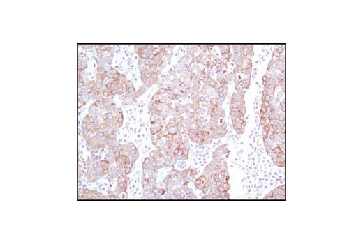
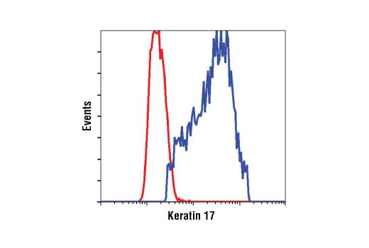
| Keratin 17 (D73C7) Rabbit mAb | 4543 | 20 µl | 48 kDa | Rabbit IgG |
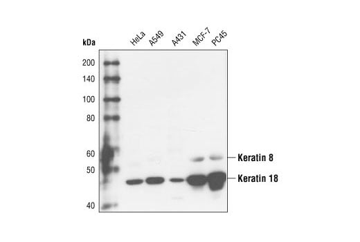
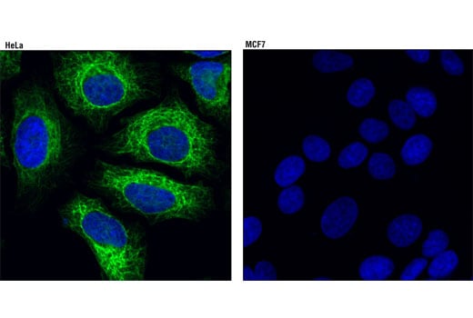
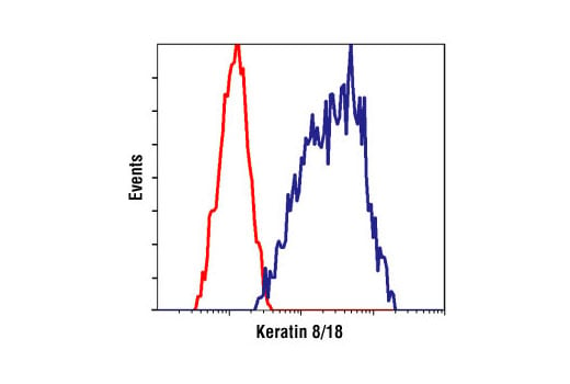
| Keratin 8/18 (C51) Mouse mAb | 4546 | 20 µl | 46 Keratin 18. 55 Keratin 8. kDa | Mouse IgG1 |
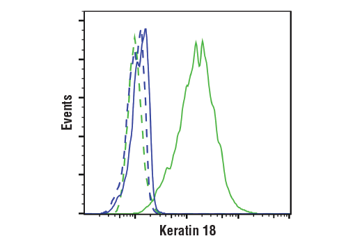
| Keratin 18 (DC10) Mouse mAb | 4548 | 20 µl | 46 kDa | Mouse IgG1 |
| Keratin 19 (BA17) Mouse mAb | 4558 | 20 µl | 40 kDa | Mouse IgG1 |
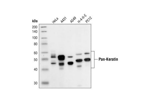
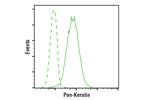
| Pan-Keratin (C11) Mouse mAb | 4545 | 20 µl | 46-58 kDa | Mouse IgG1 |
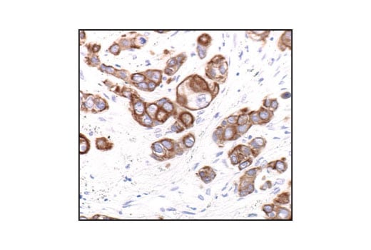
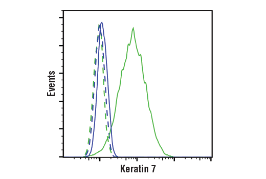
| Keratin 7 (D1E4) XP® Rabbit mAb | 4465 | 20 µl | 52 kDa | Rabbit IgG |
| Anti-mouse IgG, HRP-linked Antibody | 7076 | 100 µl | Horse | |
| Anti-rabbit IgG, HRP-linked Antibody | 7074 | 100 µl | Goat |
Please visit cellsignal.com for individual component applications, species cross-reactivity, dilutions, protocols, and additional product information.
Description
Storage
Background
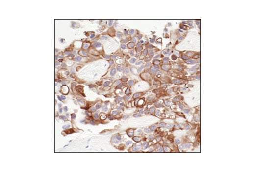
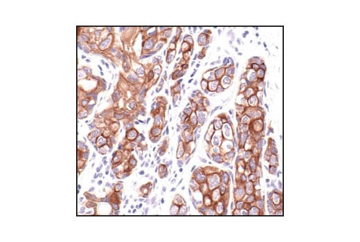
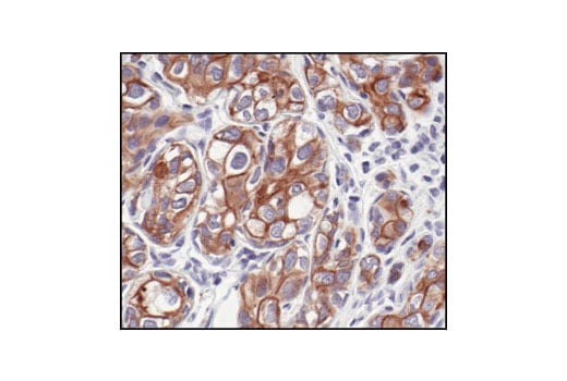
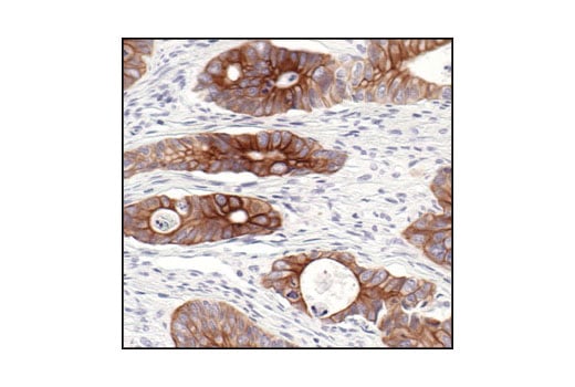
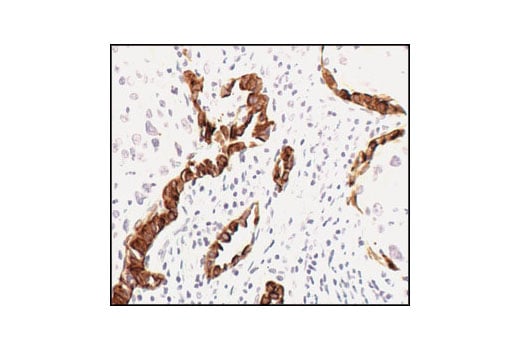
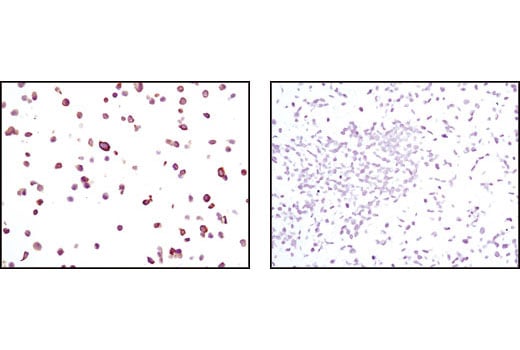
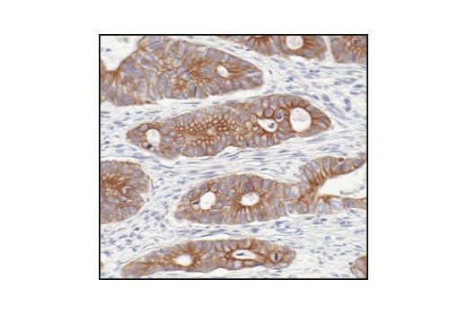
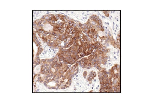
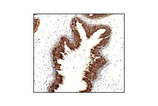
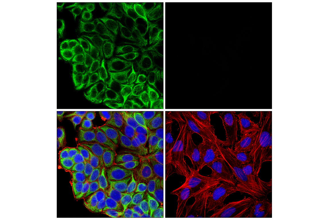
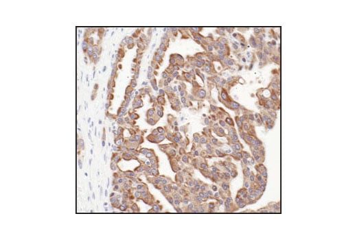
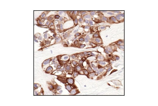
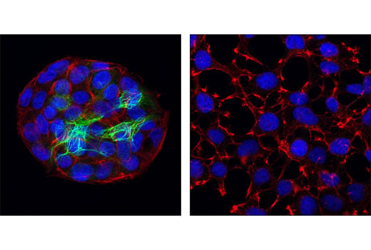
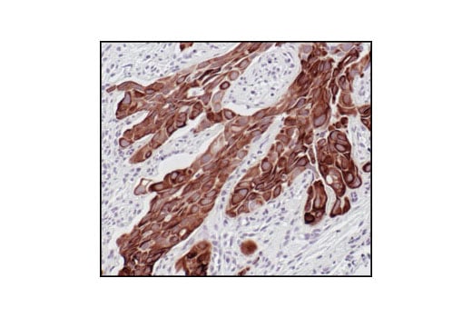
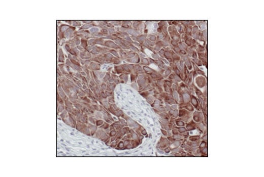
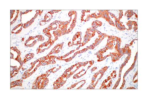
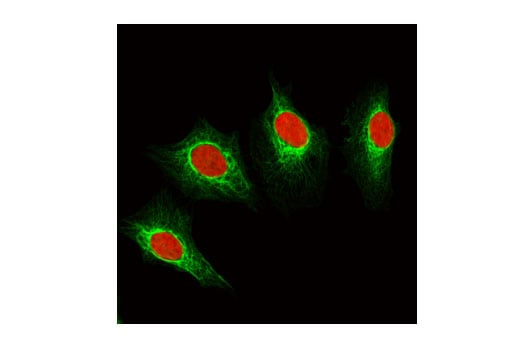
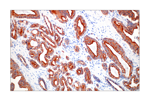
Dysregulation/mutations in keratin genes can lead to a variety of disorders affecting the skin, hair, nails, and other epithelial tissues (3). While expression of keratins can be variable, immunohistochemical staining of keratins is widely used to help in the identification and classification of epithelial tumors, and may also provide prognostic information.
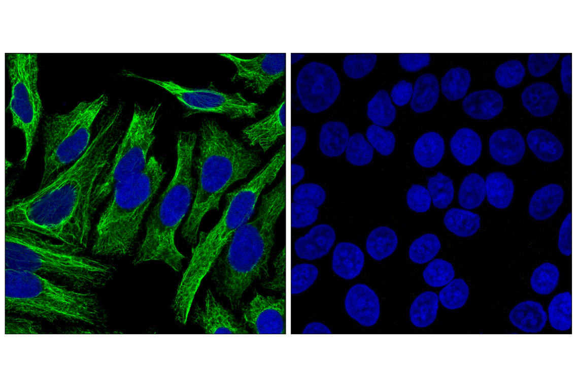
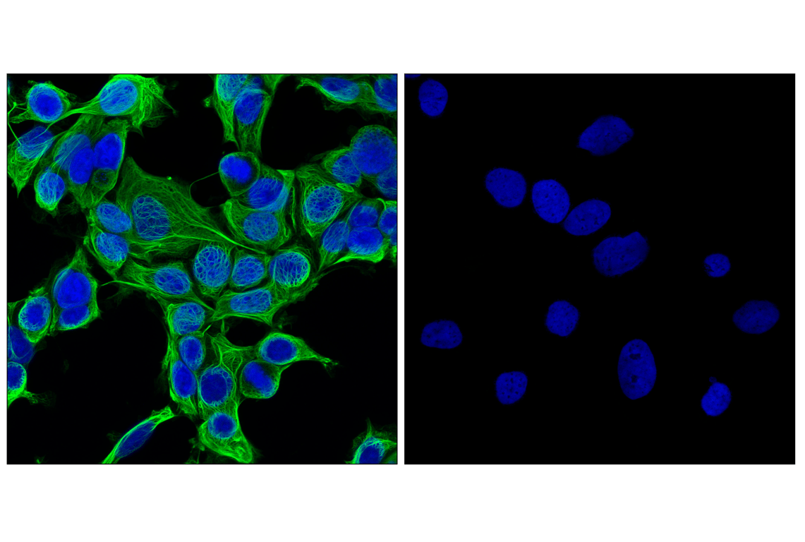
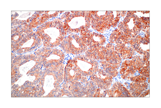
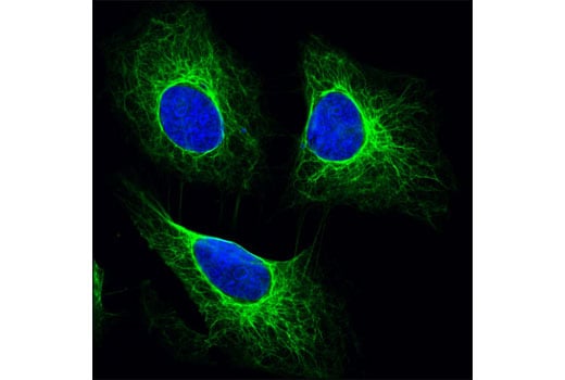
Keratins 8 and 18 (K8/K18) are expressed in simple epithelia of normal tissue, as well as in adenocarcinomas of the breast, lung, ovary, and gastrointestinal tract. Keratin 17 is expressed in basal keratinocytes of stratified epithelia, hair follicles, and sebaceous glands. Onset of keratin 17 expression coincides with the definition of major epithelial lineages during skin development (4). Keratin 14 (K14) is expressed in basal cells of stratified epithelia, and in basal-like subtypes of breast cancer and squamous cell carcinomas. Keratin 19 (K19) is expressed in glandular epithelia, including the liver, gallbladder, and pancreas, as well as in adenocarcinomas of the breast, thyroid, and bile duct. Keratin 20 (K20) is expressed in gastrointestinal epithelium, urothelium, and Merkel cells in the skin, as well as in colorectal carcinomas and some urothelial carcinomas. Keratin 5/6 (K5/6) is expressed in basal cells of stratified epithelia, including the skin, prostate, and breast, as well as in basal-like breast cancers, squamous cell carcinomas, and some lung carcinomas. Keratin 7 (K7) is expressed in glandular epithelia, such as those in the lung, breast, and female reproductive tract, as well as in adenocarcinomas of the lung, breast, and ovary (5,6).
Keratins, particularly K8, K18, and K19, serve as biomarkers for identification of circulating tumor cells (CTCs) (5).
Post-translational modifications, including phosphorylation, acetylation, ubiquitylation, sumoylation, glycosylation, and transamidation, have been shown to affect the functions of keratins in normal and disease states (6). Understanding the molecular mechanisms underlying these PTMs may provide insights into cancer pathogenesis.
Background References
- Chang, L. and Goldman, R.D. (2004) Nat Rev Mol Cell Biol 5, 601-13.
- Schweizer, J. et al. (2006) J Cell Biol 174, 169-74.
- Sarma, A. (2022) Int J Biol Macromol 219, 395-413.
- McGowan, K.M. and Coulombe, P.A. (1998) J Cell Biol 143, 469-86.
- Werner, S. et al. (2020) Mol Aspects Med 72, 100817.
- Dmello, C. et al. (2019) J Biosci 44, 33.
Trademarks and Patents
Cell Signaling Technology is a trademark of Cell Signaling Technology, Inc.
All other trademarks are the property of their respective owners. Visit cellsignal.com/trademarks for more information.
Limited Uses
Except as otherwise expressly agreed in a writing signed by a legally authorized representative of CST, the following terms apply to Products provided by CST, its affiliates or its distributors. Any Customer's terms and conditions that are in addition to, or different from, those contained herein, unless separately accepted in writing by a legally authorized representative of CST, are rejected and are of no force or effect.
Products are labeled with For Research Use Only or a similar labeling statement and have not been approved, cleared, or licensed by the FDA or other regulatory foreign or domestic entity, for any purpose. Customer shall not use any Product for any diagnostic or therapeutic purpose, or otherwise in any manner that conflicts with its labeling statement. Products sold or licensed by CST are provided for Customer as the end-user and solely for research and development uses. Any use of Product for diagnostic, prophylactic or therapeutic purposes, or any purchase of Product for resale (alone or as a component) or other commercial purpose, requires a separate license from CST. Customer shall (a) not sell, license, loan, donate or otherwise transfer or make available any Product to any third party, whether alone or in combination with other materials, or use the Products to manufacture any commercial products, (b) not copy, modify, reverse engineer, decompile, disassemble or otherwise attempt to discover the underlying structure or technology of the Products, or use the Products for the purpose of developing any products or services that would compete with CST products or services, (c) not alter or remove from the Products any trademarks, trade names, logos, patent or copyright notices or markings, (d) use the Products solely in accordance with CST Product Terms of Sale and any applicable documentation, and (e) comply with any license, terms of service or similar agreement with respect to any third party products or services used by Customer in connection with the Products.
Revision 1

Revision 1


Revision 1


Revision 1
Revision 1
Revision 1
Revision 1
Revision 1
Revision 1
Revision 1
Revision 1
Revision 1
Revision 1