WB, IP
H M R
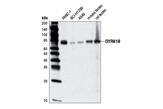
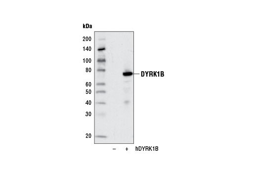
Endogenous
70-80
Rabbit IgG
#Q9Y463
9149
Product Information
Product Usage Information
| Application | Dilution |
|---|---|
| Western Blotting | 1:1000 |
| Immunoprecipitation | 1:100 |
Storage
Specificity / Sensitivity
Species Reactivity:
Human, Mouse, Rat
Species predicted to react based on 100% sequence homology
The antigen sequence used to produce this antibody shares
100% sequence homology with the species listed here, but
reactivity has not been tested or confirmed to work by CST.
Use of this product with these species is not covered under
our
Product Performance Guarantee.
Monkey
Source / Purification
Monoclonal antibody is produced by immunizing animals with a synthetic peptide corresponding to residues near the carboxy terminus of human DYRK1B protein.
Background
The DYRK family includes several dual-specificity tyrosine-phosphorylated and regulated kinases capable of phosphorylating proteins at both Tyr and Ser/Thr residues (1). The DYRK family was identified based on homology to the yeast Yak1 (2) and the Drosophila minibrain (mnb) kinases (3). Seven mammalian isoforms have been discovered, including DYRK1A, DYRK1B, DYRK1C, DYRK2, DYRK3, DYRK4, and DYRK4B. Differences in substrate specificity, expression, and subcellular localization are seen across the DYRK family (4,5). All DYRK proteins have a Tyr-X-Tyr motif in the catalytic domain activation loop; phosphorylation of the second Tyr residue (e.g. Tyr312 of DYRK1A) is necessary for kinase activity. DYRKs typically autophosphorylate the Tyr residue within their activation loop, but phosphorylate substrates at Ser and Thr residues (1,6).
In contrast to the ubiquitous DYRK1A, DYRK1B exhibits relatively restricted expression with highest levels found in the testis and muscle (7,8). Three major DYRK1B splice variants demonstrate distinct expression patterns and functional properties (9). DYRK1B plays a critical role in myoblast differentiation by affecting cell motility, transcription, cell cycle progression, and survival (10,11). In addition, DYRK1B contributes to the survival of certain cancer cells (7,12,13).
- Becker, W. and Joost, H.G. (1999) Prog. Nucleic Acid Res. Mol. Biol. 62, 1-17.
- Garrett, S. and Broach, J. (1989) Genes Dev. 3, 1336-1348.
- Tejedor, F. et al. (1995) Neuron 14, 287-301.
- Kentrup, H. et al. (1996) J. Biol. Chem. 271, 3488-3495.
- Becker, W. et al. (1998) J. Biol. Chem. 273, 25893-25902.
- Lochhead, P.A. et al. (2005) Cell 121, 925-936.
- Leder, S. et al. (1999) Biochem Biophys Res Commun 254, 474-9.
- Lee, K. et al. (2000) Cancer Res 60, 3631-7.
- Leder, S. et al. (2003) Biochem J 372, 881-8.
- Mercer, S.E. and Friedman, E. (2006) Cell Biochem Biophys 45, 303-15.
- Deng, X. et al. (2003) J Biol Chem 278, 41347-54.
- Deng, X. et al. (2006) Cancer Res 66, 4149-58.
- Mercer, S.E. et al. (2006) Cancer Res 66, 5143-50.
Species Reactivity
Species reactivity is determined by testing in at least one approved application (e.g., western blot).
Western Blot Buffer
IMPORTANT: For western blots, incubate membrane with diluted primary antibody in 5% w/v BSA, 1X TBS, 0.1% Tween® 20 at 4°C with gentle shaking, overnight.
Applications Key
WB: Western Blotting IP: Immunoprecipitation
Cross-Reactivity Key
H: human M: mouse R: rat Hm: hamster Mk: monkey Vir: virus Mi: mink C: chicken Dm: D. melanogaster X: Xenopus Z: zebrafish B: bovine Dg: dog Pg: pig Sc: S. cerevisiae Ce: C. elegans Hr: horse GP: Guinea Pig Rab: rabbit All: all species expected
Trademarks and Patents
Limited Uses
Except as otherwise expressly agreed in a writing signed by a legally authorized representative of CST, the following terms apply to Products provided by CST, its affiliates or its distributors. Any Customer's terms and conditions that are in addition to, or different from, those contained herein, unless separately accepted in writing by a legally authorized representative of CST, are rejected and are of no force or effect.
Products are labeled with For Research Use Only or a similar labeling statement and have not been approved, cleared, or licensed by the FDA or other regulatory foreign or domestic entity, for any purpose. Customer shall not use any Product for any diagnostic or therapeutic purpose, or otherwise in any manner that conflicts with its labeling statement. Products sold or licensed by CST are provided for Customer as the end-user and solely for research and development uses. Any use of Product for diagnostic, prophylactic or therapeutic purposes, or any purchase of Product for resale (alone or as a component) or other commercial purpose, requires a separate license from CST. Customer shall (a) not sell, license, loan, donate or otherwise transfer or make available any Product to any third party, whether alone or in combination with other materials, or use the Products to manufacture any commercial products, (b) not copy, modify, reverse engineer, decompile, disassemble or otherwise attempt to discover the underlying structure or technology of the Products, or use the Products for the purpose of developing any products or services that would compete with CST products or services, (c) not alter or remove from the Products any trademarks, trade names, logos, patent or copyright notices or markings, (d) use the Products solely in accordance with CST Product Terms of Sale and any applicable documentation, and (e) comply with any license, terms of service or similar agreement with respect to any third party products or services used by Customer in connection with the Products.