WB, W-S, IP, IHC-P, IF-IC, FC-FP, ChIP, ChIP-seq, C&R, C&T, eCLIP
H M R Mk
Endogenous
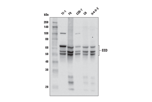
50-70
Rabbit IgG
#O75530
8726
Product Information
Product Usage Information
For optimal ChIP and ChIP-seq results, use 5 μl of antibody and 10 μg of chromatin (approximately 4 x 10⁶ cells) per IP. This antibody has been validated using SimpleChIP® Enzymatic Chromatin IP Kits.
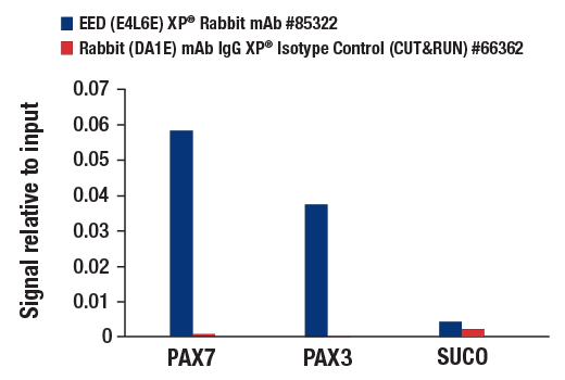
The CUT&RUN dilution was determined using CUT&RUN Assay Kit #86652.
The CUT&Tag dilution was determined using CUT&Tag Assay Kit #77552.
| Application | Dilution |
|---|---|
| Western Blotting | 1:1000 |
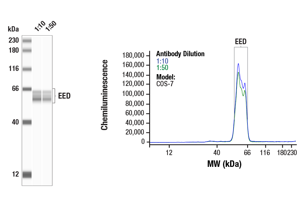
| Simple Western™ | 1:10 - 1:50 |
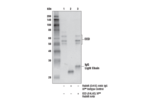
| Immunoprecipitation | 1:50 |
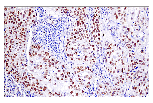
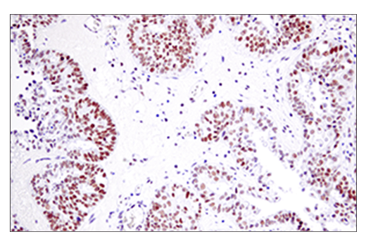
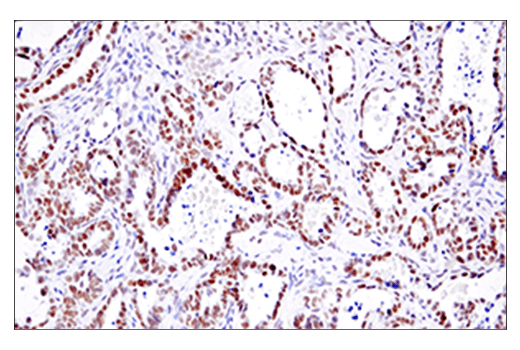
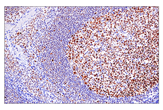
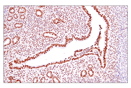
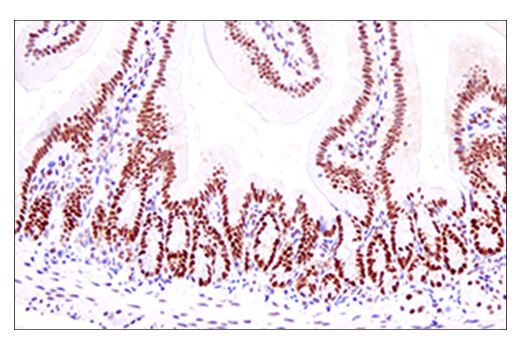
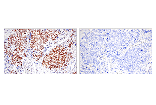
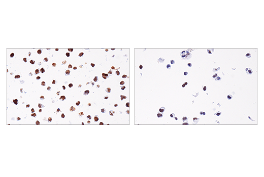
| Immunohistochemistry (Paraffin) | 1:50 - 1:200 |
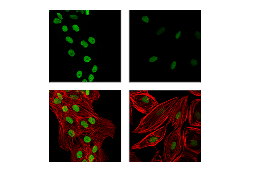
| Immunofluorescence (Immunocytochemistry) | 1:50 - 1:200 |
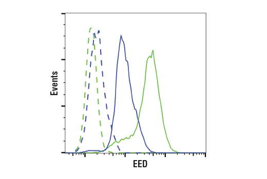
| Flow Cytometry (Fixed/Permeabilized) | 1:50 - 1:200 |
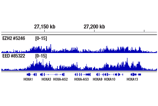
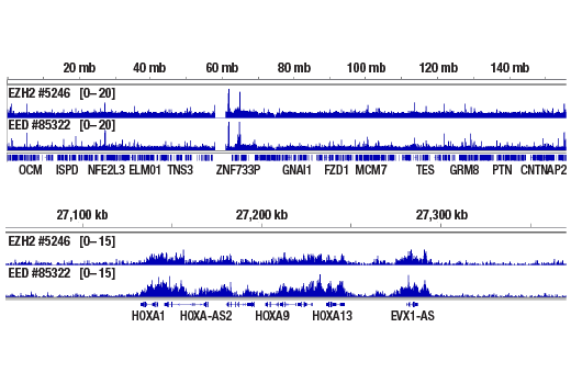
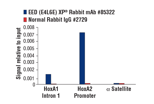
| Chromatin IP | 1:100 |
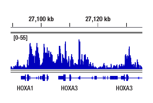
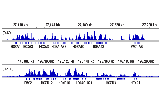
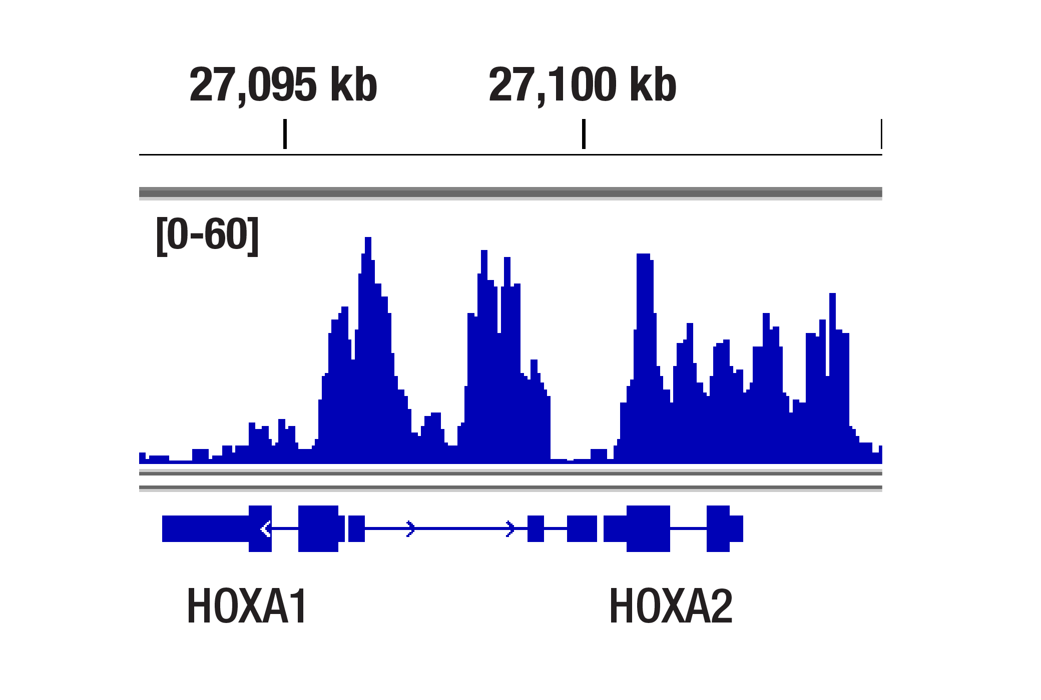
| Chromatin IP-seq | 1:100 |
| CUT&RUN | 1:100 |
| CUT&Tag | 1:100 |
| eCLIP | 1:200 |
For more information about the RBP-eCLIP service please visit Eclipsebio.
Storage
Specificity / Sensitivity
Species Reactivity:
Human, Mouse, Rat, Monkey
Species predicted to react based on 100% sequence homology
The antigen sequence used to produce this antibody shares
100% sequence homology with the species listed here, but
reactivity has not been tested or confirmed to work by CST.
Use of this product with these species is not covered under
our
Product Performance Guarantee.
Bovine, Pig
Source / Purification
Monoclonal antibody is produced by immunizing animals with a synthetic peptide corresponding to residues near the amino terminus of human EED protein.
Background
The polycomb group (PcG) proteins contribute to the maintenance of cell identity, stem cell self-renewal, cell cycle regulation and oncogenesis by maintaining the silenced state of genes that promote cell lineage specification, cell death and cell-cycle arrest (1-4). PcG proteins exist in two complexes that cooperate to maintain long-term gene silencing through epigenetic chromatin modifications. The first complex, EED-EZH2, is recruited to genes by DNA-binding transcription factors and methylates histone H3 on Lys27. Methylation of Lys27 facilitates the recruitment of the second complex, PRC1, which ubiquitinylates histone H2A on Lys119 (5). Embryonic ectoderm development protein (EED) is a component of the PRC2 complex, which together with Ezh2 and SUZ12 is absolutely required for histone methyl-transferase activity (6). EED, SUZ12 and EZH2 are overexpressed in various types of cancer, including tumors of the colon, breast, prostate and ovary (7-9).
- Boyer, L.A. et al. (2006) Nature 441, 349-53.
- Lee, T.I. et al. (2006) Cell 125, 301-13.
- Cao, R. et al. (2002) Science 298, 1039-43.
- Müller, J. et al. (2002) Cell 111, 197-208.
- Wang, H. et al. (2004) Nature 431, 873-8.
- Cao, R. and Zhang, Y. (2004) Mol Cell 15, 57-67.
- Kirmizis, A. et al. (2003) Mol Cancer Ther 2, 113-21.
- Kirmizis, A. et al. (2004) Genes Dev 18, 1592-605.
- Yu, H. et al. (2012) PLoS One 7, e51239.
Species Reactivity
Species reactivity is determined by testing in at least one approved application (e.g., western blot).
Western Blot Buffer
IMPORTANT: For western blots, incubate membrane with diluted primary antibody in 5% w/v BSA, 1X TBS, 0.1% Tween® 20 at 4°C with gentle shaking, overnight.
Applications Key
WB: Western Blotting W-S: Simple Western™ IP: Immunoprecipitation IHC-P: Immunohistochemistry (Paraffin) IF-IC: Immunofluorescence (Immunocytochemistry) FC-FP: Flow Cytometry (Fixed/Permeabilized) ChIP: Chromatin IP ChIP-seq: Chromatin IP-seq C&R: CUT&RUN C&T: CUT&Tag eCLIP: eCLIP
Cross-Reactivity Key
H: human M: mouse R: rat Hm: hamster Mk: monkey Vir: virus Mi: mink C: chicken Dm: D. melanogaster X: Xenopus Z: zebrafish B: bovine Dg: dog Pg: pig Sc: S. cerevisiae Ce: C. elegans Hr: horse GP: Guinea Pig Rab: rabbit All: all species expected
Trademarks and Patents
Limited Uses
Except as otherwise expressly agreed in a writing signed by a legally authorized representative of CST, the following terms apply to Products provided by CST, its affiliates or its distributors. Any Customer's terms and conditions that are in addition to, or different from, those contained herein, unless separately accepted in writing by a legally authorized representative of CST, are rejected and are of no force or effect.
Products are labeled with For Research Use Only or a similar labeling statement and have not been approved, cleared, or licensed by the FDA or other regulatory foreign or domestic entity, for any purpose. Customer shall not use any Product for any diagnostic or therapeutic purpose, or otherwise in any manner that conflicts with its labeling statement. Products sold or licensed by CST are provided for Customer as the end-user and solely for research and development uses. Any use of Product for diagnostic, prophylactic or therapeutic purposes, or any purchase of Product for resale (alone or as a component) or other commercial purpose, requires a separate license from CST. Customer shall (a) not sell, license, loan, donate or otherwise transfer or make available any Product to any third party, whether alone or in combination with other materials, or use the Products to manufacture any commercial products, (b) not copy, modify, reverse engineer, decompile, disassemble or otherwise attempt to discover the underlying structure or technology of the Products, or use the Products for the purpose of developing any products or services that would compete with CST products or services, (c) not alter or remove from the Products any trademarks, trade names, logos, patent or copyright notices or markings, (d) use the Products solely in accordance with CST Product Terms of Sale and any applicable documentation, and (e) comply with any license, terms of service or similar agreement with respect to any third party products or services used by Customer in connection with the Products.