| Products Included | No. | Volume | Applicaton | Dilution | Reactivity | Homology† |
|---|---|---|---|---|---|---|
| Primary Cocktail | 7972 | 100 µl | Immunofluorescence (Immunocytochemistry), Immunofluorescence (Paraffin)‡ | 1:100 | Human, Monkey | Mouse |
| Detection Cocktail | 7973 | 100 µl | Immunofluorescence (Immunocytochemistry), Immunofluorescence (Paraffin)‡ | 1:100 | N/A | N/A |
†Species predicted to react based on 100% sequence homology.
‡Immunofluorescence (Paraffin) protocol recommended unmasking buffer: EDTA
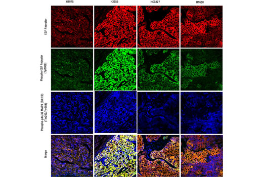
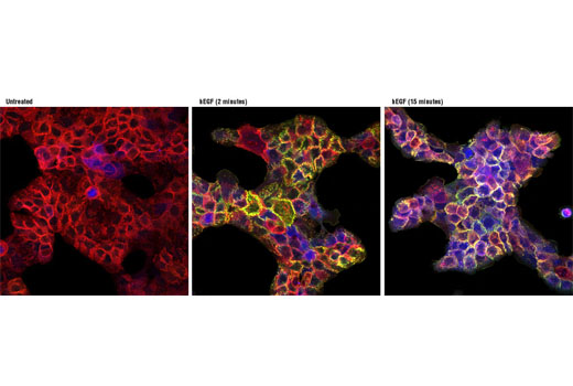
| Kit Analytes | Detection Dye | Ex(max) (nm) | Em(max) (nm) |
|---|---|---|---|
| EGF Receptor | Alexa Fluor® 555 | 555 | 565 |
| Phospho-EGF Receptor (Tyr1068) | Alexa Fluor® 488 | 495 | 519 |
| Phospho-p44/42 MAPK (Erk1/2) (Thr202/Tyr204) | Alexa Fluor® 647 | 650 | 665 |
IF-P, IF-IC
#P27361, #P28482, #P00533
5595, 5594, 1956
Product Information
Storage
Specificity / Sensitivity
Species Reactivity:
Human, Monkey
Source / Purification
Monoclonal antibodies were produced by immunizing animals with synthetic phosphopeptides corresponding to residues surrounding Thr202/Tyr204 of human p44 MAP kinase and Tyr1068 of human EGF receptor, or with a fusion protein containing the cytoplasmic domain of human EGF receptor.
Product Description
| MW (kDa) | n/a |
Background
The epidermal growth factor receptor (EGFR) is a 170 kDa transmembrane receptor tyrosine kinase that belongs to the HER/ErbB protein family that includes HER2/ErbB2/neu, HER3/ErbB3, and HER4/ErbB4 (1,2). Ligand binding results in receptor homo- and heterodimerization which stimulates its intrinsic tyrosine kinase activity. Autophosphorylation at residues Tyr992, Tyr1045, Tyr1068, Tyr1148, and Tyr1173, as well as c-Src mediated phosphorylation at Tyr845 and Tyr1101 promote docking of SH2 domain-bearing signaling proteins resulting in cellular responses to EGFR activation (2-6). For example, phosphorylation of Tyr1068 facilitates recruitment of Grb2 which, via association with Sos, stimulates the GTP binding activity of Ras, leading to the activation of MAP kinase and other signaling cascades (7). Following activation, EGFR is rapidly endocytosed and either recycled back to the plasma membrane or targeted for lysosomal degradation (8). Dysregulation of EGFR signaling through activating mutations or gene amplification has been implicated in the pathogenesis of many human malignancies, leading to intense clinical study of this pathway (9-13).
- Hackel, P.O. et al. (1999) Curr Opin Cell Biol 11, 184-9.
- Zwick, E. et al. (1999) Trends Pharmacol Sci 20, 408-12.
- Cooper, J.A. and Howell, B. (1993) Cell 73, 1051-4.
- Emlet, D.R. et al. (1997) J Biol Chem 272, 4079-86.
- Emlet, D.R. et al. (1997) J Biol Chem 272, 4079-86.
- Levkowitz, G. et al. (1999) Mol Cell 4, 1029-40.
- Rojas, M. et al. (1996) J Biol Chem 271, 27456-61.
- Lai, W.H. et al. (1989) J Cell Biol 109, 2741-9.
- Yarden, Y. and Sliwkowski, M.X. (2001) Nat Rev Mol Cell Biol 2, 127-37.
- Press, M.F. and Lenz, H.J. (2007) Drugs 67, 2045-75.
- Baselga, J. (2002) Oncologist 7 Suppl 4, 2-8.
- Foon, K.A. et al. (2004) Int J Radiat Oncol Biol Phys 58, 984-90.
- Sridhar, S.S. et al. (2003) Lancet Oncol 4, 397-406.
Species Reactivity
Species reactivity is determined by testing in at least one approved application (e.g., western blot).
Applications Key
IF-P: Immunofluorescence (Paraffin) IF-IC: Immunofluorescence (Immunocytochemistry)
Cross-Reactivity Key
H: human M: mouse R: rat Hm: hamster Mk: monkey Vir: virus Mi: mink C: chicken Dm: D. melanogaster X: Xenopus Z: zebrafish B: bovine Dg: dog Pg: pig Sc: S. cerevisiae Ce: C. elegans Hr: horse GP: Guinea Pig Rab: rabbit All: all species expected
Trademarks and Patents
Limited Uses
Except as otherwise expressly agreed in a writing signed by a legally authorized representative of CST, the following terms apply to Products provided by CST, its affiliates or its distributors. Any Customer's terms and conditions that are in addition to, or different from, those contained herein, unless separately accepted in writing by a legally authorized representative of CST, are rejected and are of no force or effect.
Products are labeled with For Research Use Only or a similar labeling statement and have not been approved, cleared, or licensed by the FDA or other regulatory foreign or domestic entity, for any purpose. Customer shall not use any Product for any diagnostic or therapeutic purpose, or otherwise in any manner that conflicts with its labeling statement. Products sold or licensed by CST are provided for Customer as the end-user and solely for research and development uses. Any use of Product for diagnostic, prophylactic or therapeutic purposes, or any purchase of Product for resale (alone or as a component) or other commercial purpose, requires a separate license from CST. Customer shall (a) not sell, license, loan, donate or otherwise transfer or make available any Product to any third party, whether alone or in combination with other materials, or use the Products to manufacture any commercial products, (b) not copy, modify, reverse engineer, decompile, disassemble or otherwise attempt to discover the underlying structure or technology of the Products, or use the Products for the purpose of developing any products or services that would compete with CST products or services, (c) not alter or remove from the Products any trademarks, trade names, logos, patent or copyright notices or markings, (d) use the Products solely in accordance with CST Product Terms of Sale and any applicable documentation, and (e) comply with any license, terms of service or similar agreement with respect to any third party products or services used by Customer in connection with the Products.