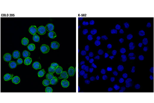
WB, IP, IHC-P, IF-IC
H M R
Endogenous
130
Rabbit IgG
#P29323
2048
Product Information
Product Usage Information
| Application | Dilution |
|---|---|
| Western Blotting | 1:1000 |
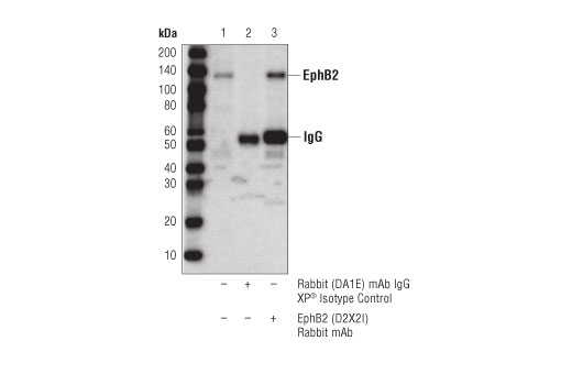
| Immunoprecipitation | 1:50 |
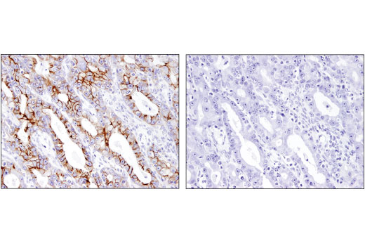
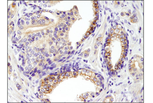
| Immunohistochemistry (Paraffin) | 1:200 |
| Immunofluorescence (Immunocytochemistry) | 1:600 |
Storage
Specificity / Sensitivity
Species Reactivity:
Human, Mouse, Rat
Source / Purification
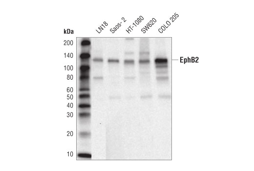
Monoclonal antibody is produced by immunizing animals with a synthetic peptide corresponding to residues surrounding Ala573 of human EphB2 protein.
Background
The ephrin receptor B2 (EphB2) is an ephrin family receptor tyrosine kinase that plays an important role in regulating growth and development of multiple tissues and organs (1,2). The EphB2 transmembrane receptor protein contains a kinase domain, a PDZ motif, and a SAM domain within a conserved cytoplasmic domain. A ligand binding domain, a cysteine-rich domain, and fibronectin type III repeats comprise the conserved EphB2 extracellular domain (3). EphB2 binds with high affinity to ephrin B ligands, and to some ephrin A proteins, to initiate bidirectional signaling between neighboring cells (1,2). Upon binding, EphB2-Ephrin B2 dimers form a heterotetramer and position the receptor-ligand complex on the cell membrane to facilitate bidirectional signal transduction (3). In addition to associating with ephrin ligands, EphB2 also regulates a number of biological processes through interaction with focal adhesion kinase (FAK), NMDA receptor (NMDAR), the Rac1 guanine nucleotide exchange factor Tiam1, and p21-activated kinase (PAK1) (4-7). While some studies support a role for EphB2 as a pro-oncogenic kinase, other research suggests that EphB2 acts as a tumor suppressor (1,2,4,8).
- Pasquale, E.B. (2008) Cell 133, 38-52.
- Klein, R. (2009) Nat Neurosci 12, 15-20.
- Himanen, J.P. et al. (2001) Nature 414, 933-8.
- Wang, S.D. et al. (2012) Oncogene 31, 5132-43.
- Nolt, M.J. et al. (2011) J Neurosci 31, 5353-64.
- Tolias, K.F. et al. (2007) Proc Natl Acad Sci U S A 104, 7265-70.
- Srivastava, N. et al. (2013) Mol Cell Neurosci 52, 106-16.
- Blume-Jensen, P. and Hunter, T. (2001) Nature 411, 355-65.
Species Reactivity
Species reactivity is determined by testing in at least one approved application (e.g., western blot).
Western Blot Buffer
IMPORTANT: For western blots, incubate membrane with diluted primary antibody in 5% w/v BSA, 1X TBS, 0.1% Tween® 20 at 4°C with gentle shaking, overnight.
Applications Key
WB: Western Blotting IP: Immunoprecipitation IHC-P: Immunohistochemistry (Paraffin) IF-IC: Immunofluorescence (Immunocytochemistry)
Cross-Reactivity Key
H: human M: mouse R: rat Hm: hamster Mk: monkey Vir: virus Mi: mink C: chicken Dm: D. melanogaster X: Xenopus Z: zebrafish B: bovine Dg: dog Pg: pig Sc: S. cerevisiae Ce: C. elegans Hr: horse GP: Guinea Pig Rab: rabbit All: all species expected
Trademarks and Patents
Limited Uses
Except as otherwise expressly agreed in a writing signed by a legally authorized representative of CST, the following terms apply to Products provided by CST, its affiliates or its distributors. Any Customer's terms and conditions that are in addition to, or different from, those contained herein, unless separately accepted in writing by a legally authorized representative of CST, are rejected and are of no force or effect.
Products are labeled with For Research Use Only or a similar labeling statement and have not been approved, cleared, or licensed by the FDA or other regulatory foreign or domestic entity, for any purpose. Customer shall not use any Product for any diagnostic or therapeutic purpose, or otherwise in any manner that conflicts with its labeling statement. Products sold or licensed by CST are provided for Customer as the end-user and solely for research and development uses. Any use of Product for diagnostic, prophylactic or therapeutic purposes, or any purchase of Product for resale (alone or as a component) or other commercial purpose, requires a separate license from CST. Customer shall (a) not sell, license, loan, donate or otherwise transfer or make available any Product to any third party, whether alone or in combination with other materials, or use the Products to manufacture any commercial products, (b) not copy, modify, reverse engineer, decompile, disassemble or otherwise attempt to discover the underlying structure or technology of the Products, or use the Products for the purpose of developing any products or services that would compete with CST products or services, (c) not alter or remove from the Products any trademarks, trade names, logos, patent or copyright notices or markings, (d) use the Products solely in accordance with CST Product Terms of Sale and any applicable documentation, and (e) comply with any license, terms of service or similar agreement with respect to any third party products or services used by Customer in connection with the Products.