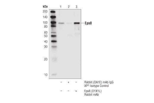
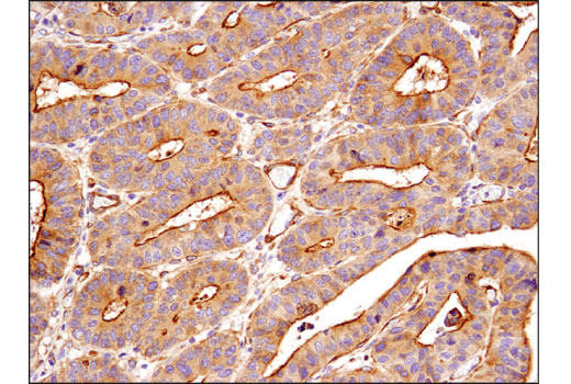
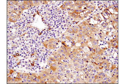
WB, IP, IHC-P
H M R
Endogenous
95
Rabbit IgG
#Q12929
2059
Product Information
Product Usage Information
| Application | Dilution |
|---|---|
| Western Blotting | 1:1000 |
| Immunoprecipitation | 1:50 |
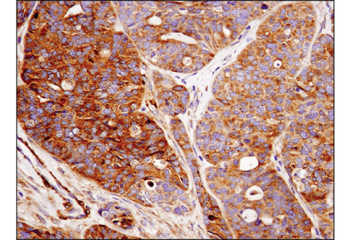
| Immunohistochemistry (Paraffin) | 1:100 |
Storage
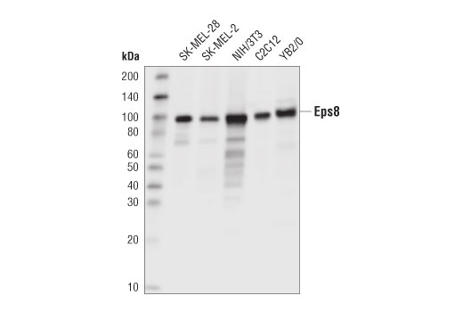
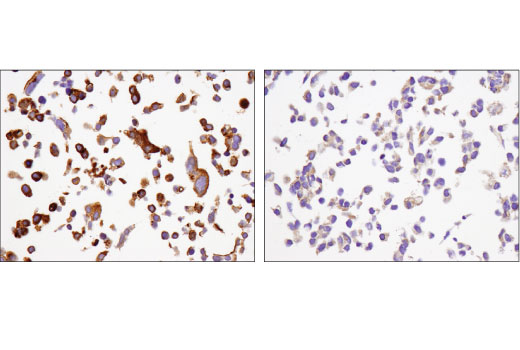
Specificity / Sensitivity
Species Reactivity:
Human, Mouse, Rat
Source / Purification
Monoclonal antibody is produced by immunizing animals with a synthetic peptide corresponding to residues surrounding Arg250 of human Eps8 protein.
Background
Epidermal growth factor receptor pathway substrate 8 (Eps8) is an adaptor protein and can be phosphorylated by several receptor tyrosine kinases including EGFR and Src (1,2). Eps8 is composed of an N-terminal PTB domain, followed by an SH3 domain and a C-terminal effector domain. Eps8 controls actin-based motility by capping the barbed end of actin and bundling actin subunits through its C-terminal effector domain (3,4). The C-terminal α hexlical structure of Eps8 interacts directly with actin to exert these capping and bundling functions (5). The actin capping activity requires the release of Eps8 autoinhibitory binding through SH3 domain interaction with an adaptor molecule, such as Abi-1 (6). This SH3 domain of Eps8 also binds to RN-tre to regulate the down stream Rab5-mediated endocytosis pathway (6). Eps8 functions by binding several receptor tyrosine kinases, such as EGFR or FGFR, to enhance receptor mediated mitogenic Rac signaling and Rab5 endocytosis (6,7). The effector region of Eps8 is necessary for this process. By association with Abi-1 and forming the Eps8/Abi-1/Sos-1 complex, Eps8 couples initial growth factor stimulation to actin motility and the Rac activation pathway (8,9). Eps8 has been shown to be important in the cellular function of filopodial protrusions, cell migration, microvilli formation, and focal adhesion (10-13). Research studies have demonstrated that through its involvement in actin related cellular functions, Eps8 plays a role in cancer cell growth, survival, motility, and invasiveness (14-18).
- Fazioli, F. et al. (1993) EMBO J 12, 3799-808.
- Gallo, R. et al. (1997) Oncogene 15, 1929-36.
- Disanza, A. et al. (2004) Nat Cell Biol 6, 1180-8.
- Disanza, A. et al. (2006) Nat Cell Biol 8, 1337-47.
- Hertzog, M. et al. (2010) PLoS Biol 8, e1000387.
- Lanzetti, L. et al. (2000) Nature 408, 374-7.
- Auciello, G. et al. (2013) J Cell Sci 126, 613-24.
- Scita, G. et al. (1999) Nature 401, 290-3.
- Innocenti, M. et al. (2003) J Cell Biol 160, 17-23.
- Welsch, T. et al. (2007) Cancer Lett 255, 205-18.
- Frittoli, E. et al. (2011) Immunity 35, 388-99.
- Zwaenepoel, I. et al. (2012) Mol Biol Cell 23, 1080-94.
- Maa, M.C. et al. (2007) J Biol Chem 282, 19399-409.
- Xu, M. et al. (2009) Endocrinology 150, 2064-71.
- Chen, Y.J. et al. (2008) Mol Cancer Ther 7, 1376-85.
- Yap, L.F. et al. (2009) Oncogene 28, 2524-34.
- Chen, H. et al. (2010) Cancer Res 70, 9979-90.
- Funato, Y. et al. (2004) Cancer Res 64, 5237-44.
Species Reactivity
Species reactivity is determined by testing in at least one approved application (e.g., western blot).
Western Blot Buffer
IMPORTANT: For western blots, incubate membrane with diluted primary antibody in 5% w/v BSA, 1X TBS, 0.1% Tween® 20 at 4°C with gentle shaking, overnight.
Applications Key
WB: Western Blotting IP: Immunoprecipitation IHC-P: Immunohistochemistry (Paraffin)
Cross-Reactivity Key
H: human M: mouse R: rat Hm: hamster Mk: monkey Vir: virus Mi: mink C: chicken Dm: D. melanogaster X: Xenopus Z: zebrafish B: bovine Dg: dog Pg: pig Sc: S. cerevisiae Ce: C. elegans Hr: horse GP: Guinea Pig Rab: rabbit All: all species expected
Trademarks and Patents
Limited Uses
Except as otherwise expressly agreed in a writing signed by a legally authorized representative of CST, the following terms apply to Products provided by CST, its affiliates or its distributors. Any Customer's terms and conditions that are in addition to, or different from, those contained herein, unless separately accepted in writing by a legally authorized representative of CST, are rejected and are of no force or effect.
Products are labeled with For Research Use Only or a similar labeling statement and have not been approved, cleared, or licensed by the FDA or other regulatory foreign or domestic entity, for any purpose. Customer shall not use any Product for any diagnostic or therapeutic purpose, or otherwise in any manner that conflicts with its labeling statement. Products sold or licensed by CST are provided for Customer as the end-user and solely for research and development uses. Any use of Product for diagnostic, prophylactic or therapeutic purposes, or any purchase of Product for resale (alone or as a component) or other commercial purpose, requires a separate license from CST. Customer shall (a) not sell, license, loan, donate or otherwise transfer or make available any Product to any third party, whether alone or in combination with other materials, or use the Products to manufacture any commercial products, (b) not copy, modify, reverse engineer, decompile, disassemble or otherwise attempt to discover the underlying structure or technology of the Products, or use the Products for the purpose of developing any products or services that would compete with CST products or services, (c) not alter or remove from the Products any trademarks, trade names, logos, patent or copyright notices or markings, (d) use the Products solely in accordance with CST Product Terms of Sale and any applicable documentation, and (e) comply with any license, terms of service or similar agreement with respect to any third party products or services used by Customer in connection with the Products.