Revision 1
Cell Signaling Technology

Orders: 877-616-CELL (2355) [email protected]

Support: 877-678-TECH (8324)

Web: [email protected] cellsignal.com

3 Trask LaneDanversMassachusetts01923USA
For Research Use Only. Not for Use in Diagnostic Procedures.
:

:

:

:

UniProt ID:

#P03372

Entrez-Gene Id:

2099

Please visit cellsignal.com for individual component applications, species cross-reactivity, dilutions, protocols, and additional product information.

Description

PhosphoPlus® Duets from Cell Signaling Technology (CST) provide a means to assess protein activation status. Each Duet contains an activation-state and total protein antibody to your target of interest. These antibodies have been selected from CST's product offering based upon superior performance in specified applications.

Storage

Supplied in 10 mM sodium HEPES (pH 7.5), 150 mM NaCl, 100 µg/ml BSA, 50% glycerol and less than 0.02% sodium azide. Store at –20°C. Do not aliquot the antibody.

Background

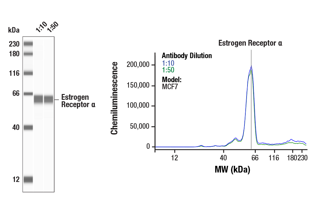
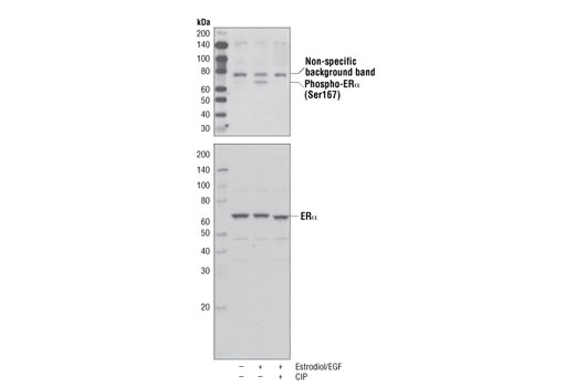
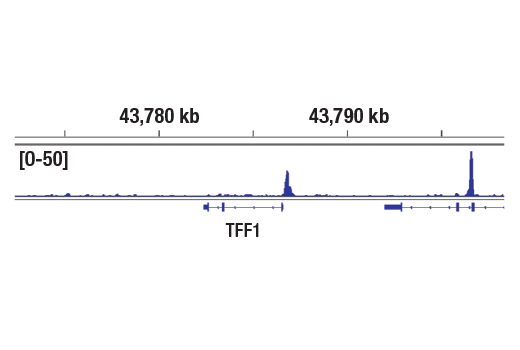
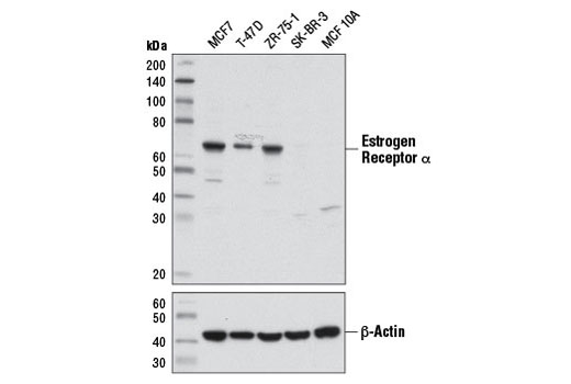
Estrogen receptor α (ERα), a member of the steroid receptor superfamily, contains highly conserved DNA binding and ligand binding domains (1). Through its estrogen-independent and estrogen-dependent activation domains (AF-1 and AF-2, respectively), ERα regulates transcription by recruiting coactivator proteins and interacting with general transcriptional machinery (2). Phosphorylation at multiple sites provides an important mechanism to regulate ERα activity (3-5). Ser104, 106, 118, and 167 are located in the amino-terminal transcription activation function domain AF-1, and phosphorylation of these serine residues plays an important role in regulating ERα activity. Ser118 may be the substrate of the transcription regulatory kinase CDK7 (5). Ser167 may be phosphorylated by p90RSK and Akt (4,6). According to the research literature, phosphorylation at Ser167 may confer tamoxifen resistance in breast cancer patients (4).

  1. Mangelsdorf, D.J. et al. (1995) Cell 83, 835-9.
  2. Glass, C.K. and Rosenfeld, M.G. (2000) Genes Dev 14, 121-41.
  3. Chen, D. et al. (1999) Mol Cell Biol 19, 1002-15.
  4. Campbell, R.A. et al. (2001) J Biol Chem 276, 9817-24.
  5. Chen, D. et al. (2000) Mol Cell 6, 127-37.
  6. Joel, P.B. et al. (1998) Mol Cell Biol 18, 1978-84.

Background References

    Trademarks and Patents

    Cell Signaling Technology is a trademark of Cell Signaling Technology, Inc.
    PhosphoPlus is a registered trademark of Cell Signaling Technology, Inc.
    All other trademarks are the property of their respective owners. Visit cellsignal.com/trademarks for more information.

    Limited Uses

    Except as otherwise expressly agreed in a writing signed by a legally authorized representative of CST, the following terms apply to Products provided by CST, its affiliates or its distributors. Any Customer's terms and conditions that are in addition to, or different from, those contained herein, unless separately accepted in writing by a legally authorized representative of CST, are rejected and are of no force or effect.

    Products are labeled with For Research Use Only or a similar labeling statement and have not been approved, cleared, or licensed by the FDA or other regulatory foreign or domestic entity, for any purpose. Customer shall not use any Product for any diagnostic or therapeutic purpose, or otherwise in any manner that conflicts with its labeling statement. Products sold or licensed by CST are provided for Customer as the end-user and solely for research and development uses. Any use of Product for diagnostic, prophylactic or therapeutic purposes, or any purchase of Product for resale (alone or as a component) or other commercial purpose, requires a separate license from CST. Customer shall (a) not sell, license, loan, donate or otherwise transfer or make available any Product to any third party, whether alone or in combination with other materials, or use the Products to manufacture any commercial products, (b) not copy, modify, reverse engineer, decompile, disassemble or otherwise attempt to discover the underlying structure or technology of the Products, or use the Products for the purpose of developing any products or services that would compete with CST products or services, (c) not alter or remove from the Products any trademarks, trade names, logos, patent or copyright notices or markings, (d) use the Products solely in accordance with CST Product Terms of Sale and any applicable documentation, and (e) comply with any license, terms of service or similar agreement with respect to any third party products or services used by Customer in connection with the Products.

    Revision 1
    #12108

    PhosphoPlus® Estrogen Receptor α (Ser167) Antibody Duet

    PhosphoPlus® Estrogen Receptor α (Ser167) Antibody Duet: Image 1 Expand Image
    Simple Western™ analysis of lysates (1 mg/mL) from MCF7 cells using Estrogen Receptor α (D8H8) Rabbit mAb #8644. The virtual lane view (left) shows the target band (as indicated) at 1:10 and 1:50 dilutions of primary antibody. The corresponding electropherogram view (right) plots chemiluminescence by molecular weight along the capillary at 1:10 (blue line) and 1:50 (green line) dilutions of primary antibody. This experiment was performed under reducing conditions on the Jess™ ​​​​​​​ Simple Western instrument from ProteinSimple, a BioTechne brand, using the 12-230 kDa separation module.
    PhosphoPlus® Estrogen Receptor α (Ser167) Antibody Duet: Image 2 Expand Image
    Western analysis of extracts from MCF7 cells, untreated or treated with Estrodiol/EGF (100nM each, together for 30 min) and further treated with calf intestinal phosphatase (CIP), using Phospho-Estrogen Receptor α (Ser167) (D1A3) Rabbit mAb (upper) or Estrogen Receptor α (D8H8) Rabbit mAb #8644 (lower).
    PhosphoPlus® Estrogen Receptor α (Ser167) Antibody Duet: Image 3 Expand Image
    Chromatin immunoprecipitations were performed with cross-linked chromatin from MCF7 cells grown in phenol red free medium and 5% charcoal stripped FBS for 4 d then treated with β-estradiol (10 nM) for 45 minutes and Estrogen Receptor α (D8H8) Rabbit mAb, using SimpleChIP® Plus Enzymatic Chromatin IP Kit (Magnetic Beads) #9005. DNA Library Prep Kit for Illumina® (ChIP-seq, CUT&RUN) #56795. The figure shows binding across TFF1/pS2, a known target gene of Estrogen Receptor α (see additional figure containing ChIP-qPCR data).
    PhosphoPlus® Estrogen Receptor α (Ser167) Antibody Duet: Image 4 Expand Image
    Western blot analysis of extracts from ER-positive cell lines (MCF7, T-47D, ZR-75-1) and ER-negative cell lines (SK-BR-3 and MCF 10A) using Estrogen Receptor α (D8H8) Rabbit mAb (upper) or β-Actin (D6A8) Rabbit mAb #8457 (lower).
    PhosphoPlus® Estrogen Receptor α (Ser167) Antibody Duet: Image 5 Expand Image
    CUT&RUN was performed with MCF7 cells grown in phenol red free medium and 5% charcoal stripped FBS for 4 days, then treated with β-estradiol (10 nM) for 45 minutes and either Estrogen Receptor a (D8H8) Rabbit mAb or Estrogen Receptor α (D6R2W) Rabbit mAb #13258, using CUT&RUN Assay Kit #86652. DNA Libraries were prepared using DNA Library Prep Kit for Illumina® #56795. The figure shows binding across TFF1 gene.
    PhosphoPlus® Estrogen Receptor α (Ser167) Antibody Duet: Image 6 Expand Image
    Chromatin immunoprecipitations were performed with cross-linked chromatin from MCF7 cells grown in phenol red free medium and 5% charcoal stripped FBS for 4 d then treated with β-estradiol (10 nM) for 45 minutes and 5 μl of Estrogen Receptor α (D8H8) Rabbit mAb, using SimpleChIP® Plus Enzymatic Chromatin IP Kit (Magnetic Beads) #9005. DNA Library Prep Kit for Illumina® (ChIP-seq, CUT&RUN) #56795. The figure shows binding across chromosome 21 (upper), including TFF1/pS2 (lower), a known target gene of Estrogen Receptor α (see additional figure containing ChIP-qPCR data).
    PhosphoPlus® Estrogen Receptor α (Ser167) Antibody Duet: Image 7 Expand Image
    CUT&RUN was performed with MCF7 cells grown in phenol red free medium and 5% charcoal stripped FBS for 4 days, then treated with β-estradiol (10 nM) for 45 minutes and either Estrogen Receptor a (D8H8) Rabbit mAb or Estrogen Receptor α (D6R2W) Rabbit mAb #13258, using CUT&RUN Assay Kit #86652. DNA Libraries were prepared using DNA Library Prep Kit for Illumina® #56795. The figures show binding across chromosome 21 (upper), including TFF1 gene (lower).
    PhosphoPlus® Estrogen Receptor α (Ser167) Antibody Duet: Image 8 Expand Image
    Chromatin immunoprecipitations were performed with cross-linked chromatin from MCF7 cells grown in phenol red free medium and 5% charcoal stripped FBS for 4 d then treated with β-estradiol (10 nM) for 45 minutes and either Estrogen Receptor α (D8H8) Rabbit mAb or Normal Rabbit IgG #2729 using SimpleChIP® Enzymatic Chromatin IP Kit (Magnetic Beads) #9003. The enriched DNA was quantified by real-time PCR using SimpleChIP® Human ESR1 Promoter Primers #9673, SimpleChIP® Human pS2 Promoter Primers #9702, and SimpleChIP® Human α Satellite Repeat Primers #4486. The amount of immunoprecipitated DNA in each sample is represented as signal relative to the total amount of input chromatin, which is equivalent to one.
    PhosphoPlus® Estrogen Receptor α (Ser167) Antibody Duet: Image 9 Expand Image
    CUT&RUN was performed with MCF7 cells grown in phenol red free medium and 5% charcoal stripped FBS for 4 days, then treated with β-estradiol (10 nM) for 45 minutes and either Estrogen Receptor a (D8H8) Rabbit mAb or Rabbit (DA1E) mAb IgG XP® Isotype Control (CUT&RUN) #66362, using CUT&RUN Assay Kit #86652. The enriched DNA was quantified by real-time PCR using SimpleChIP® Human PS2 Promoter Primers #9702 and SimpleChIP® Human α Satellite Repeat Primers #4486. The amount of immunoprecipitated DNA in each sample is represented as signal relative to the total amount of input chromatin, which is equivalent to one.