WB, W-S, IP, IHC-Bond, IHC-P, IF-IC
H
Endogenous
125
Rabbit IgG
#Q05397
5747
Product Information
Product Usage Information
| Application | Dilution |
|---|---|
| Western Blotting | 1:1000 |
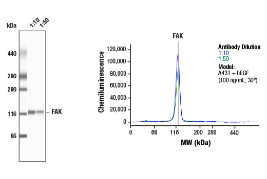
| Simple Western™ | 1:10 - 1:50 |
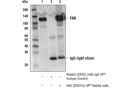
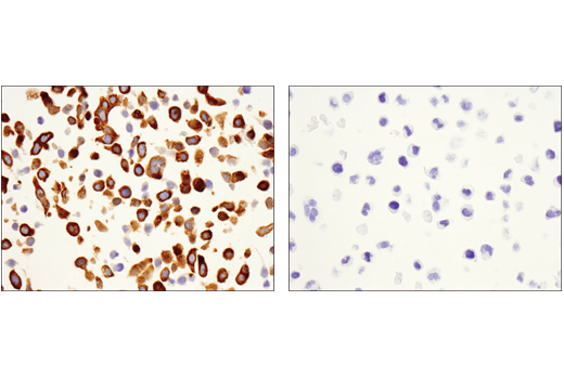
| Immunoprecipitation | 1:50 |
| IHC Leica Bond | 1:150 - 1:600 |
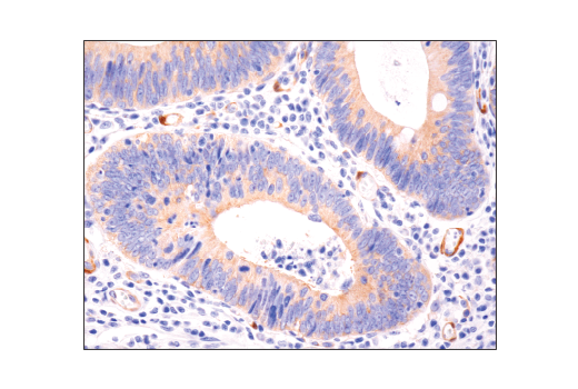
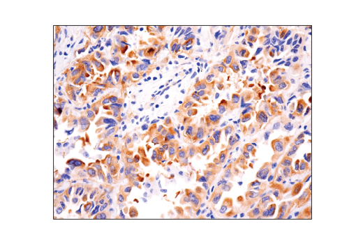
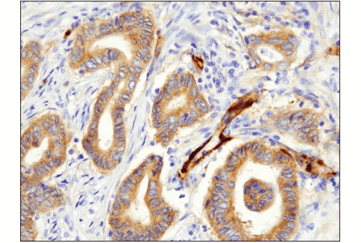
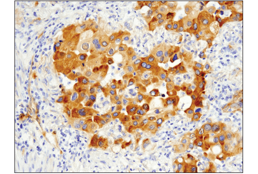
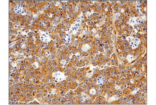
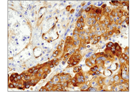
| Immunohistochemistry (Paraffin) | 1:300 - 1:1200 |
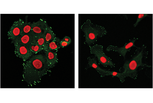
| Immunofluorescence (Immunocytochemistry) | 1:50 - 1:100 |
Storage
Supplied in 10 mM sodium HEPES (pH 7.5), 150 mM NaCl, 100 µg/ml BSA, 50% glycerol and less than 0.02% sodium azide. Store at –20°C. Do not aliquot the antibody.
For a carrier-free (BSA and azide free) version of this product see product #60557.
Specificity / Sensitivity
Species Reactivity:
Human
Source / Purification
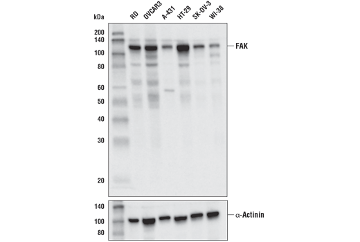
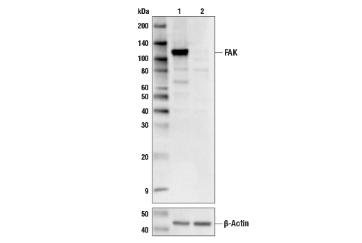
Monoclonal antibody is produced by immunizing animals with a synthetic peptide corresponding to residues surrounding Val793 of human FAK protein.
Background
Focal adhesion kinase (FAK) is a widely expressed cytoplasmic protein tyrosine kinase involved in integrin-mediated signal transduction. It plays an important role in the control of several biological processes, including cell spreading, migration, and survival (1). Activation of FAK by integrin clustering leads to autophosphorylation at Tyr397, which is a binding site for the Src family kinases PI3K and PLCγ (2-5). Recruitment of Src family kinases results in the phosphorylation of Tyr407, Tyr576, and Tyr577 in the catalytic domain, and Tyr871 and Tyr925 in the carboxy-terminal region of FAK (6,7).
- Parsons, J.T. et al. (2000) Oncogene 19, 5606-13.
- Schaller, M.D. et al. (1994) Mol Cell Biol 14, 1680-8.
- Cobb, B.S. et al. (1994) Mol Cell Biol 14, 147-55.
- Chen, H.C. et al. (1996) J Biol Chem 271, 26329-34.
- Zhang, X. et al. (1999) Proc Natl Acad Sci U S A 96, 9021-6.
- Calalb, M.B. et al. (1995) Mol Cell Biol 15, 954-63.
- Schlaepfer, D.D. et al. (1994) Nature 372, 786-791.
Species Reactivity
Species reactivity is determined by testing in at least one approved application (e.g., western blot).
Western Blot Buffer
IMPORTANT: For western blots, incubate membrane with diluted primary antibody in 5% w/v BSA, 1X TBS, 0.1% Tween® 20 at 4°C with gentle shaking, overnight.
Applications Key
WB: Western Blotting W-S: Simple Western™ IP: Immunoprecipitation IHC-Bond: IHC Leica Bond IHC-P: Immunohistochemistry (Paraffin) IF-IC: Immunofluorescence (Immunocytochemistry)
Cross-Reactivity Key
H: human M: mouse R: rat Hm: hamster Mk: monkey Vir: virus Mi: mink C: chicken Dm: D. melanogaster X: Xenopus Z: zebrafish B: bovine Dg: dog Pg: pig Sc: S. cerevisiae Ce: C. elegans Hr: horse GP: Guinea Pig Rab: rabbit All: all species expected
Trademarks and Patents
Limited Uses
Except as otherwise expressly agreed in a writing signed by a legally authorized representative of CST, the following terms apply to Products provided by CST, its affiliates or its distributors. Any Customer's terms and conditions that are in addition to, or different from, those contained herein, unless separately accepted in writing by a legally authorized representative of CST, are rejected and are of no force or effect.
Products are labeled with For Research Use Only or a similar labeling statement and have not been approved, cleared, or licensed by the FDA or other regulatory foreign or domestic entity, for any purpose. Customer shall not use any Product for any diagnostic or therapeutic purpose, or otherwise in any manner that conflicts with its labeling statement. Products sold or licensed by CST are provided for Customer as the end-user and solely for research and development uses. Any use of Product for diagnostic, prophylactic or therapeutic purposes, or any purchase of Product for resale (alone or as a component) or other commercial purpose, requires a separate license from CST. Customer shall (a) not sell, license, loan, donate or otherwise transfer or make available any Product to any third party, whether alone or in combination with other materials, or use the Products to manufacture any commercial products, (b) not copy, modify, reverse engineer, decompile, disassemble or otherwise attempt to discover the underlying structure or technology of the Products, or use the Products for the purpose of developing any products or services that would compete with CST products or services, (c) not alter or remove from the Products any trademarks, trade names, logos, patent or copyright notices or markings, (d) use the Products solely in accordance with CST Product Terms of Sale and any applicable documentation, and (e) comply with any license, terms of service or similar agreement with respect to any third party products or services used by Customer in connection with the Products.