WB, IP, IHC-Bond, IHC-P, FC-FP
M
Endogenous
60-80
Rabbit IgG
#P08101
14130
Product Information
Product Usage Information
| Application | Dilution |
|---|---|
| Western Blotting | 1:1000 |
| Immunoprecipitation | 1:50 |
| IHC Leica Bond | 1:100 |
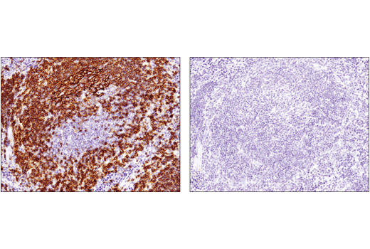
| Immunohistochemistry (Paraffin) | 1:100 |
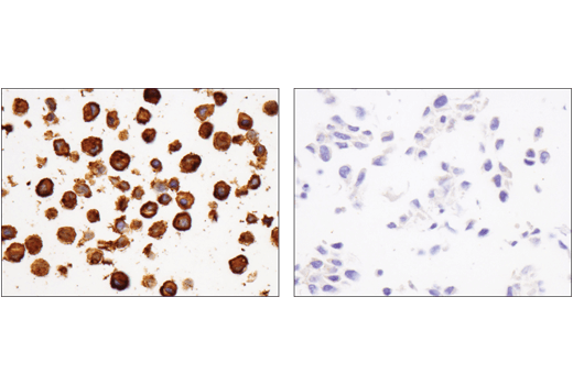
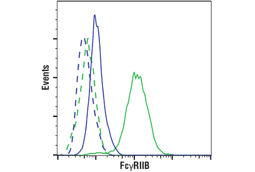
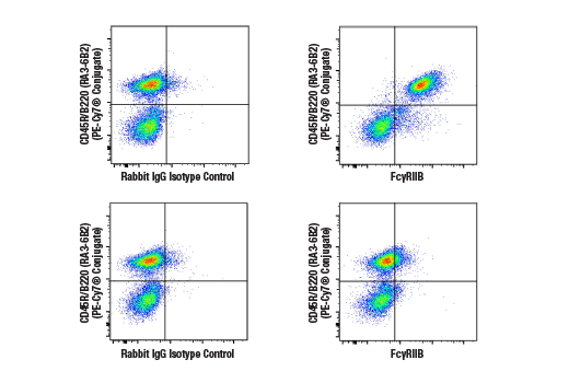
| Flow Cytometry (Fixed/Permeabilized) | 1:400 |
Storage
For a carrier free (BSA and azide free) version of this product see product #81563.
Specificity / Sensitivity
Species Reactivity:
Mouse
Source / Purification
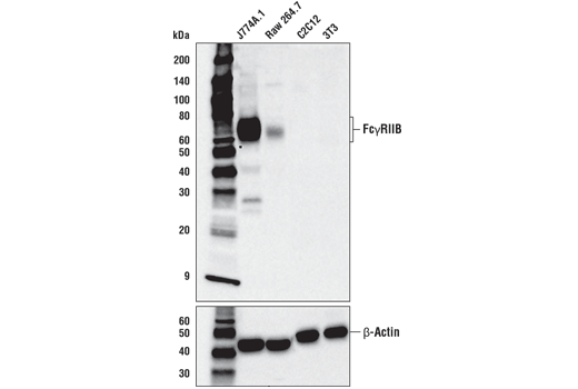
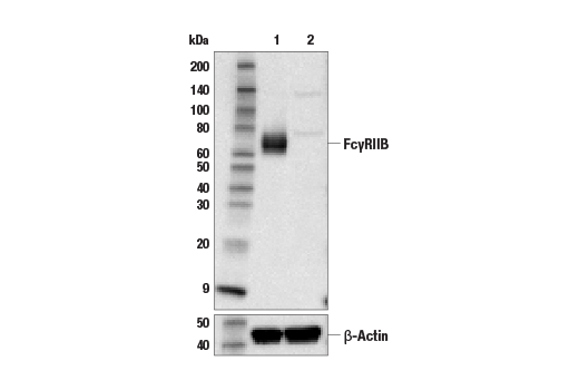
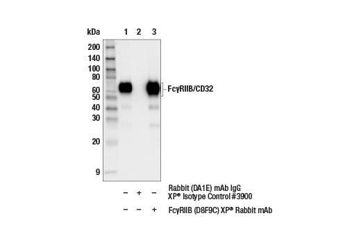
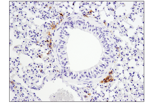
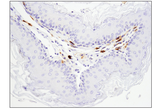
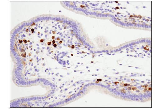
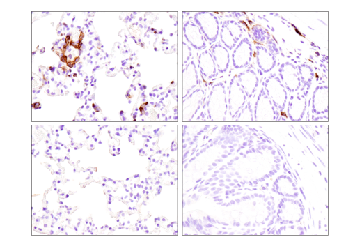
Monoclonal antibody is produced by immunizing animals with a synthetic peptide corresponding to residues surrounding Pro284 of mouse FcγRIIB protein.
Background
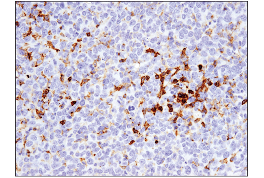
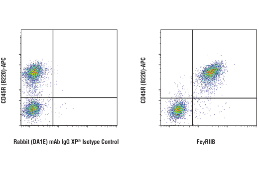
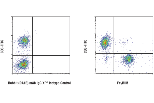
FcγRIIB (CD32B) is a low affinity, IgG Fc-binding receptor expressed on B cells, monocytes, macrophages, and dendritic cells (DCs) (1-3). It is the inhibitory Fc receptor and signals through an immunoreceptor tyrosine-based inhibitory motif (ITIM) within its carboxy-terminal cytoplasmic tail (2). Binding of immune complexes to FcγRIIB results in tyrosine phosphorylation of the ITIM motif at Tyr292 and recruitment of the phosphatase SHIP, which mediates inhibitory effects on immune cell activation (2,4). In this way, FcγRIIB suppresses the effects of activating Fc-binding receptors (3). For example, mice deficient for FcγRIIB have greater T cell and DC responses following injection of immune complexes (5,6). In addition, FcγRIIB plays a role in B cell affinity maturation (7). Signaling through FcγRIIB in the absence of signaling through the B cell receptor (BCR) is proapoptotic, while signaling through FcγRIIB and the BCR simultaneously attenuates the apoptotic signal and results in selection of B cells with higher antigen affinity (7).
- Tridandapani, S. et al. (2002) J. Biol. Chem. 277, 5082-89.
- Tridandapani, S. et al. (1997) Mol. Cell. Biol. 17, 4305-11.
- Guilliams, M. et al. (2014) Nat Rev Immunol 14, 94-108.
- Bruhns, P. et al. (2000) J. Biol. Chem. 275, 37357-64.
- Kalergis, A.M. and Ravetch, J.V. (2002) J Exp Med 195, 1653-9.
- Desai, D.D. et al. (2007) J Immunol 178, 6217-26.
- Pearse, R.N. et al. (1999) Immunity 10, 753-60.
Species Reactivity
Species reactivity is determined by testing in at least one approved application (e.g., western blot).
Western Blot Buffer
IMPORTANT: For western blots, incubate membrane with diluted primary antibody in 5% w/v BSA, 1X TBS, 0.1% Tween® 20 at 4°C with gentle shaking, overnight.
Applications Key
WB: Western Blotting IP: Immunoprecipitation IHC-Bond: IHC Leica Bond IHC-P: Immunohistochemistry (Paraffin) FC-FP: Flow Cytometry (Fixed/Permeabilized)
Cross-Reactivity Key
H: human M: mouse R: rat Hm: hamster Mk: monkey Vir: virus Mi: mink C: chicken Dm: D. melanogaster X: Xenopus Z: zebrafish B: bovine Dg: dog Pg: pig Sc: S. cerevisiae Ce: C. elegans Hr: horse GP: Guinea Pig Rab: rabbit All: all species expected
Trademarks and Patents
Limited Uses
Except as otherwise expressly agreed in a writing signed by a legally authorized representative of CST, the following terms apply to Products provided by CST, its affiliates or its distributors. Any Customer's terms and conditions that are in addition to, or different from, those contained herein, unless separately accepted in writing by a legally authorized representative of CST, are rejected and are of no force or effect.
Products are labeled with For Research Use Only or a similar labeling statement and have not been approved, cleared, or licensed by the FDA or other regulatory foreign or domestic entity, for any purpose. Customer shall not use any Product for any diagnostic or therapeutic purpose, or otherwise in any manner that conflicts with its labeling statement. Products sold or licensed by CST are provided for Customer as the end-user and solely for research and development uses. Any use of Product for diagnostic, prophylactic or therapeutic purposes, or any purchase of Product for resale (alone or as a component) or other commercial purpose, requires a separate license from CST. Customer shall (a) not sell, license, loan, donate or otherwise transfer or make available any Product to any third party, whether alone or in combination with other materials, or use the Products to manufacture any commercial products, (b) not copy, modify, reverse engineer, decompile, disassemble or otherwise attempt to discover the underlying structure or technology of the Products, or use the Products for the purpose of developing any products or services that would compete with CST products or services, (c) not alter or remove from the Products any trademarks, trade names, logos, patent or copyright notices or markings, (d) use the Products solely in accordance with CST Product Terms of Sale and any applicable documentation, and (e) comply with any license, terms of service or similar agreement with respect to any third party products or services used by Customer in connection with the Products.