| Product Includes | Product # | Quantity | Mol. Wt | Isotype/Source |
|---|---|---|---|---|
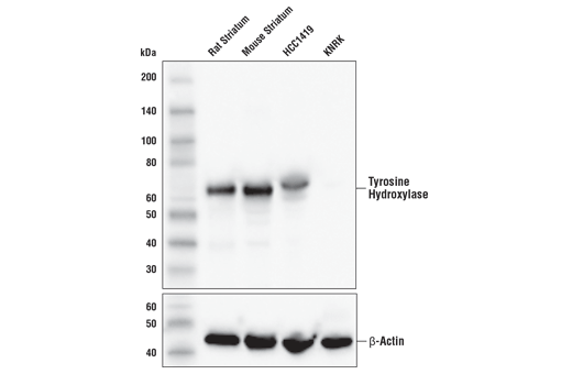
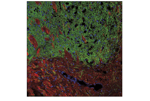
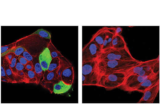
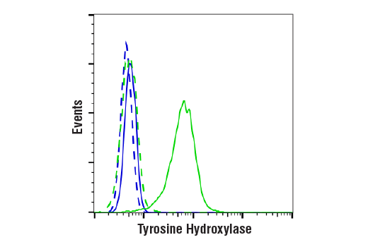
| Tyrosine Hydroxylase (E2L6M) Rabbit mAb | 58844 | 20 µl | 55-60 kDa | Rabbit IgG |
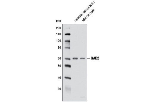
| GAD2 (D5G2) XP® Rabbit mAb | 5843 | 20 µl | 60 kDa | Rabbit IgG |
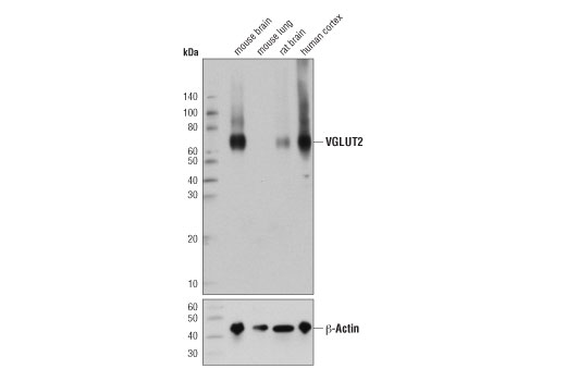
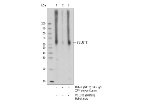
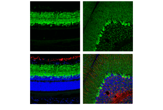
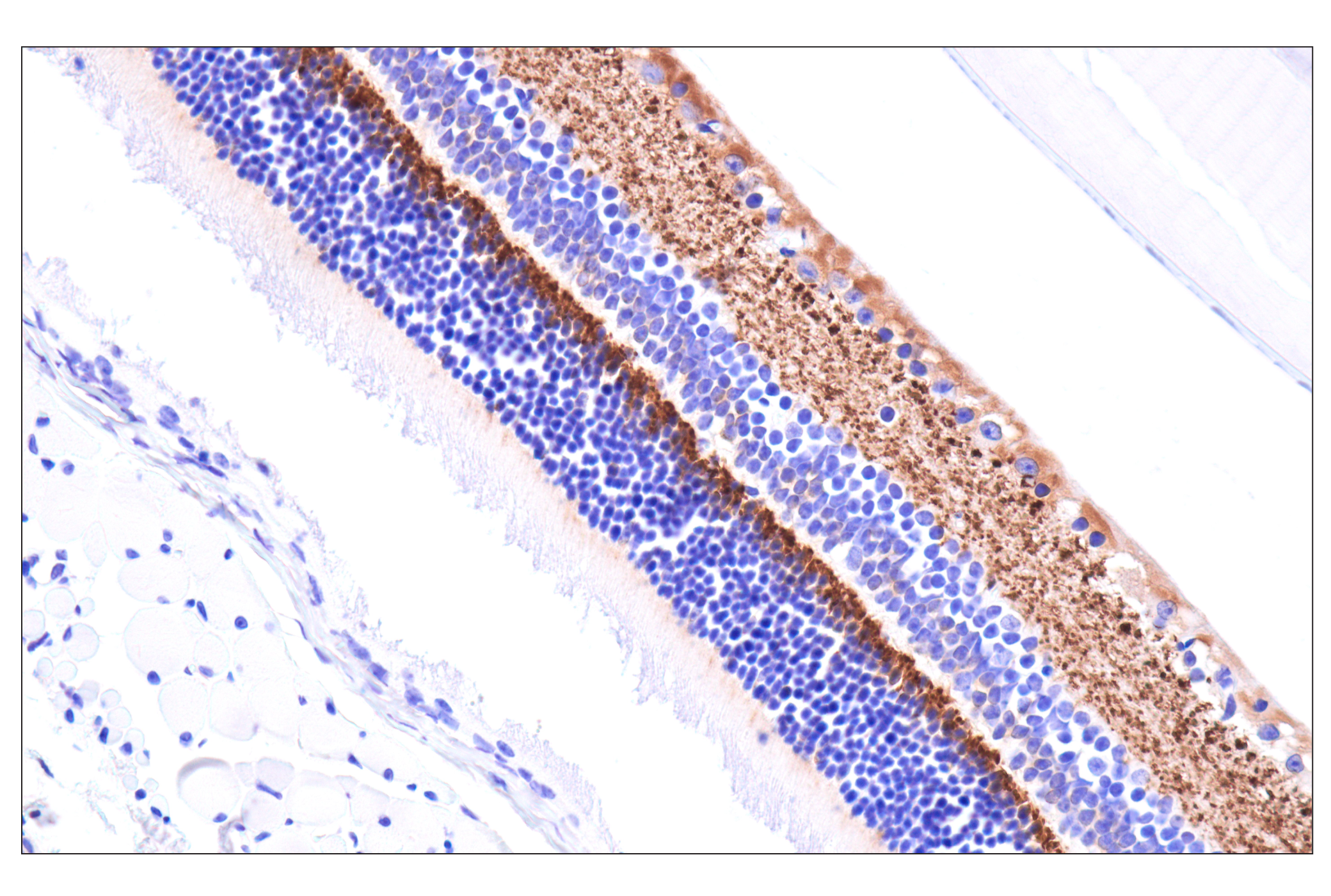
| VGLUT2 (D7D2H) Rabbit mAb | 71555 | 20 µl | 65-70 kDa | Rabbit IgG |
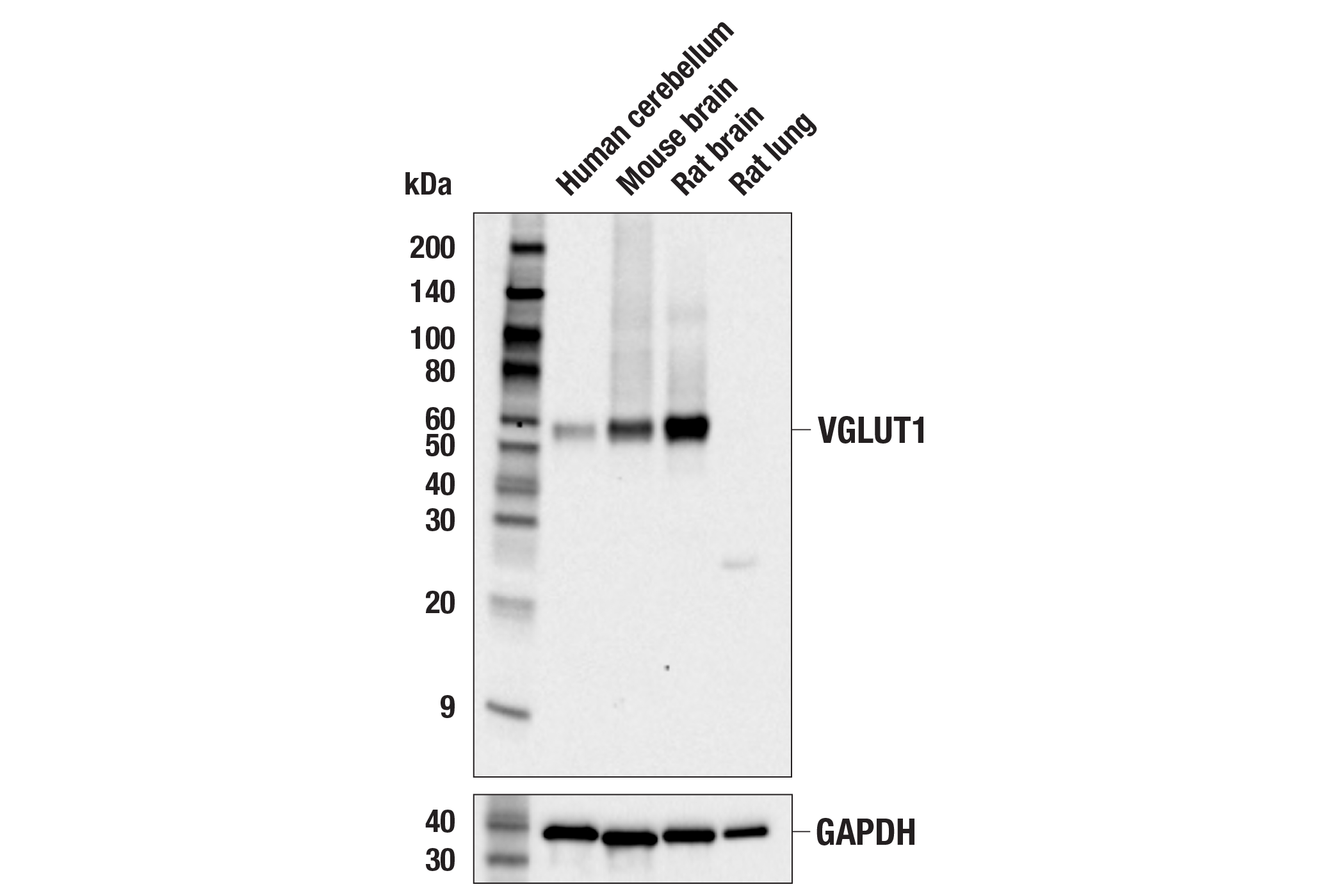
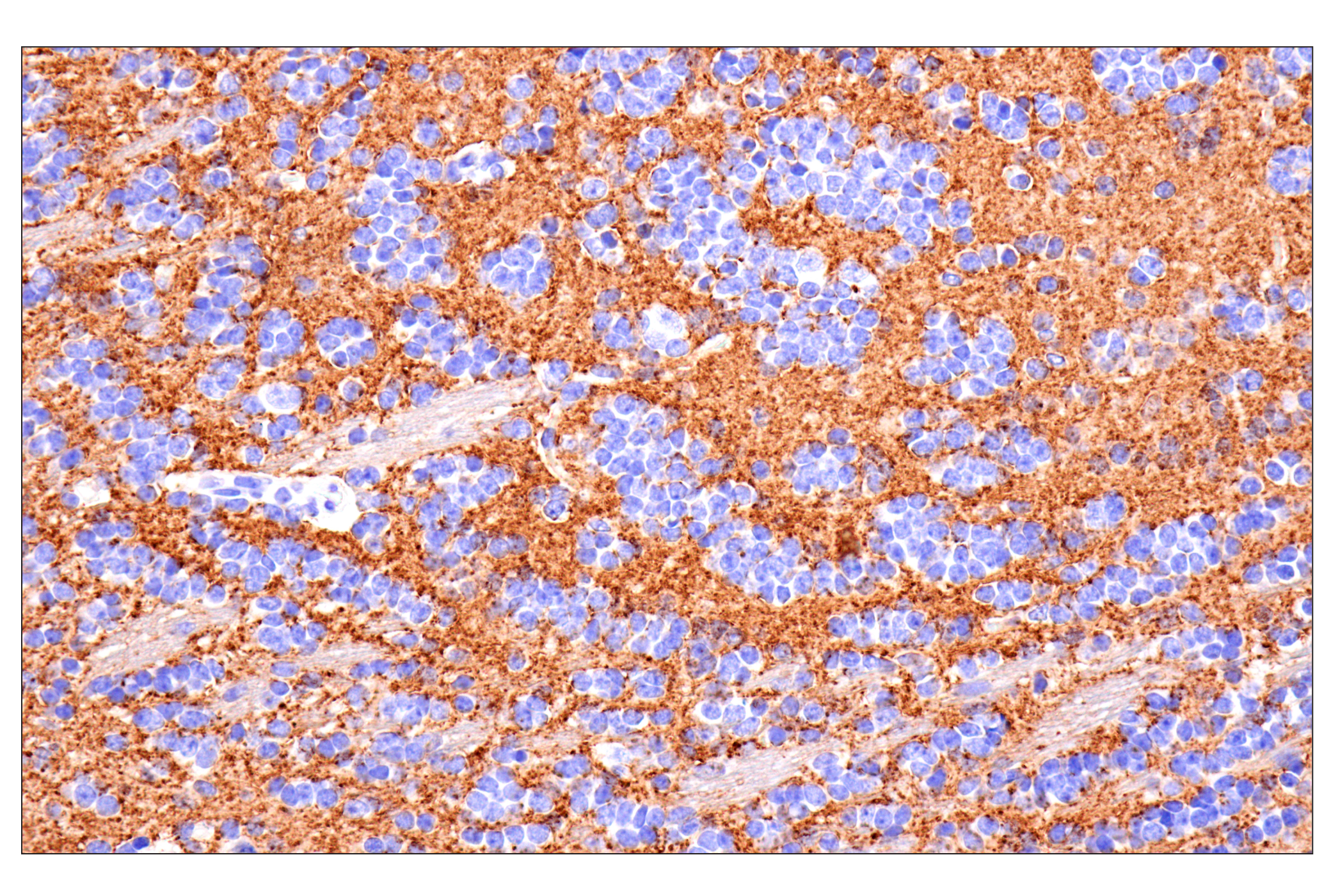
| VGLUT1 (E8L5B) Rabbit mAb | 47181 | 20 µl | 62 kDa | Rabbit IgG |
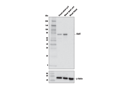
| ChAT (E4F9G) Rabbit mAb | 27269 | 20 µl | 71 kDa | Rabbit IgG |
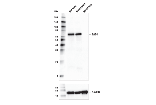
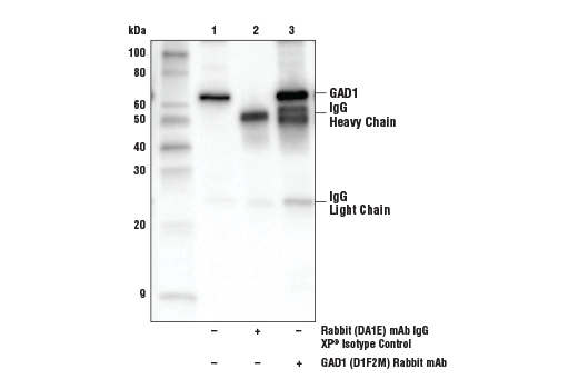
| GAD1 (D1F2M) Rabbit mAb | 41318 | 20 µl | 67 kDa | Rabbit IgG |
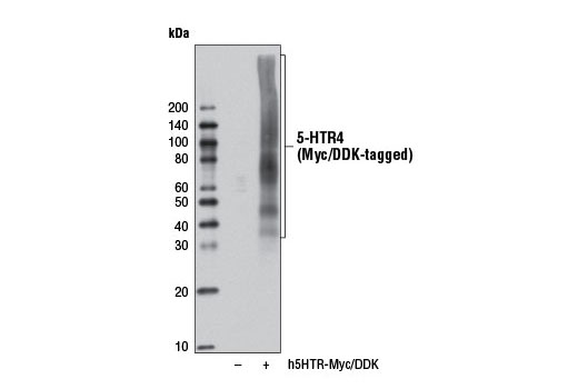
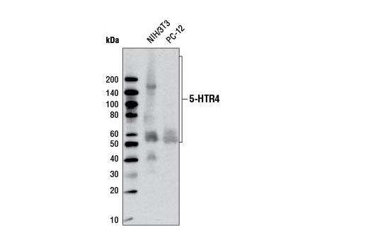
| 5-HTR4 (D8O5K) Rabbit mAb | 13690 | 20 µl | 40-140 kDa | Rabbit IgG |
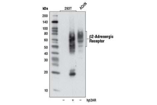
| β2-Adrenergic Receptor (D6H2) Rabbit mAb | 8513 | 20 µl | 50-100 kDa | Rabbit IgG |
| Anti-rabbit IgG, HRP-linked Antibody | 7074 | 100 µl | Goat |
Please visit cellsignal.com for individual component applications, species cross-reactivity, dilutions, protocols, and additional product information.
Description
The Functional Neuron Marker Antibody Sampler Kit provides an economical means of detecting markers that facilitate phenotyping neurons by function. The kit includes enough antibodies to perform at least two western blot experiments with each primary antibody.
Storage
Background
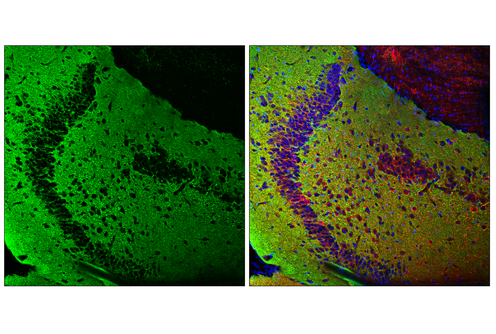
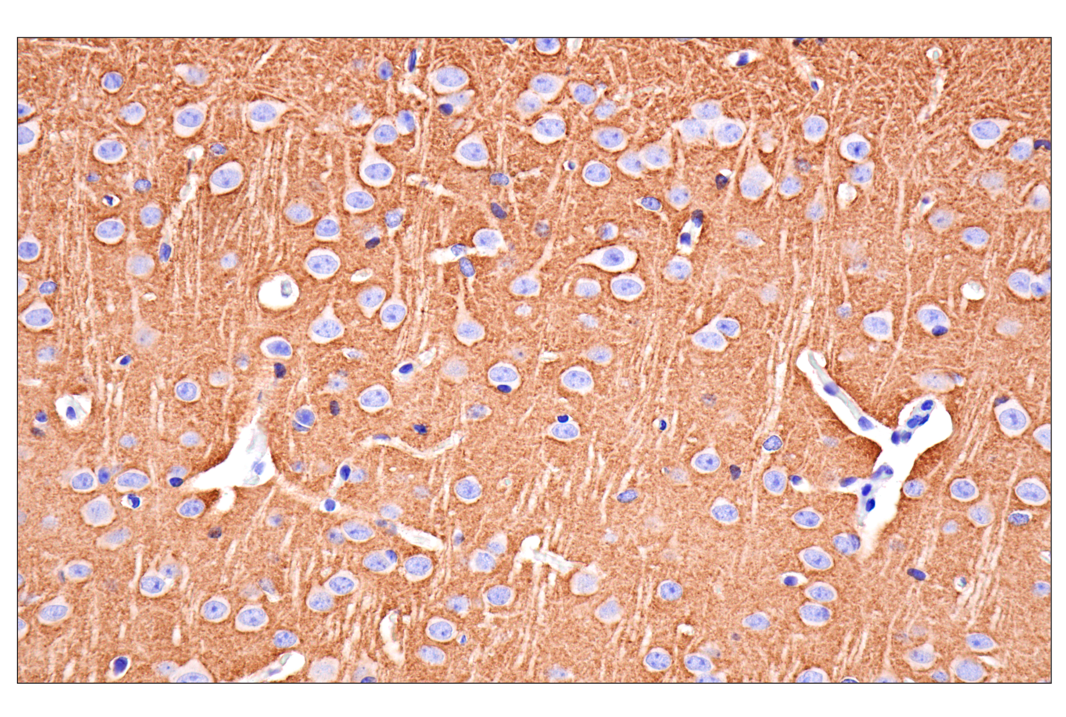
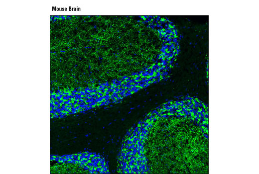
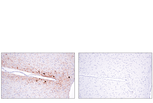
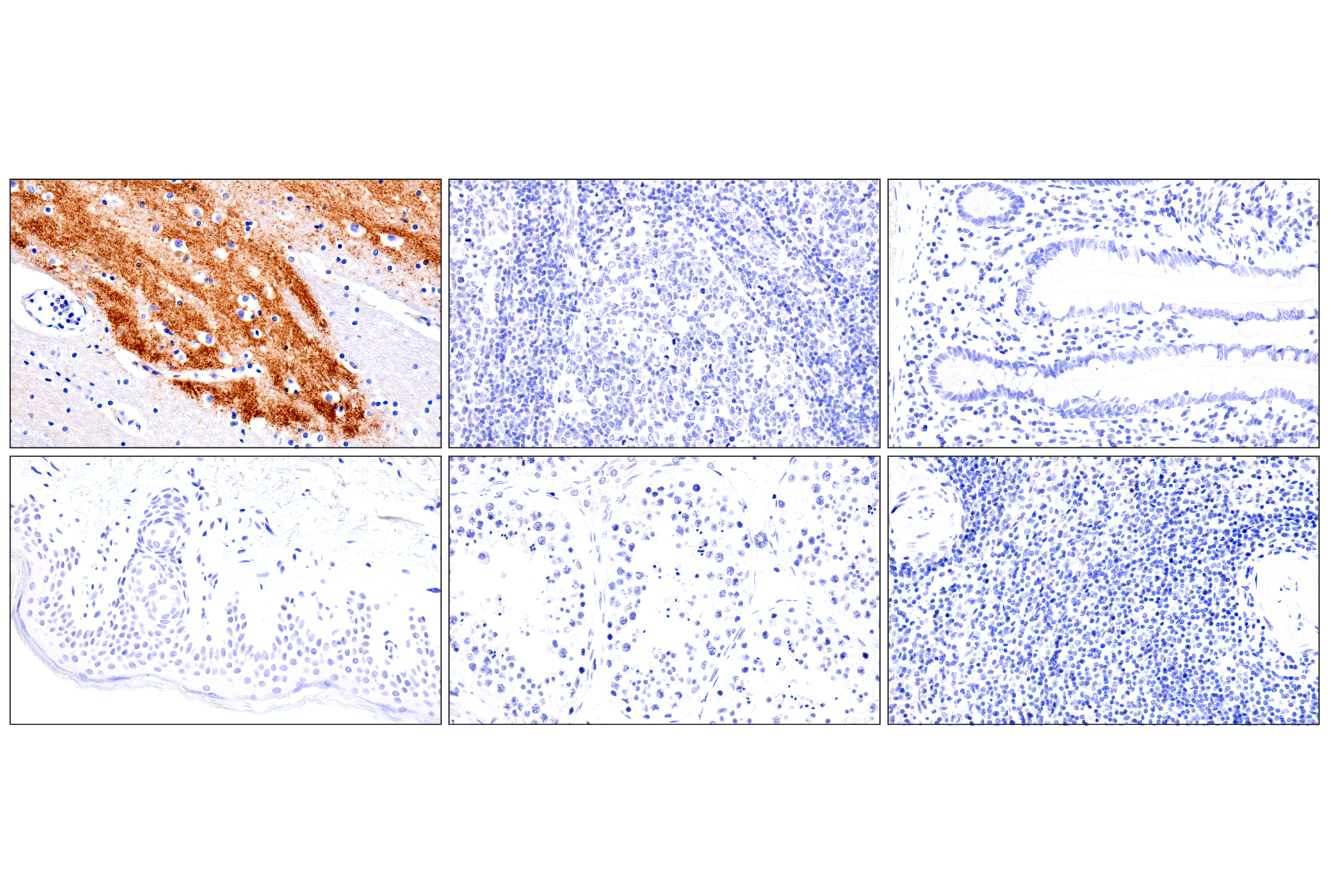
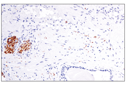
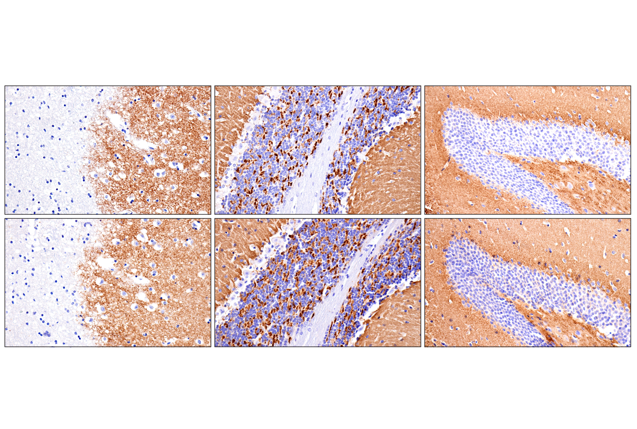
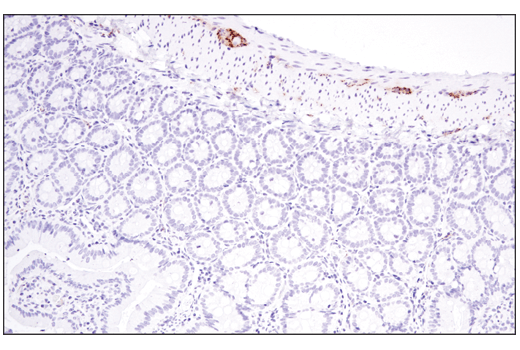
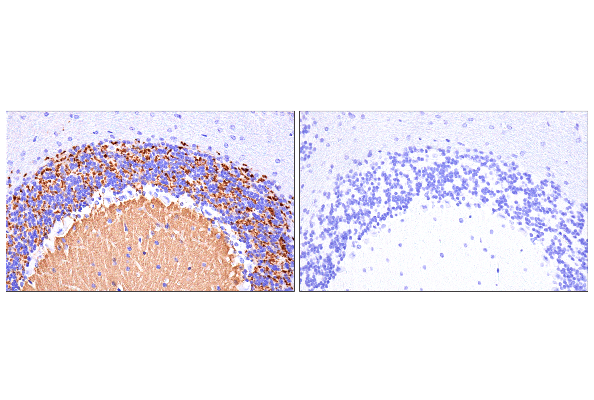
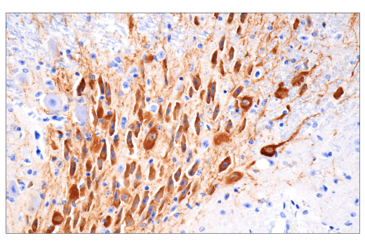
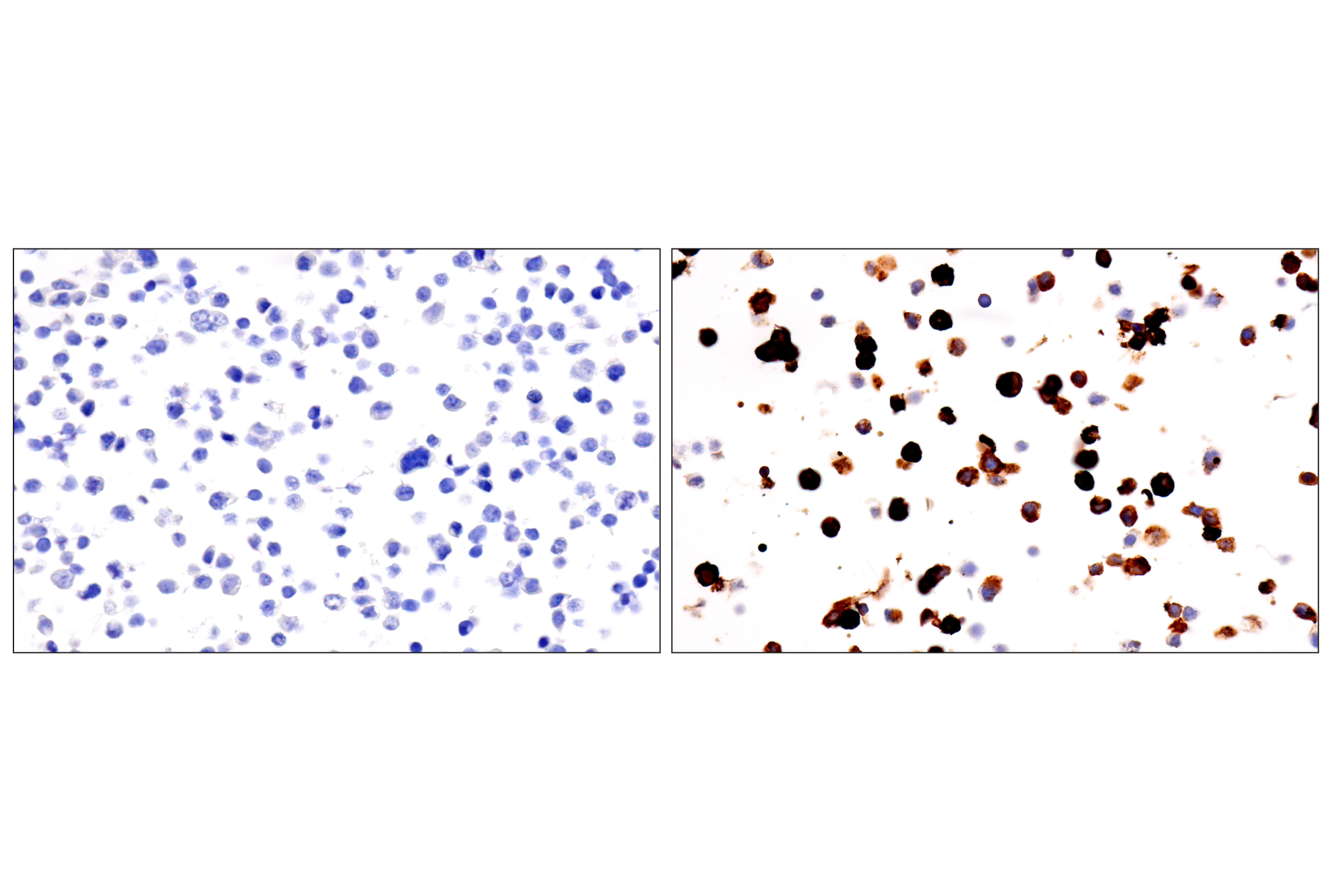
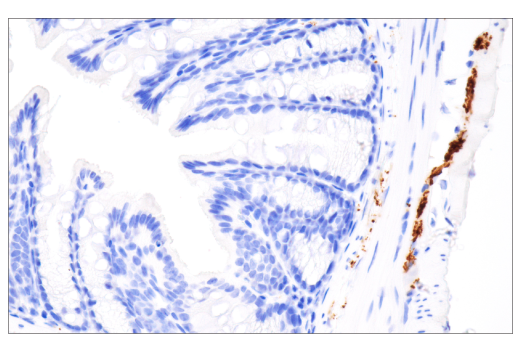
Tyrosine hydroxylase (TH) catalyzes the rate-limiting step in the synthesis of the neurotransmitter dopamine and other catecholamines, and acts as a marker for dopaminergic neurons (1,2). Choline O-acetyltransferase (ChAT) is the enzyme that catalyzes synthesis of acetylcholine (ACh) in the central and peripheral nervous system. ChAT is found in high levels within cholinergic neurons and can be used to measure their functional states (3). Vesicular glutamate transporters 1 and 2 (VGLUT1 and VGLUT2) are responsible for transporting the excitatory neurotransmitter glutamate into synaptic vesicles of glutamatergic neurons. VGLUT1 and VGLUT2 are complimentarily expressed and act as markers for glutamatergic neurons (4). Glutamate decarboxylase (GAD) is the main enzyme that synthesizes GABA from glutamate. GABA producing neurons, called GABAergic neurons, utilize GABA as their major inhibitory neurotransmitter with both isoforms of GAD, GAD1, and GAD2, acting as functional markers for these neurons (5). β2-adrenergic receptor (β2AR) is a G protein-coupled receptor (GPCR) that mediates the actions of catecholamines, mainly through stimulation by epinephrine (adrenaline), in the central and peripheral nervous system (6,7). Serotonin receptor 4 (5-HTR4) is an excitatory GPCR that activates the cyclic AMP (cAMP)-PKA pathway (8,9). 5-HTR4 is located post-synaptically on serotonergic neurons (10).
- Kumer, S.C. and Vrana, K.E. (1996) J Neurochem 67, 443-62.
- Weihe, E. et al. Cell Mol Neurobiol 26, 659-78.
- Oda, Y. (1999) Pathol Int 49, 921-37.
- Ziegler, D.R. et al. (2002) J Comp Neurol 448, 217-29.
- Le, T.N. et al. (2017) J Neurosci 37, 8816-8829.
- Wu, Y. et al. (2021) Biomolecules 11, 936. doi: 10.3390/biom11070936.
- Wallukat, G. (2002) Herz 27, 683-90.
- Nichols, D.E. and Nichols, C.D. (2008) Chem Rev 108, 1614-41.
- Karayol, R. et al. (2021) Mol Psychiatry 26, 2334-2349.
- Samuels, B.A. et al. (2016) Neuroscientist 22, 26-45.
Background References
Trademarks and Patents
Limited Uses
Except as otherwise expressly agreed in a writing signed by a legally authorized representative of CST, the following terms apply to Products provided by CST, its affiliates or its distributors. Any Customer's terms and conditions that are in addition to, or different from, those contained herein, unless separately accepted in writing by a legally authorized representative of CST, are rejected and are of no force or effect.
Products are labeled with For Research Use Only or a similar labeling statement and have not been approved, cleared, or licensed by the FDA or other regulatory foreign or domestic entity, for any purpose. Customer shall not use any Product for any diagnostic or therapeutic purpose, or otherwise in any manner that conflicts with its labeling statement. Products sold or licensed by CST are provided for Customer as the end-user and solely for research and development uses. Any use of Product for diagnostic, prophylactic or therapeutic purposes, or any purchase of Product for resale (alone or as a component) or other commercial purpose, requires a separate license from CST. Customer shall (a) not sell, license, loan, donate or otherwise transfer or make available any Product to any third party, whether alone or in combination with other materials, or use the Products to manufacture any commercial products, (b) not copy, modify, reverse engineer, decompile, disassemble or otherwise attempt to discover the underlying structure or technology of the Products, or use the Products for the purpose of developing any products or services that would compete with CST products or services, (c) not alter or remove from the Products any trademarks, trade names, logos, patent or copyright notices or markings, (d) use the Products solely in accordance with CST Product Terms of Sale and any applicable documentation, and (e) comply with any license, terms of service or similar agreement with respect to any third party products or services used by Customer in connection with the Products.