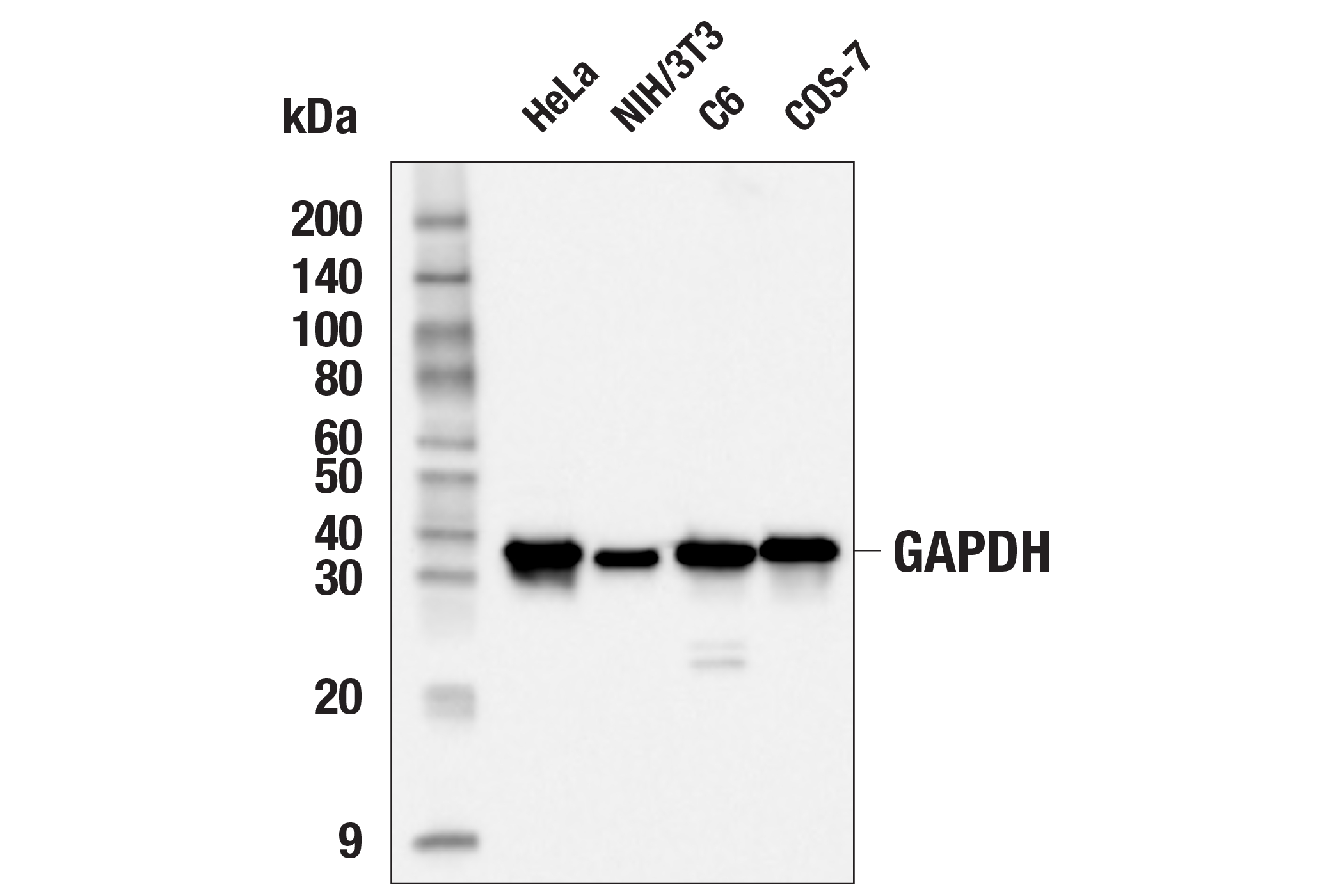
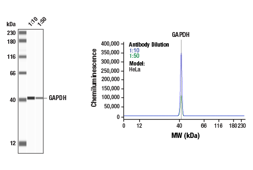
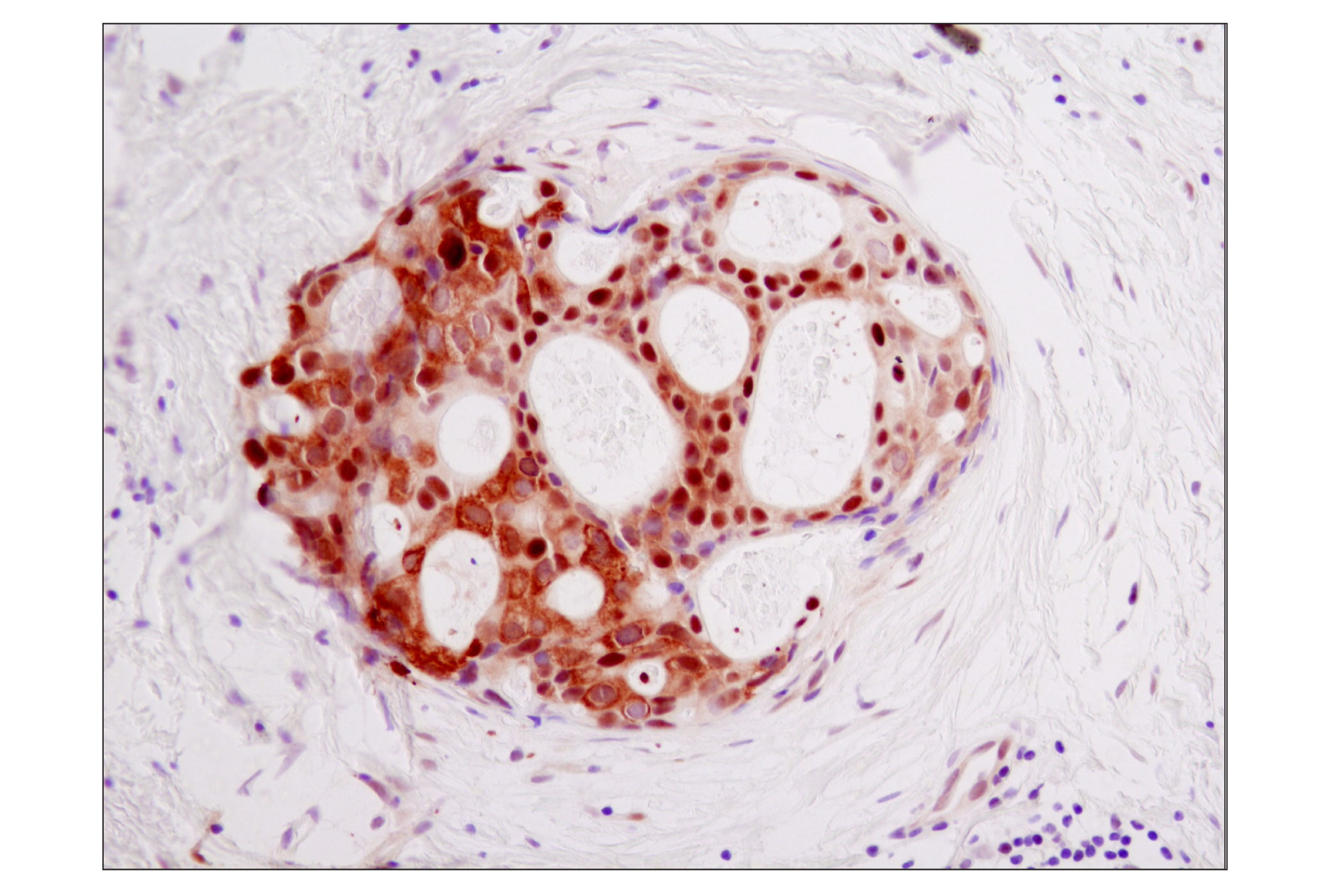
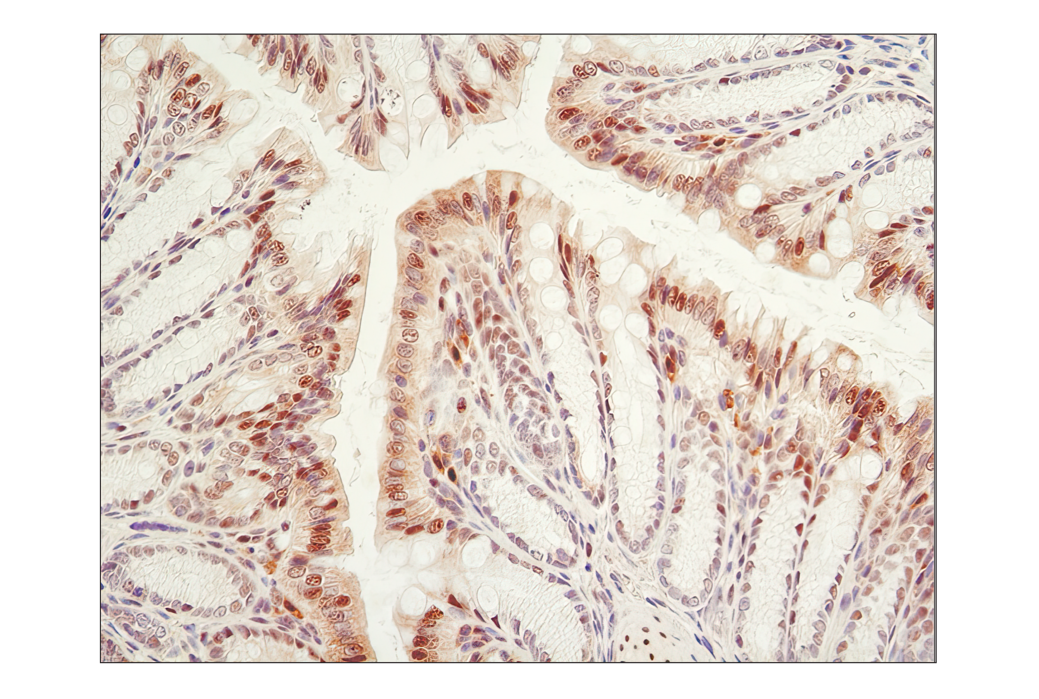
WB, W-S, IHC-P, IF-IC
H M R Mk
Endogenous
37
Rabbit IgG
#P04406
2597
Product Information
Product Usage Information
| Application | Dilution |
|---|---|
| Western Blotting | 1:1000 |
| Simple Western™ | 1:10 - 1:50 |
| Immunohistochemistry (Paraffin) | 1:400 - 1:1600 |
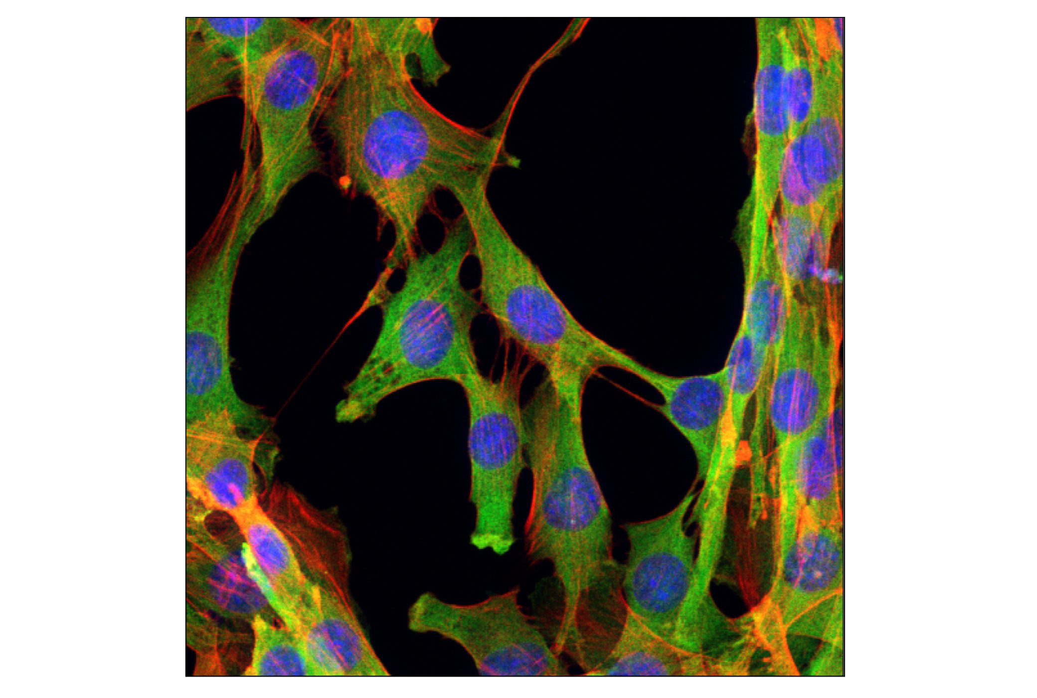
| Immunofluorescence (Immunocytochemistry) | 1:100 - 1:400 |
Storage
For a carrier free (BSA and azide free) version of this product see product #92310.
Specificity / Sensitivity
Species Reactivity:
Human, Mouse, Rat, Monkey
Species predicted to react based on 100% sequence homology
The antigen sequence used to produce this antibody shares
100% sequence homology with the species listed here, but
reactivity has not been tested or confirmed to work by CST.
Use of this product with these species is not covered under
our
Product Performance Guarantee.
Pig
Source / Purification
Monoclonal antibody is produced by immunzing animals with a synthetic peptide corresponding to residues surrounding Lys260 of human GAPDH protein.
Background
Glyceraldehyde-3-phosphate dehydrogenase (GAPDH) catalyzes the phosphorylation of glyceraldehyde-3-phosphate during glycolysis. Though differentially expressed from tissue to tissue (1), GAPDH is thought to be a constitutively expressed housekeeping protein. For this reason, GAPDH mRNA and protein levels are often measured as controls in experiments quantifying specific changes in expression of other targets. Recent work has elucidated roles for GAPDH in apoptosis (2), gene expression (3), and nuclear transport (4). GAPDH may also play a role in neurodegenerative pathologies such as Huntington and Alzheimer's diseases (4,5).
- Barber, R.D. et al. (2005) Physiol. Genomics 21, 389-95.
- Hara, M.R. and Snyder, S.H. (2006) Cell Mol. Neurobiol. 26, 527-38.
- Zheng, L. et al. (2003) Cell 114, 255-66.
- Bae, B.I. et al. (2006) Proc. Natl. Acad. Sci. USA 103, 3405-9.
- Wang, Q. et al. (2005) FASEB J. 19, 869-71.
- Kozako, T. et al. (2015) Sci Rep 5, 11345.
Species Reactivity
Species reactivity is determined by testing in at least one approved application (e.g., western blot).
Western Blot Buffer
IMPORTANT: For western blots, incubate membrane with diluted primary antibody in 5% w/v nonfat dry milk, 1X TBS, 0.1% Tween® 20 at 4°C with gentle shaking, overnight.
Applications Key
WB: Western Blotting W-S: Simple Western™ IHC-P: Immunohistochemistry (Paraffin) IF-IC: Immunofluorescence (Immunocytochemistry)
Cross-Reactivity Key
H: human M: mouse R: rat Hm: hamster Mk: monkey Vir: virus Mi: mink C: chicken Dm: D. melanogaster X: Xenopus Z: zebrafish B: bovine Dg: dog Pg: pig Sc: S. cerevisiae Ce: C. elegans Hr: horse GP: Guinea Pig Rab: rabbit All: all species expected
Trademarks and Patents
Limited Uses
Except as otherwise expressly agreed in a writing signed by a legally authorized representative of CST, the following terms apply to Products provided by CST, its affiliates or its distributors. Any Customer's terms and conditions that are in addition to, or different from, those contained herein, unless separately accepted in writing by a legally authorized representative of CST, are rejected and are of no force or effect.
Products are labeled with For Research Use Only or a similar labeling statement and have not been approved, cleared, or licensed by the FDA or other regulatory foreign or domestic entity, for any purpose. Customer shall not use any Product for any diagnostic or therapeutic purpose, or otherwise in any manner that conflicts with its labeling statement. Products sold or licensed by CST are provided for Customer as the end-user and solely for research and development uses. Any use of Product for diagnostic, prophylactic or therapeutic purposes, or any purchase of Product for resale (alone or as a component) or other commercial purpose, requires a separate license from CST. Customer shall (a) not sell, license, loan, donate or otherwise transfer or make available any Product to any third party, whether alone or in combination with other materials, or use the Products to manufacture any commercial products, (b) not copy, modify, reverse engineer, decompile, disassemble or otherwise attempt to discover the underlying structure or technology of the Products, or use the Products for the purpose of developing any products or services that would compete with CST products or services, (c) not alter or remove from the Products any trademarks, trade names, logos, patent or copyright notices or markings, (d) use the Products solely in accordance with CST Product Terms of Sale and any applicable documentation, and (e) comply with any license, terms of service or similar agreement with respect to any third party products or services used by Customer in connection with the Products.