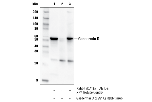
WB, IP
H M R
Endogenous
53, 30
Rabbit IgG
#Q9D8T2
69146
Product Information
Product Usage Information
| Application | Dilution |
|---|---|
| Western Blotting | 1:1000 |
| Immunoprecipitation | 1:50 |
Storage
Specificity / Sensitivity
Species Reactivity:
Human, Mouse, Rat
Source / Purification
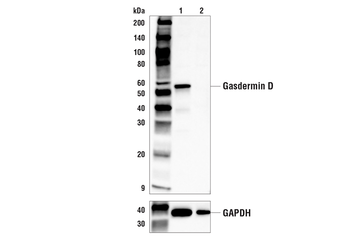
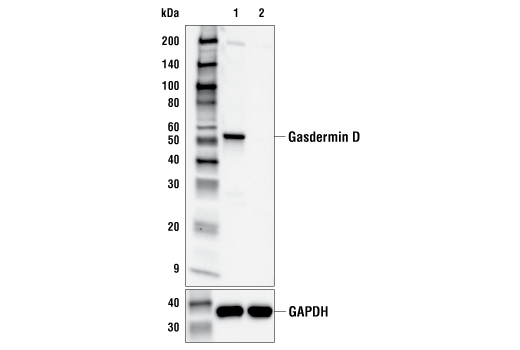
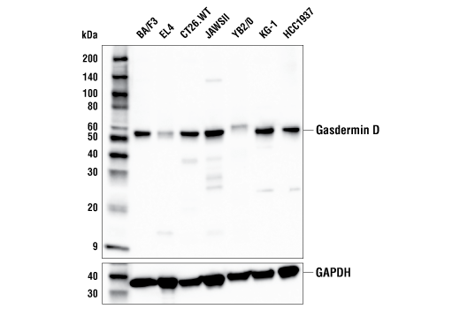
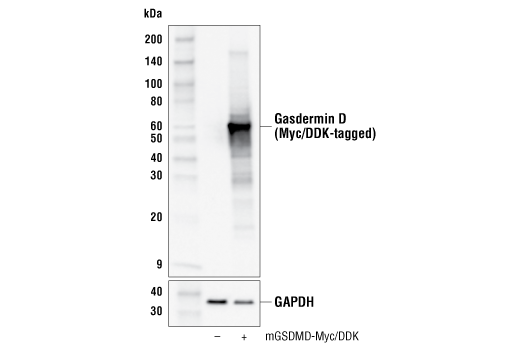
Monoclonal antibody is produced by immunizing animals with a synthetic peptide corresponding to residues surrounding Leu60 of mouse Gasdermin D protein.
Background
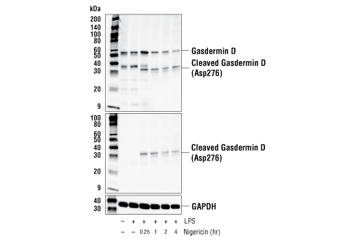
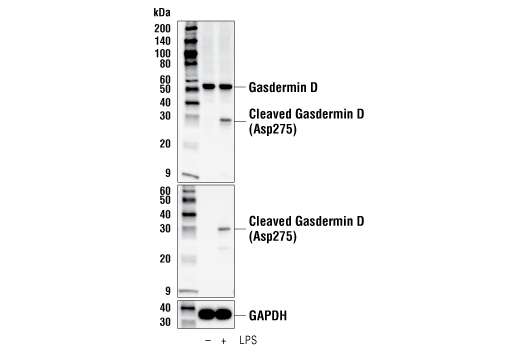
Gasdermin D (GSDMD), a member of the gasdermin family that includes GSDMA, GSDMB, and GSDMC, has been reported to have a critical role as a downstream effector of pyroptosis (1,2). Pyroptosis is a lytic type of cell death triggered by inflammasomes, multiprotein complexes assembled in response to pathogen-associated molecular patterns (PAMPs) or danger-associated molecular patterns (DAMPs) that result in the activation of caspase-1 and subsequent cleavage of pro-inflammatory cytokines IL-1β and IL-18 (3). Gasdermin D was identified by two independent groups as a substrate of inflammatory caspases, caspase-1 and caspase-11/4/5, producing two fragments: GSDMD-N and GSDMD-C. Cleavage results in release of an intramolecular inhibitory interaction between the N- and C-terminal domains, allowing the N-terminal fragment GSDMD-N to initiate pyroptosis through the formation of pores on the plasma membrane (4-7).
- Kayagaki, N. et al. (2015) Nature 526, 666-71.
- Shi, J. et al. (2015) Nature 526, 660-5.
- Broz, P. and Dixit, V.M. (2016) Nat Rev Immunol 16, 407-20.
- Aglietti, R.A. et al. (2016) Proc Natl Acad Sci U S A 113, 7858-63.
- Ding, J. et al. (2016) Nature 535, 111-6.
- Liu, X. et al. (2016) Nature 535, 153-8.
- Sborgi, L. et al. (2016) EMBO J 35, 1766-78.
Species Reactivity
Species reactivity is determined by testing in at least one approved application (e.g., western blot).
Western Blot Buffer
IMPORTANT: For western blots, incubate membrane with diluted primary antibody in 5% w/v BSA, 1X TBS, 0.1% Tween® 20 at 4°C with gentle shaking, overnight.
Applications Key
WB: Western Blotting IP: Immunoprecipitation
Cross-Reactivity Key
H: human M: mouse R: rat Hm: hamster Mk: monkey Vir: virus Mi: mink C: chicken Dm: D. melanogaster X: Xenopus Z: zebrafish B: bovine Dg: dog Pg: pig Sc: S. cerevisiae Ce: C. elegans Hr: horse GP: Guinea Pig Rab: rabbit All: all species expected
Trademarks and Patents
Limited Uses
Except as otherwise expressly agreed in a writing signed by a legally authorized representative of CST, the following terms apply to Products provided by CST, its affiliates or its distributors. Any Customer's terms and conditions that are in addition to, or different from, those contained herein, unless separately accepted in writing by a legally authorized representative of CST, are rejected and are of no force or effect.
Products are labeled with For Research Use Only or a similar labeling statement and have not been approved, cleared, or licensed by the FDA or other regulatory foreign or domestic entity, for any purpose. Customer shall not use any Product for any diagnostic or therapeutic purpose, or otherwise in any manner that conflicts with its labeling statement. Products sold or licensed by CST are provided for Customer as the end-user and solely for research and development uses. Any use of Product for diagnostic, prophylactic or therapeutic purposes, or any purchase of Product for resale (alone or as a component) or other commercial purpose, requires a separate license from CST. Customer shall (a) not sell, license, loan, donate or otherwise transfer or make available any Product to any third party, whether alone or in combination with other materials, or use the Products to manufacture any commercial products, (b) not copy, modify, reverse engineer, decompile, disassemble or otherwise attempt to discover the underlying structure or technology of the Products, or use the Products for the purpose of developing any products or services that would compete with CST products or services, (c) not alter or remove from the Products any trademarks, trade names, logos, patent or copyright notices or markings, (d) use the Products solely in accordance with CST Product Terms of Sale and any applicable documentation, and (e) comply with any license, terms of service or similar agreement with respect to any third party products or services used by Customer in connection with the Products.