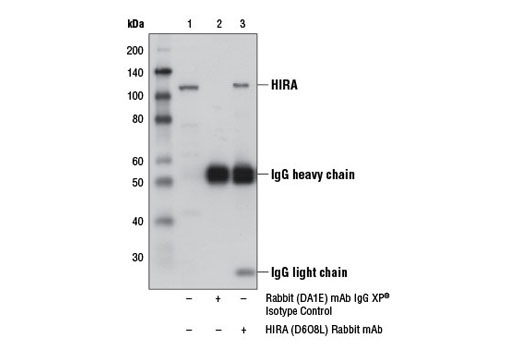
WB, IP
H M R Mk
Endogenous
112
Rabbit IgG
#P54198
7290
Product Information
Product Usage Information
| Application | Dilution |
|---|---|
| Western Blotting | 1:1000 |
| Immunoprecipitation | 1:50 |
Storage
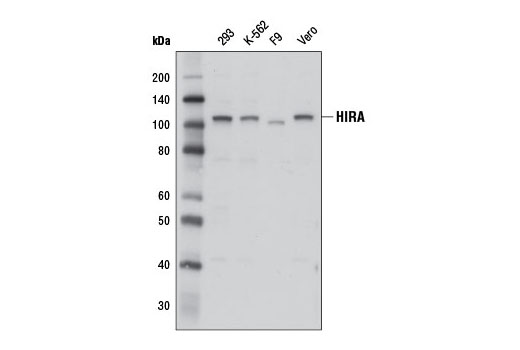
Specificity / Sensitivity
Species Reactivity:
Human, Mouse, Rat, Monkey
Source / Purification
Monoclonal antibody is produced by immunizing animals with a recombinant protein specific to the carboxy terminus of human HIRA protein.
Background
Histone cell cycle regulation defective homolog A (HIRA), also known as TUP1-like enhancer of split protein 1 (TUPLE1), is the mammalian homolog of the yeast HIR1 and HIR2 transcriptional repressor proteins (1). HIRA interacts with UBN1, CABIN, and ASF1A in the cell nucleus to form the evolutionarily conserved HUCA histone chaperone complex that deposits the variant histone H3.3 into chromatin in a DNA-replication independent manner (2). HIRA is required for deposition of histone H3.3 at the transcription start sites of genes, where incorporation of histone H3.3 facilitates nucleosome destabilization and contributes to transcriptional activation (3-5). Histone H3.3 is also linked to gene silencing and is incorporated into regions of the genome thought to be transcriptionally inactive (5-7). While some incorporation of H3.3 into heterochromatin is facilitated by a different histone chaperone complex that contains ATRX and DAXX (ie. telomeric incorporation of H3.3), HIRA is required for incorporation of histone H3.3 and formation of senescence-associated heterochromatin foci (SAHF) during cellular senescence (5-8). HIRA is ubiquitously expressed during mouse embryonic development (9). In the adult mouse, HIRA is expressed at high levels in the kidney, skeletal muscle, and pancreas, but it is expressed at lower levels in the heart, lung, placenta, brain, and liver (9). A missing copy of the HIRA gene on human chromosome region 22q11.2 is a common characteristic of DiGeorge syndrome patients and insufficient production of the HIRA protein may disrupt normal embryonic development (9).
- Lamour, V. et al. (1995) Hum Mol Genet 4, 791-9.
- Rai, T.S. et al. (2011) Mol Cell Biol 31, 4107-18.
- Jin, C. et al. (2009) Nat Genet 41, 941-5.
- Jin, C. and Felsenfeld, G. (2007) Genes Dev 21, 1519-29.
- Goldberg, A.D. et al. (2010) Cell 140, 678-91.
- Wong, L.H. et al. (2010) Genome Res 20, 351-60.
- Wong, L.H. et al. (2009) Genome Res 19, 404-14.
- Zhang, R. et al. (2007) Mol Cell Biol 27, 2343-58.
- Wilming, L.G. et al. (1997) Hum Mol Genet 6, 247-58.
Species Reactivity
Species reactivity is determined by testing in at least one approved application (e.g., western blot).
Western Blot Buffer
IMPORTANT: For western blots, incubate membrane with diluted primary antibody in 5% w/v nonfat dry milk, 1X TBS, 0.1% Tween® 20 at 4°C with gentle shaking, overnight.
Applications Key
WB: Western Blotting IP: Immunoprecipitation
Cross-Reactivity Key
H: human M: mouse R: rat Hm: hamster Mk: monkey Vir: virus Mi: mink C: chicken Dm: D. melanogaster X: Xenopus Z: zebrafish B: bovine Dg: dog Pg: pig Sc: S. cerevisiae Ce: C. elegans Hr: horse GP: Guinea Pig Rab: rabbit All: all species expected
Trademarks and Patents
Limited Uses
Except as otherwise expressly agreed in a writing signed by a legally authorized representative of CST, the following terms apply to Products provided by CST, its affiliates or its distributors. Any Customer's terms and conditions that are in addition to, or different from, those contained herein, unless separately accepted in writing by a legally authorized representative of CST, are rejected and are of no force or effect.
Products are labeled with For Research Use Only or a similar labeling statement and have not been approved, cleared, or licensed by the FDA or other regulatory foreign or domestic entity, for any purpose. Customer shall not use any Product for any diagnostic or therapeutic purpose, or otherwise in any manner that conflicts with its labeling statement. Products sold or licensed by CST are provided for Customer as the end-user and solely for research and development uses. Any use of Product for diagnostic, prophylactic or therapeutic purposes, or any purchase of Product for resale (alone or as a component) or other commercial purpose, requires a separate license from CST. Customer shall (a) not sell, license, loan, donate or otherwise transfer or make available any Product to any third party, whether alone or in combination with other materials, or use the Products to manufacture any commercial products, (b) not copy, modify, reverse engineer, decompile, disassemble or otherwise attempt to discover the underlying structure or technology of the Products, or use the Products for the purpose of developing any products or services that would compete with CST products or services, (c) not alter or remove from the Products any trademarks, trade names, logos, patent or copyright notices or markings, (d) use the Products solely in accordance with CST Product Terms of Sale and any applicable documentation, and (e) comply with any license, terms of service or similar agreement with respect to any third party products or services used by Customer in connection with the Products.