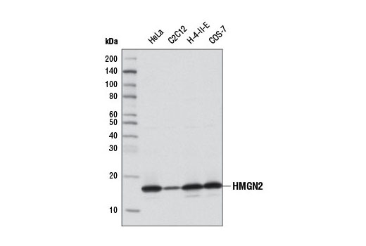
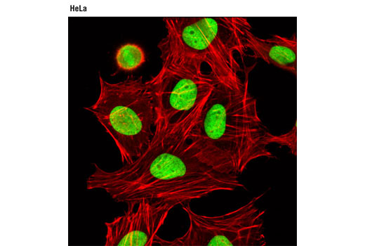
WB, IP, IF-IC
H M R Mk
Endogenous
17
Rabbit IgG
#P05204
3151
Product Information
Product Usage Information
| Application | Dilution |
|---|---|
| Western Blotting | 1:1000 |
| Immunoprecipitation | 1:200 |
| Immunofluorescence (Immunocytochemistry) | 1:6400 |
Storage
Specificity / Sensitivity
Species Reactivity:
Human, Mouse, Rat, Monkey
Species predicted to react based on 100% sequence homology
The antigen sequence used to produce this antibody shares
100% sequence homology with the species listed here, but
reactivity has not been tested or confirmed to work by CST.
Use of this product with these species is not covered under
our
Product Performance Guarantee.
Bovine, Dog, Pig, Horse, Guinea Pig
Source / Purification
Monoclonal antibody is produced by immunizing animals with a synthetic peptide corresponding to residues surrounding Asp74 of human HMGN2 protein.
Background
High mobility group (HMG) proteins are a superfamily of abundant and ubiquitous nuclear proteins that bind DNA without sequence specificity and induce structural changes to the chromatin fiber to regulate access to the underlying DNA. The HMGN family of proteins, which includes five members (HMGN1-5), is characterized by the presence of several conserved protein domains: a positively charged domain, a nucleosome binding domain, and an acidic C-terminal chromatin-unfolding domain (1,2). HMGN proteins function in transcriptional regulation and are recruited to gene promoters by transcription factors, such as estrogen receptor α (ERα), serum responsive factor (SRF), and PITX2, where they can facilitate either gene activation or repression (3-5). HMGN proteins bind specifically to nucleosomal DNA and reduce compaction of the chromatin fiber, in part by competing with linker histone H1 for nucleosome binding (6). In addition, HMGN proteins act to modulate local levels of post-translational histone modifications, decreasing phosphorylation of histone H3 at Ser10 and histone H2A at Ser1 and increasing acetylation of histone H3 at Lys14 (7-9). HMGN proteins can also modulate the activity of several chromatin-remodeling factors and restrict nucleosome mobility (10).
HMGN2 (also known as HMG17) expression is tightly linked to cellular differentiation. HMGN2 is ubiquitous and highly expressed in all embryonic tissues. During mouse embryogenesis, expression is down-regulated throughout the embryo, except in committed, continuously renewing cell types undergoing active differentiation, such as the basal layer of the epithelium and kidney cells undergoing mesenchyme to epithelium transition (11,12). In addition to its function in regulating chromatin structure in the nucleus, HMGN2 also plays a role in innate immunity against bacterial pathogens and tumor cells. Leukocytes, which play a central role in the innate immune defense in mammals by secreting an array of antimicrobial proteins and peptides, secrete HMGN2 upon stimulation with interleukin 2 (IL-2). Following stimulation, HMGN2 translocates from the nucleus to the cytoplasm and is released into the extracellular environment (13). HMGN2, more specifically the alpha-helical domain (residues 18 to 48), shows strong antimicrobial activity towards multiple bacterial pathogens (13). In addition, the amino-terminus of HMGN2 has been shown to contain tumor homing activity, while the carboxy-terminal region inhibits tumor invasion and metastasis (14,15).
- Hock, R. et al. (2007) Trends Cell Biol 17, 72-9.
- Gerlitz, G. Biochim Biophys Acta 1799, 80-5.
- Zhu, N. and Hansen, U. (2007) Mol Cell Biol 27, 8859-73.
- Amen, M. et al. (2008) Nucleic Acids Res 36, 462-76.
- Belova, G.I. et al. (2008) J Biol Chem 283, 8080-8.
- Catez, F. et al. (2002) EMBO Rep 3, 760-6.
- Lim, J.H. et al. (2005) EMBO J 24, 3038-48.
- Lim, J.H. et al. (2004) Mol Cell 15, 573-84.
- Postnikov, Y.V. et al. (2006) Biochemistry 45, 15092-9.
- Rattner, B.P. et al. (2009) Mol Cell 34, 620-6.
- Furusawa, T. et al. (2006) Mol Cell Biol 26, 592-604.
- Lehtonen, S. and Lehtonen, E. (2001) Differentiation 67, 154-63.
- Feng, Y. et al. (2005) J Leukoc Biol 78, 1136-41.
- Porkka, K. et al. (2002) Proc Natl Acad Sci USA 99, 7444-9.
- Isoai, A. et al. (1992) Cancer Res 52, 1422-6.
Species Reactivity
Species reactivity is determined by testing in at least one approved application (e.g., western blot).
Western Blot Buffer
IMPORTANT: For western blots, incubate membrane with diluted primary antibody in 5% w/v BSA, 1X TBS, 0.1% Tween® 20 at 4°C with gentle shaking, overnight.
Applications Key
WB: Western Blotting IP: Immunoprecipitation IF-IC: Immunofluorescence (Immunocytochemistry)
Cross-Reactivity Key
H: human M: mouse R: rat Hm: hamster Mk: monkey Vir: virus Mi: mink C: chicken Dm: D. melanogaster X: Xenopus Z: zebrafish B: bovine Dg: dog Pg: pig Sc: S. cerevisiae Ce: C. elegans Hr: horse GP: Guinea Pig Rab: rabbit All: all species expected
Trademarks and Patents
Limited Uses
Except as otherwise expressly agreed in a writing signed by a legally authorized representative of CST, the following terms apply to Products provided by CST, its affiliates or its distributors. Any Customer's terms and conditions that are in addition to, or different from, those contained herein, unless separately accepted in writing by a legally authorized representative of CST, are rejected and are of no force or effect.
Products are labeled with For Research Use Only or a similar labeling statement and have not been approved, cleared, or licensed by the FDA or other regulatory foreign or domestic entity, for any purpose. Customer shall not use any Product for any diagnostic or therapeutic purpose, or otherwise in any manner that conflicts with its labeling statement. Products sold or licensed by CST are provided for Customer as the end-user and solely for research and development uses. Any use of Product for diagnostic, prophylactic or therapeutic purposes, or any purchase of Product for resale (alone or as a component) or other commercial purpose, requires a separate license from CST. Customer shall (a) not sell, license, loan, donate or otherwise transfer or make available any Product to any third party, whether alone or in combination with other materials, or use the Products to manufacture any commercial products, (b) not copy, modify, reverse engineer, decompile, disassemble or otherwise attempt to discover the underlying structure or technology of the Products, or use the Products for the purpose of developing any products or services that would compete with CST products or services, (c) not alter or remove from the Products any trademarks, trade names, logos, patent or copyright notices or markings, (d) use the Products solely in accordance with CST Product Terms of Sale and any applicable documentation, and (e) comply with any license, terms of service or similar agreement with respect to any third party products or services used by Customer in connection with the Products.