WB, IP, IF-IC, FC-FP
H Mk
Endogenous
28
Rabbit IgG
#P17482
3219
Product Information
Product Usage Information
| Application | Dilution |
|---|---|
| Western Blotting | 1:1000 |
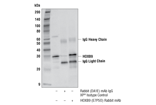
| Immunoprecipitation | 1:100 |
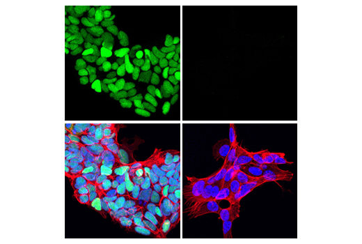
| Immunofluorescence (Immunocytochemistry) | 1:400 - 1:1600 |
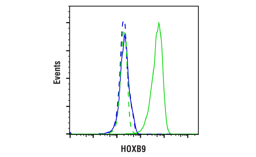
| Flow Cytometry (Fixed/Permeabilized) | 1:800 - 1:1600 |
Storage
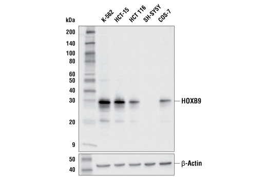
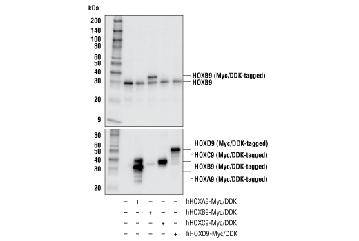
Specificity / Sensitivity
Species Reactivity:
Human, Monkey
Source / Purification
Monoclonal antibody is produced by immunizing animals with a synthetic peptide corresponding to residues surrounding Val116 of human HOXB9 protein.
Background
HOXB9 is a member of the HOXB gene cluster which, along with the HOXA, HOXC, and HOXD gene clusters, governs embryonic patterning along the cranio-caudal axis (1,2). HOXB9 protein functions to regulate formation of the rib cage and contributes to development of the forelimbs (3,4). De-regulation of HOXB9 protein expression is associated with multiple types of cancer. HOXB9 expression is downregulated in pancreatic ductal adenocarcinoma and upregulated in lung adenocarcinoma, where it is associated with poor prognosis (5,6). Overexpression of HOXB9 in breast and hepatocellular carcinomas activates the epithelial-to-mesenchymal transition (EMT) through modulation of TGF-β, promoting tumor progression and metastasis (7,8). HOXB9 overexpression in gliomas increases tumorigenicity by stimulating proliferation, migration, and sphere formation (9).
- McGinnis, W. and Krumlauf, R. (1992) Cell 68, 283-302.
- Zeltser, L. et al. (1996) Development 122, 2475-84.
- McIntyre, D.C. et al. (2007) Development 134, 2981-9.
- Xu, B. and Wellik, D.M. (2011) Proc Natl Acad Sci U S A 108, 4888-91.
- Zhan, J. et al. (2015) Cancer Lett 361, 75-85.
- Zhan, J. et al. (2015) Histopathology 66, 955-65.
- Zhussupova, A. et al. (2014) PLoS One 9, e105285.
- Sha, L. et al. (2015) Clin Exp Med 15, 55-64.
- Fang, L. et al. (2014) Biochem Biophys Res Commun 446, 272-9.
Species Reactivity
Species reactivity is determined by testing in at least one approved application (e.g., western blot).
Western Blot Buffer
IMPORTANT: For western blots, incubate membrane with diluted primary antibody in 5% w/v BSA, 1X TBS, 0.1% Tween® 20 at 4°C with gentle shaking, overnight.
Applications Key
WB: Western Blotting IP: Immunoprecipitation IF-IC: Immunofluorescence (Immunocytochemistry) FC-FP: Flow Cytometry (Fixed/Permeabilized)
Cross-Reactivity Key
H: human M: mouse R: rat Hm: hamster Mk: monkey Vir: virus Mi: mink C: chicken Dm: D. melanogaster X: Xenopus Z: zebrafish B: bovine Dg: dog Pg: pig Sc: S. cerevisiae Ce: C. elegans Hr: horse GP: Guinea Pig Rab: rabbit All: all species expected
Trademarks and Patents
Limited Uses
Except as otherwise expressly agreed in a writing signed by a legally authorized representative of CST, the following terms apply to Products provided by CST, its affiliates or its distributors. Any Customer's terms and conditions that are in addition to, or different from, those contained herein, unless separately accepted in writing by a legally authorized representative of CST, are rejected and are of no force or effect.
Products are labeled with For Research Use Only or a similar labeling statement and have not been approved, cleared, or licensed by the FDA or other regulatory foreign or domestic entity, for any purpose. Customer shall not use any Product for any diagnostic or therapeutic purpose, or otherwise in any manner that conflicts with its labeling statement. Products sold or licensed by CST are provided for Customer as the end-user and solely for research and development uses. Any use of Product for diagnostic, prophylactic or therapeutic purposes, or any purchase of Product for resale (alone or as a component) or other commercial purpose, requires a separate license from CST. Customer shall (a) not sell, license, loan, donate or otherwise transfer or make available any Product to any third party, whether alone or in combination with other materials, or use the Products to manufacture any commercial products, (b) not copy, modify, reverse engineer, decompile, disassemble or otherwise attempt to discover the underlying structure or technology of the Products, or use the Products for the purpose of developing any products or services that would compete with CST products or services, (c) not alter or remove from the Products any trademarks, trade names, logos, patent or copyright notices or markings, (d) use the Products solely in accordance with CST Product Terms of Sale and any applicable documentation, and (e) comply with any license, terms of service or similar agreement with respect to any third party products or services used by Customer in connection with the Products.