WB, IP, IHC-P, IF-IC, FC-FP, ChIP
H M R Mk
Endogenous
25
Rabbit
#P45973
23468
Product Information
Product Usage Information
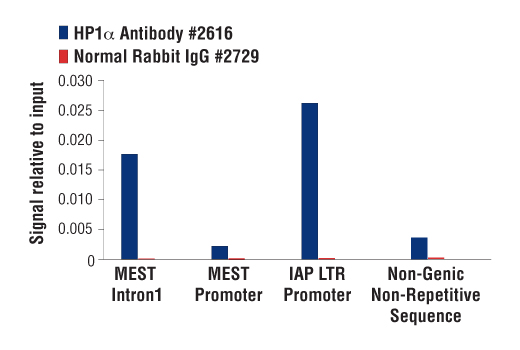
For optimal ChIP results, use 20 μl of antibody and 10 μg of chromatin (approximately 4 x 10⁶ cells) per IP. This antibody has been validated using SimpleChIP® Enzymatic Chromatin IP Kits.
| Application | Dilution |
|---|---|
| Western Blotting | 1:1000 |
| Immunoprecipitation | 1:25 |
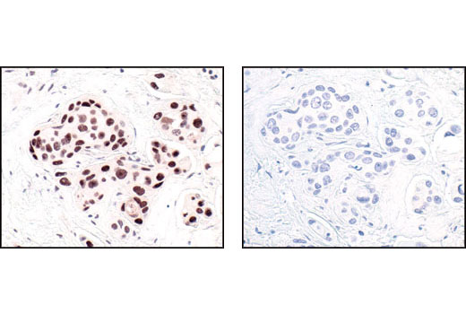
| Immunohistochemistry (Paraffin) | 1:200 |
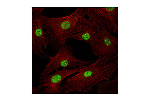
| Immunofluorescence (Immunocytochemistry) | 1:200 |
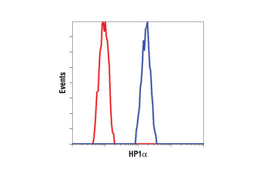
| Flow Cytometry (Fixed/Permeabilized) | 1:100 |
| Chromatin IP | 1:25 |
Storage
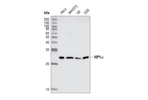
Specificity / Sensitivity
Species Reactivity:
Human, Mouse, Rat, Monkey
Species predicted to react based on 100% sequence homology
The antigen sequence used to produce this antibody shares
100% sequence homology with the species listed here, but
reactivity has not been tested or confirmed to work by CST.
Use of this product with these species is not covered under
our
Product Performance Guarantee.
Bovine
Source / Purification
Polyclonal antibodies are produced by immunizing animals with a synthetic peptide corresponding to the carboxy terminus of human HP1alpha. Antibodies are purified by protein A and peptide affinity chromatography.
Background
Heterochromatin protein 1 (HP1) is a family of heterochromatic adaptor molecules involved in both gene silencing and higher order chromatin structure (1). All three HP1 family members (α, β, and γ) are primarily associated with centromeric heterochromatin; however, HP1β and γ also localize to euchromatic sites in the genome (2,3). HP1 proteins are approximately 25 kDa in size and contain a conserved amino-terminal chromodomain, followed by a variable hinge region and a conserved carboxy-terminal chromoshadow domain. The chromodomain facilitates binding to histone H3 tri-methylated at Lys9, a histone "mark" closely associated with centromeric heterochromatin (4,5). The variable hinge region binds both RNA and DNA in a sequence-independent manner (6). The chromoshadow domain mediates the dimerization of HP1 proteins, in addition to binding multiple proteins implicated in gene silencing and heterochromatin formation, including the SUV39H histone methyltransferase, the DNMT1 and DNMT3a DNA methyltransferases, and the p150 subunit of chromatin-assembly factor-1 (CAF1) (7-9). In addition to contributing to heterochromatin formation and propagation, HP1 and SUV39H are also found complexed with retinoblastoma (Rb) and E2F6 proteins, both of which function to repress euchromatic gene transcription in quiescent cells (10,11). HP1 proteins are subject to multiple types of post-translational modifications, including phosphorylation, acetylation, methylation, ubiquitination, and sumoylation, suggesting multiple means of regulation (12-14).
- Maison, C. and Almouzni, G. (2004) Nat. Rev. Mol. Cell Biol. 5, 296-304.
- Minc, E. et al. (2000) Cytogenet. Cell Genet. 90, 279-284.
- Nielsen, A.L. et al. (2001) Mol. Cell 7, 729-739.
- Lachner, M. et al. (2001) Nature 410, 116-120.
- Bannister, A.J. et al. (2001) Nature 410, 120-124.
- Muchardt, C. et al. (2002) EMBO Rep. 3, 975-981.
- Yamamoto, K. and Sonoda, M. (2003) Biochem. Biophys. Res. Commun. 301, 287-292.
- Fuks, F. et al. (2003) Nucleic Acids Res. 31, 2305-2312.
- Murzina, N. et al. (1999) Mol. Cell 4, 529-540.
- Nielsen, S.J. et al. (2001) Nature 412, 561-565.
- Ogawa, H. et al. (2002) Science 296, 1132-1136.
- Minc, E. et al. (1999) Chromosoma 108, 220-234.
- Zhao, T. et al. (2001) J. Biol. Chem. 276, 9512-9518.
- Lomberk, G. et al. (2006) Nat. Cell Biol. 8, 407-415.
Species Reactivity
Species reactivity is determined by testing in at least one approved application (e.g., western blot).
Western Blot Buffer
IMPORTANT: For western blots, incubate membrane with diluted primary antibody in 5% w/v BSA, 1X TBS, 0.1% Tween® 20 at 4°C with gentle shaking, overnight.
Applications Key
WB: Western Blotting IP: Immunoprecipitation IHC-P: Immunohistochemistry (Paraffin) IF-IC: Immunofluorescence (Immunocytochemistry) FC-FP: Flow Cytometry (Fixed/Permeabilized) ChIP: Chromatin IP
Cross-Reactivity Key
H: human M: mouse R: rat Hm: hamster Mk: monkey Vir: virus Mi: mink C: chicken Dm: D. melanogaster X: Xenopus Z: zebrafish B: bovine Dg: dog Pg: pig Sc: S. cerevisiae Ce: C. elegans Hr: horse GP: Guinea Pig Rab: rabbit All: all species expected
Trademarks and Patents
Limited Uses
Except as otherwise expressly agreed in a writing signed by a legally authorized representative of CST, the following terms apply to Products provided by CST, its affiliates or its distributors. Any Customer's terms and conditions that are in addition to, or different from, those contained herein, unless separately accepted in writing by a legally authorized representative of CST, are rejected and are of no force or effect.
Products are labeled with For Research Use Only or a similar labeling statement and have not been approved, cleared, or licensed by the FDA or other regulatory foreign or domestic entity, for any purpose. Customer shall not use any Product for any diagnostic or therapeutic purpose, or otherwise in any manner that conflicts with its labeling statement. Products sold or licensed by CST are provided for Customer as the end-user and solely for research and development uses. Any use of Product for diagnostic, prophylactic or therapeutic purposes, or any purchase of Product for resale (alone or as a component) or other commercial purpose, requires a separate license from CST. Customer shall (a) not sell, license, loan, donate or otherwise transfer or make available any Product to any third party, whether alone or in combination with other materials, or use the Products to manufacture any commercial products, (b) not copy, modify, reverse engineer, decompile, disassemble or otherwise attempt to discover the underlying structure or technology of the Products, or use the Products for the purpose of developing any products or services that would compete with CST products or services, (c) not alter or remove from the Products any trademarks, trade names, logos, patent or copyright notices or markings, (d) use the Products solely in accordance with CST Product Terms of Sale and any applicable documentation, and (e) comply with any license, terms of service or similar agreement with respect to any third party products or services used by Customer in connection with the Products.