| Product Includes | Product # | Quantity | Mol. Wt | Isotype/Source |
|---|---|---|---|---|
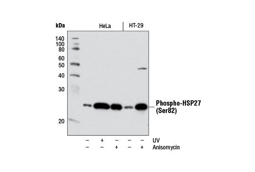
| Phospho-HSP27 (Ser82) (D1H2F6) XP® Rabbit mAb | 9709 | 20 µl | 27 kDa | Rabbit IgG |
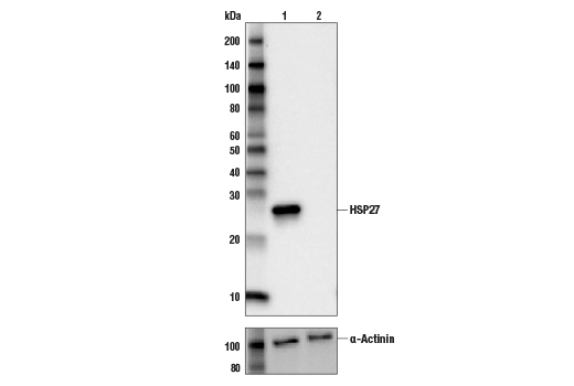
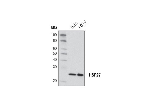
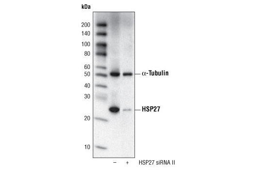
| HSP27 (G31) Mouse mAb | 2402 | 20 µl | 27 kDa | Mouse IgG1 |
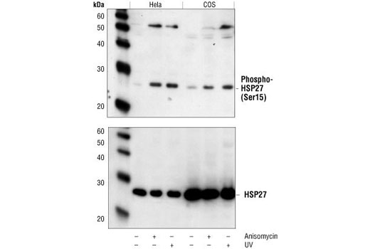
| Phospho-HSP27 (Ser15) Antibody | 2404 | 20 µl | 27 kDa | Rabbit |
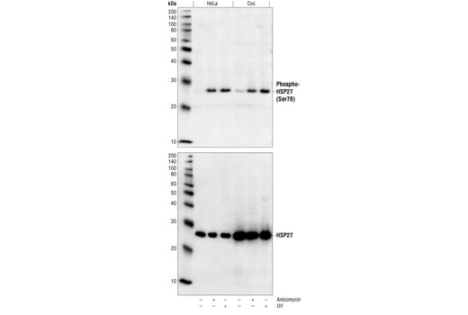
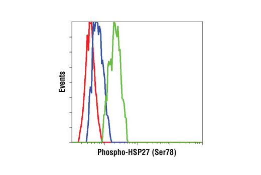
| Phospho-HSP27 (Ser78) Antibody | 2405 | 20 µl | 27 kDa | Rabbit |
| Anti-rabbit IgG, HRP-linked Antibody | 7074 | 100 µl | Goat | |
| Anti-mouse IgG, HRP-linked Antibody | 7076 | 100 µl | Horse |
Please visit cellsignal.com for individual component applications, species cross-reactivity, dilutions, protocols, and additional product information.
Description
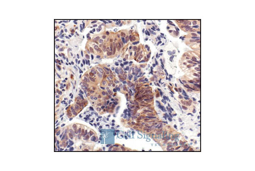
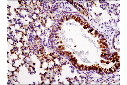
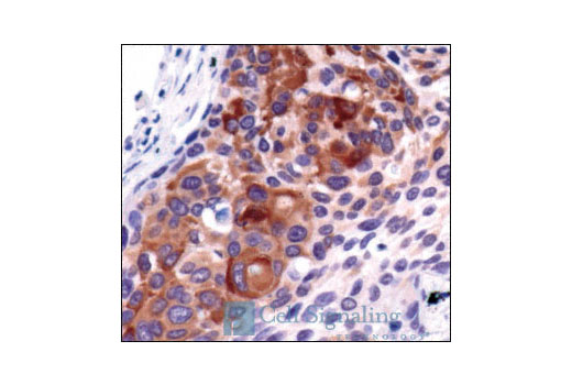
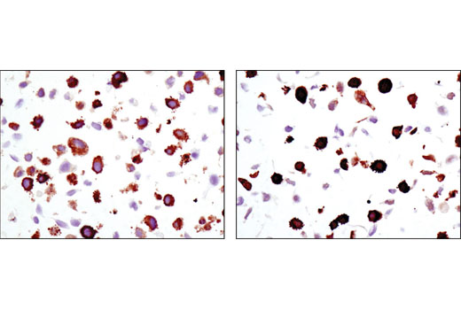
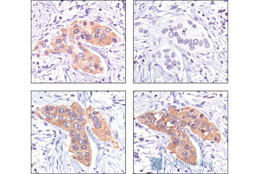
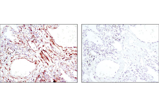
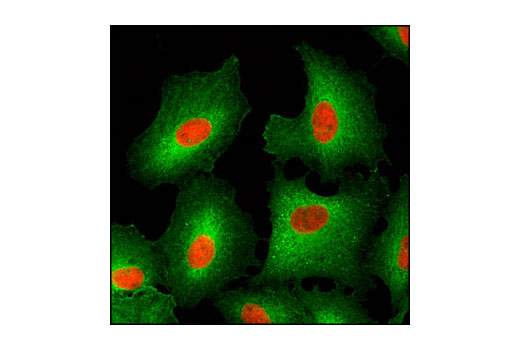
The HSP27 Antibody Kit provides an economical means to evaluate the activation status of the HSP27 protein. The kit contains enough primary antibody to perform two western blot experiments per primary antibody.
Storage
Background
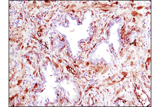
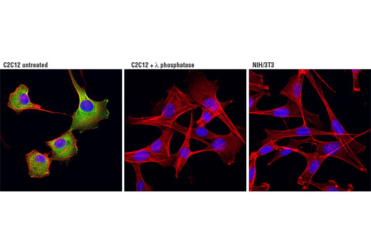
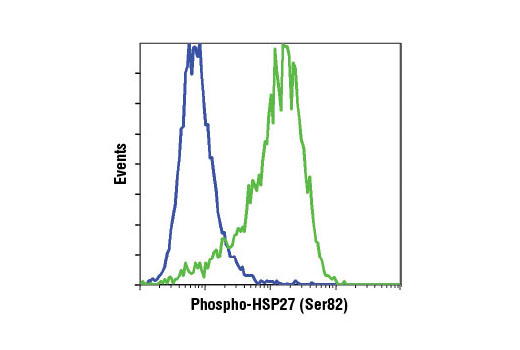
Heat shock protein (HSP) 27 is one of the small HSPs that are constitutively expressed at different levels in various cell types and tissues. Like other small HSPs, HSP27 is regulated at both the transcriptional and posttranslational levels (1). In response to stress, the HSP27 expression increases several-fold to confer cellular resistance to the adverse environmental change. HSP27 is phosphorylated at Ser15, Ser78, and Ser82 by MAPKAPK-2 as a result of the activation of the p38 MAP kinase pathway (2,3). Phosphorylation of HSP27 causes a change in its tertiary structure, which shifts from large homotypic multimers to dimers and monomers (4). It has been shown that phosphorylation and increased concentration of HSP27 modulates actin polymerization and reorganization (5,6).
Background References
Trademarks and Patents
Limited Uses
Except as otherwise expressly agreed in a writing signed by a legally authorized representative of CST, the following terms apply to Products provided by CST, its affiliates or its distributors. Any Customer's terms and conditions that are in addition to, or different from, those contained herein, unless separately accepted in writing by a legally authorized representative of CST, are rejected and are of no force or effect.
Products are labeled with For Research Use Only or a similar labeling statement and have not been approved, cleared, or licensed by the FDA or other regulatory foreign or domestic entity, for any purpose. Customer shall not use any Product for any diagnostic or therapeutic purpose, or otherwise in any manner that conflicts with its labeling statement. Products sold or licensed by CST are provided for Customer as the end-user and solely for research and development uses. Any use of Product for diagnostic, prophylactic or therapeutic purposes, or any purchase of Product for resale (alone or as a component) or other commercial purpose, requires a separate license from CST. Customer shall (a) not sell, license, loan, donate or otherwise transfer or make available any Product to any third party, whether alone or in combination with other materials, or use the Products to manufacture any commercial products, (b) not copy, modify, reverse engineer, decompile, disassemble or otherwise attempt to discover the underlying structure or technology of the Products, or use the Products for the purpose of developing any products or services that would compete with CST products or services, (c) not alter or remove from the Products any trademarks, trade names, logos, patent or copyright notices or markings, (d) use the Products solely in accordance with CST Product Terms of Sale and any applicable documentation, and (e) comply with any license, terms of service or similar agreement with respect to any third party products or services used by Customer in connection with the Products.