| Product Includes | Product # | Quantity | Mol. Wt | Isotype/Source |
|---|---|---|---|---|
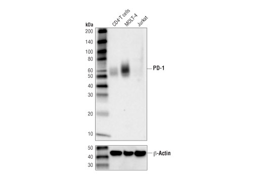
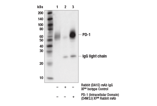
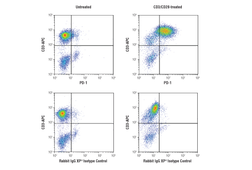
| PD-1 (Intracellular Domain) (D4W2J) XP® Rabbit mAb | 86163 | 20 µl | 52-65 kDa | Rabbit IgG |
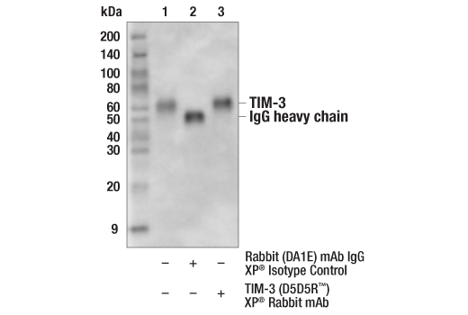
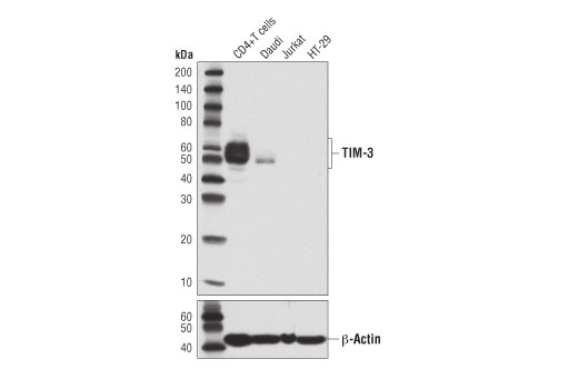
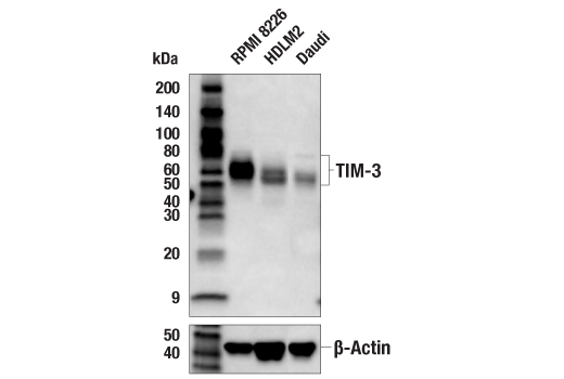
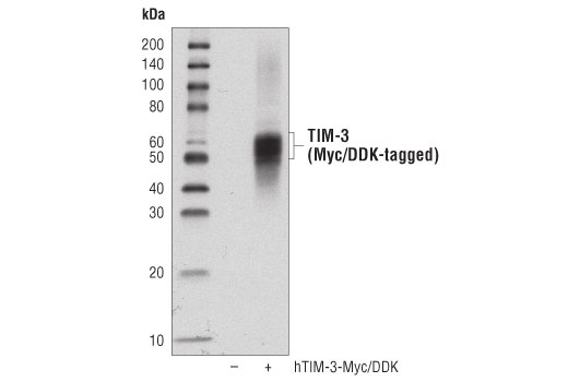
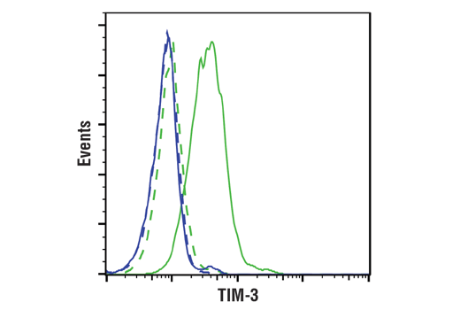
| TIM-3 (D5D5R™) XP® Rabbit mAb | 45208 | 20 µl | 45-70 kDa | Rabbit IgG |
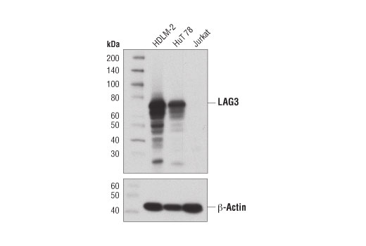
| LAG3 (D2G4O) XP® Rabbit mAb | 15372 | 20 µl | 60-80 kDa | Rabbit IgG |
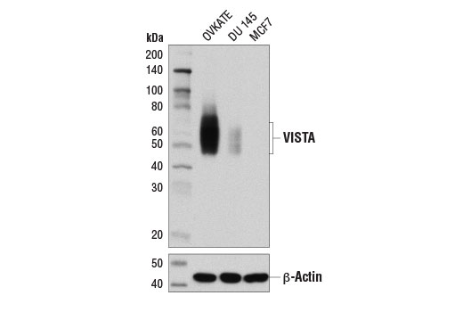
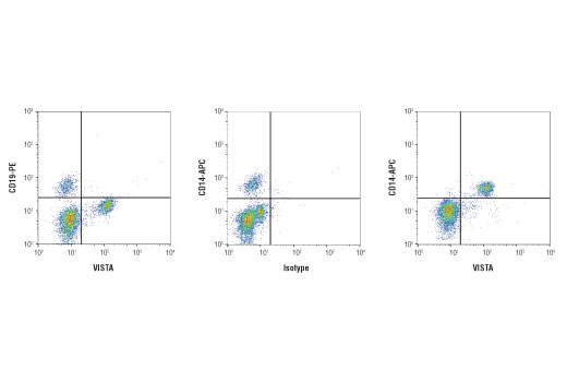
| VISTA (D1L2G™) XP® Rabbit mAb | 64953 | 20 µl | 45-65 kDa | Rabbit IgG |
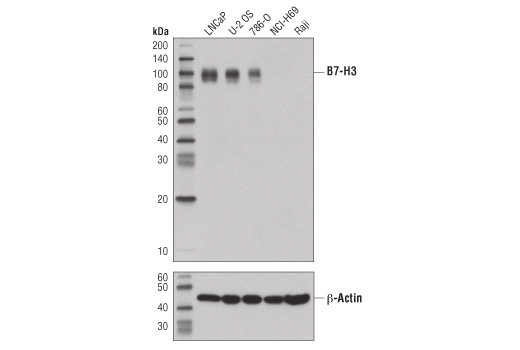
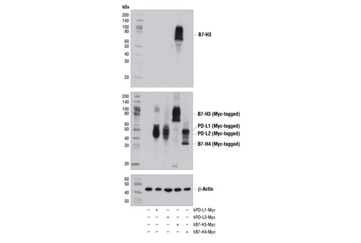
| B7-H3 (D9M2L) XP® Rabbit mAb | 14058 | 20 µl | 90 kDa | Rabbit IgG |
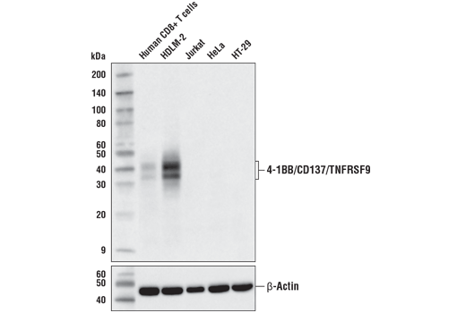
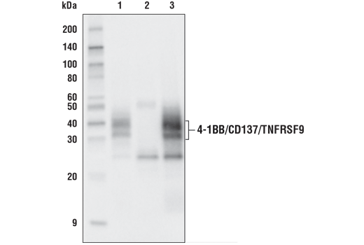
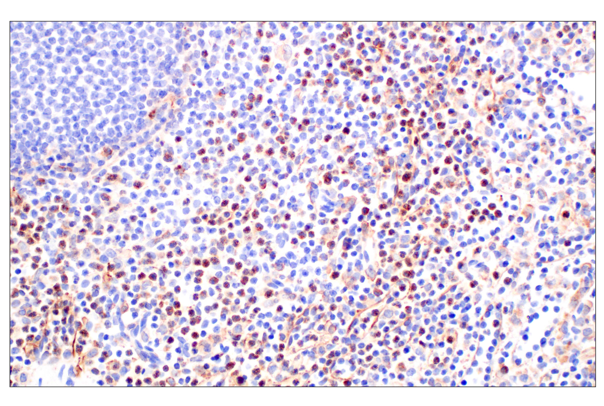
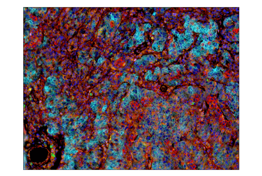
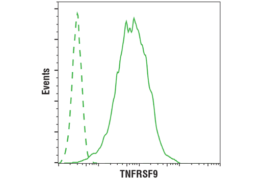
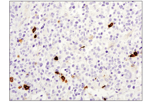
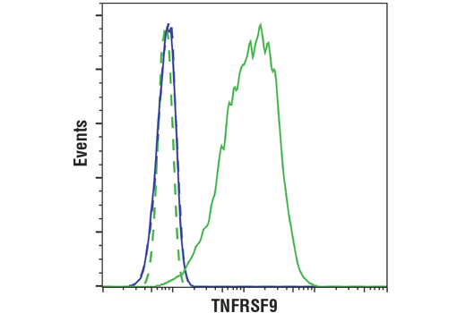
| 4-1BB/CD137/TNFRSF9 (D2Z4Y) Rabbit mAb | 34594 | 20 µl | 32, 40 kDa | Rabbit IgG |
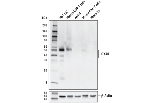
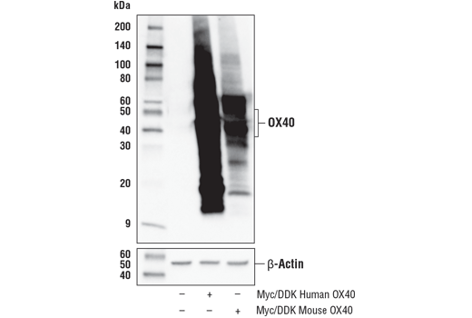
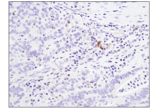
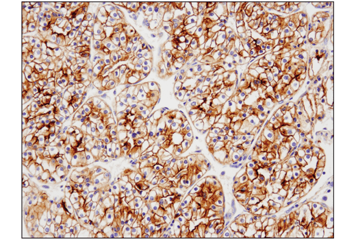
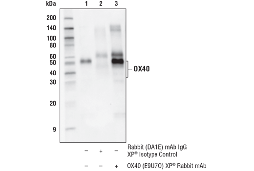
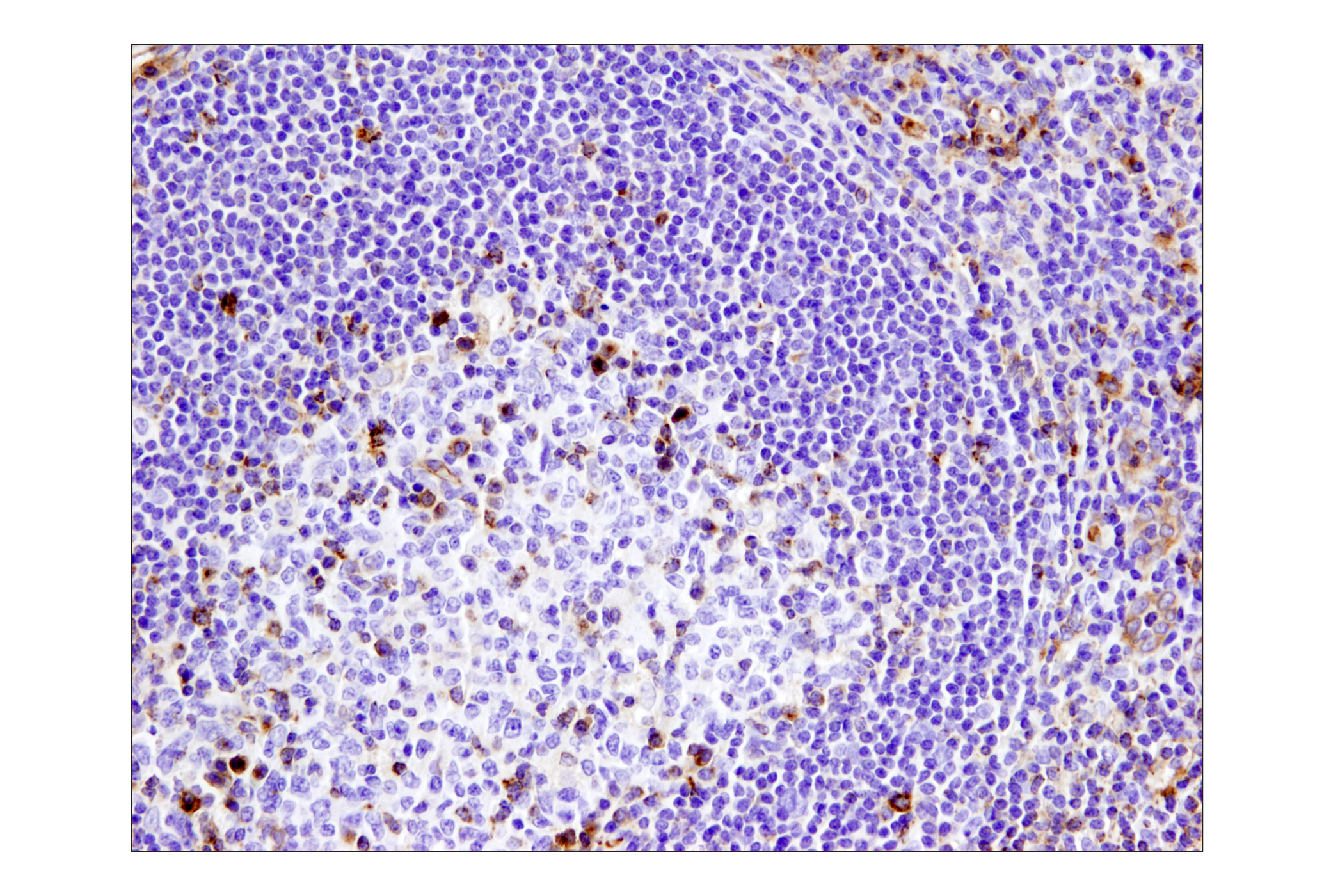
| OX40 (E9U7O) XP® Rabbit mAb | 61637 | 20 µl | 35-50 kDa | Rabbit IgG |
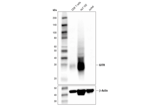
| GITR (D9I9D) Rabbit mAb | 68014 | 20 µl | 25 kDa | Rabbit IgG |
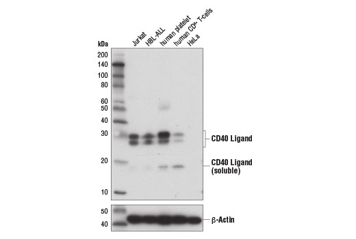
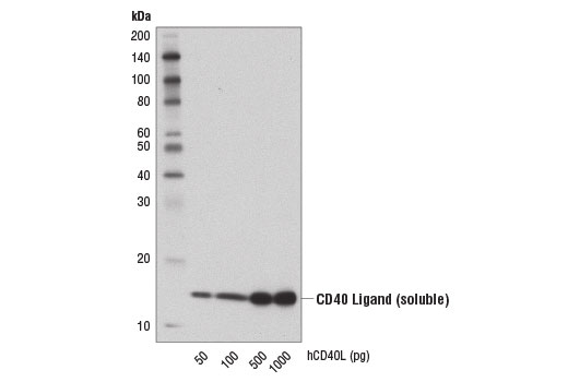
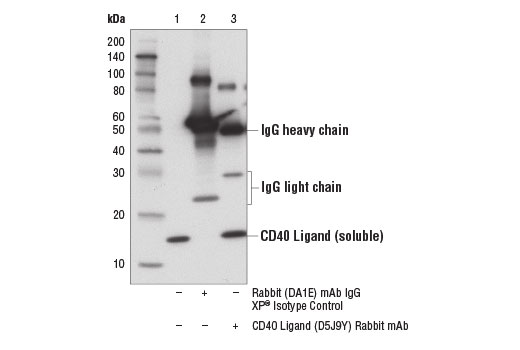
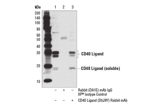
| CD40 Ligand (D5J9Y) Rabbit mAb | 15094 | 20 µl | 25-30 (membrane bound); 17 (soluble) kDa | Rabbit IgG |
Please visit cellsignal.com for individual component applications, species cross-reactivity, dilutions, protocols, and additional product information.
Description
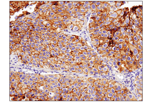
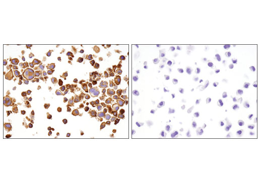
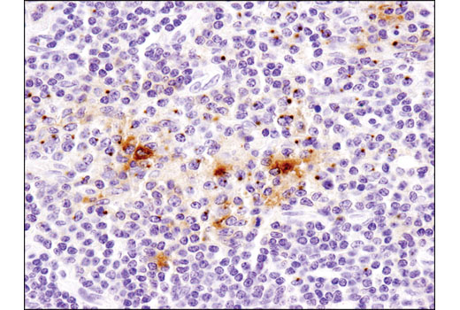
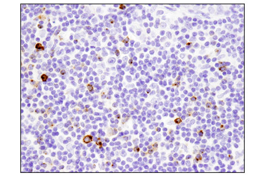
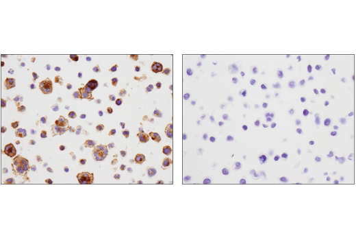
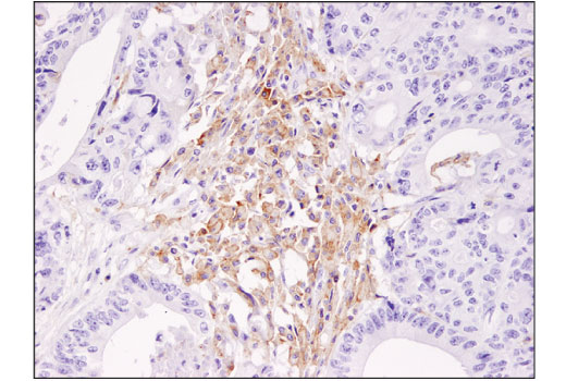
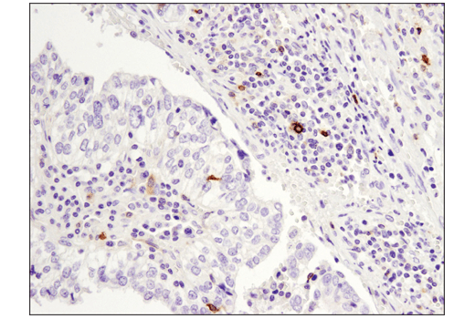
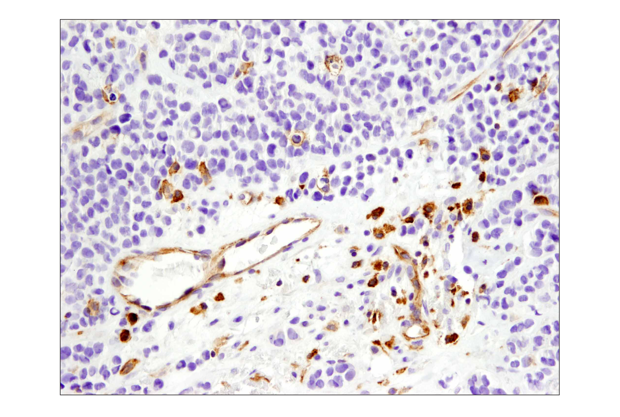
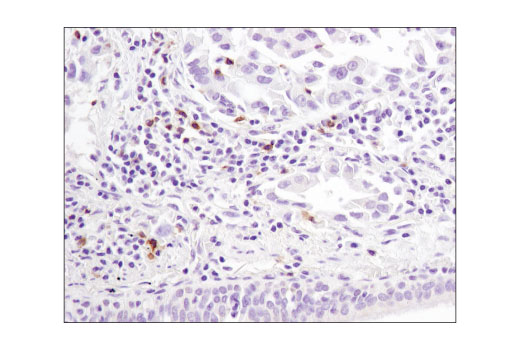
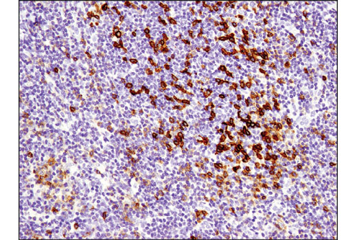
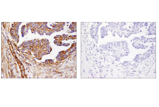
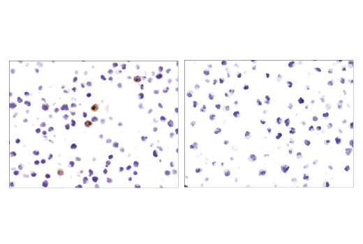
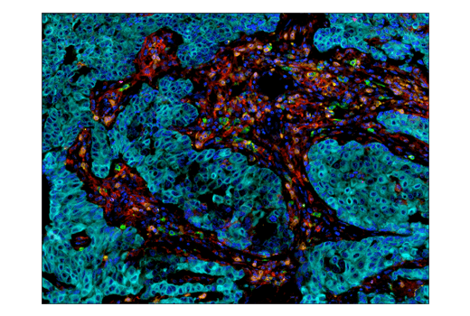
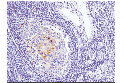
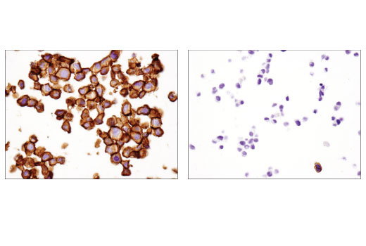
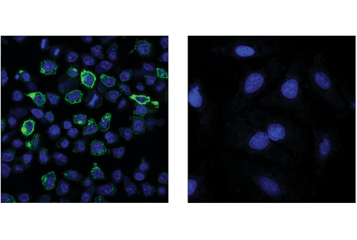
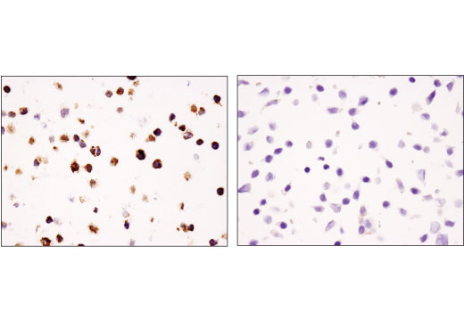
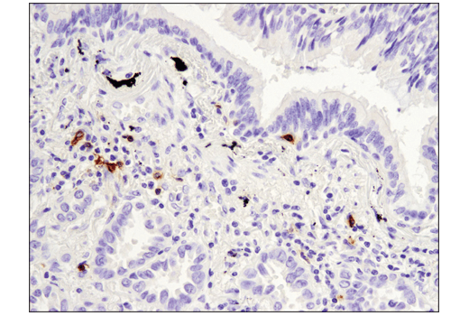
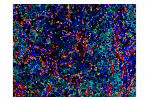
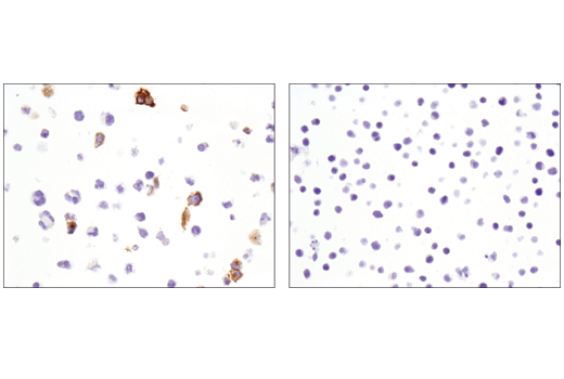
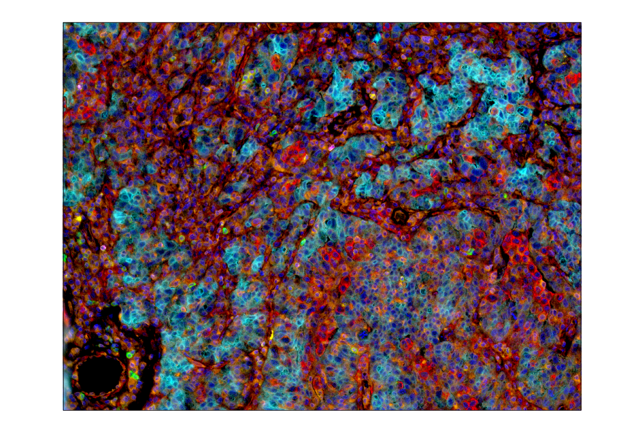
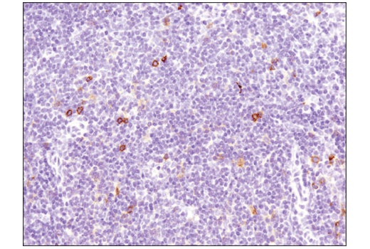
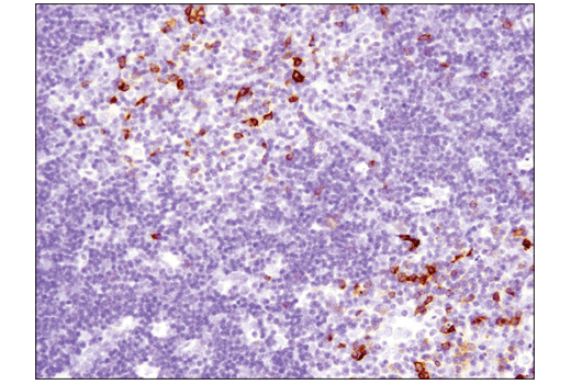
The Human T Cell Co-inhibitory and Co-stimulatory Receptor IHC Antibody Sampler Kit provides an economical means of detecting expression of receptors that modulate T cell activity in formalin-fixed, paraffin-embedded tissue samples.
Storage
Background
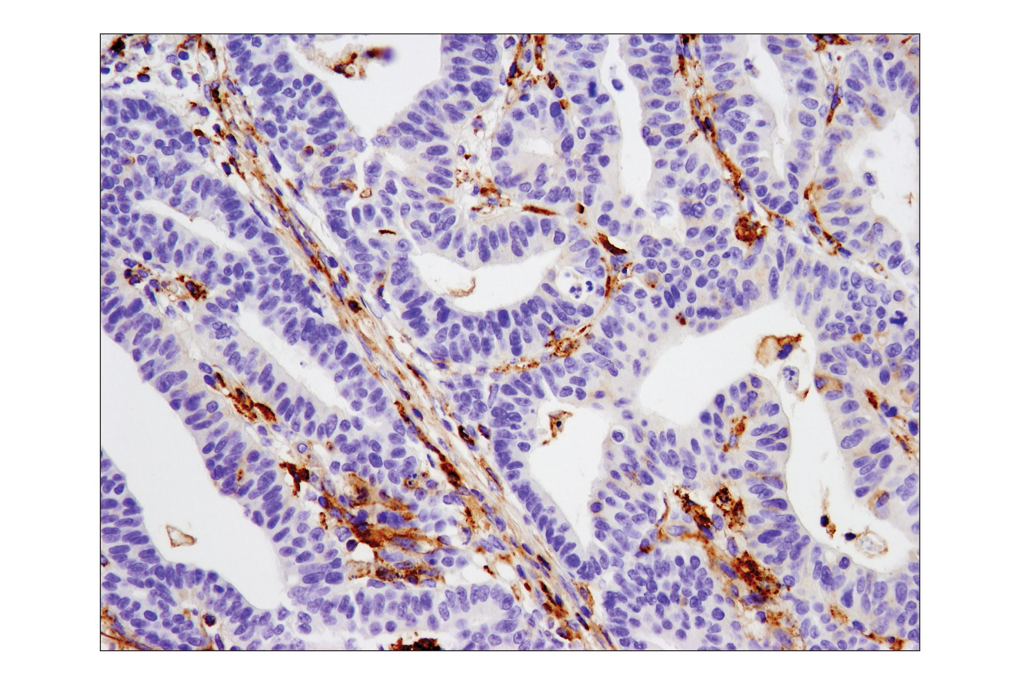
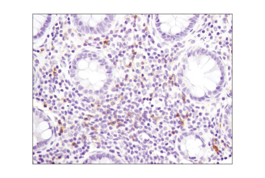
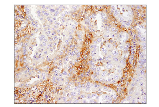
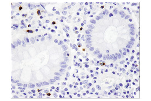
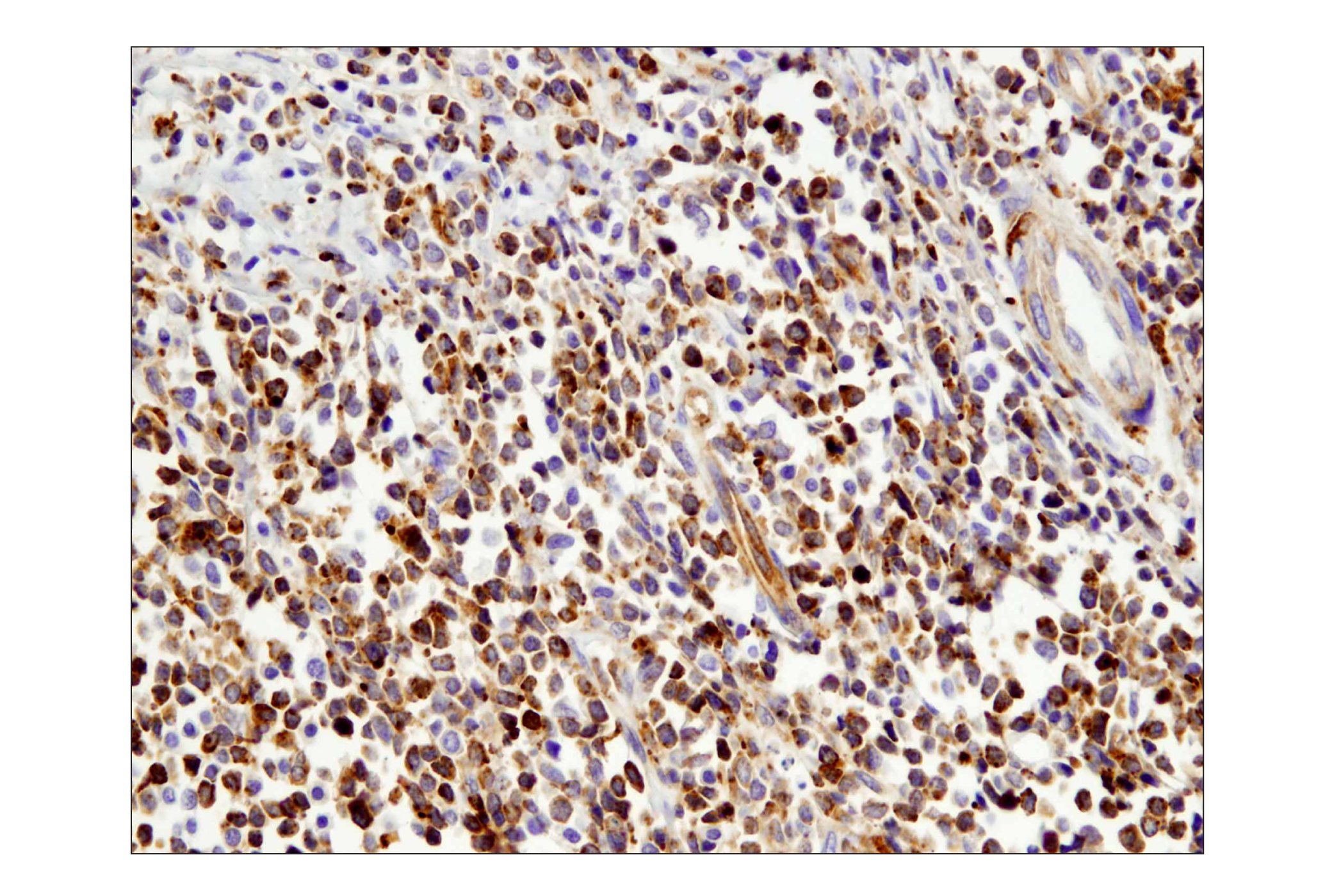
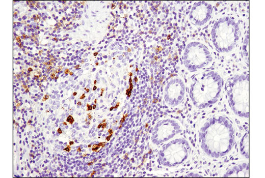
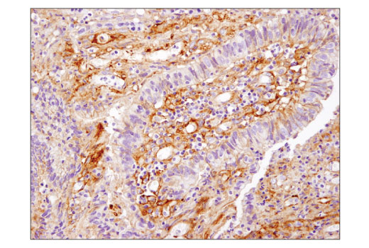
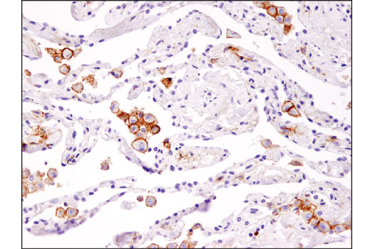
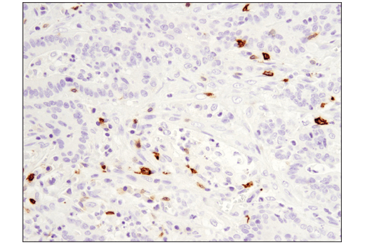
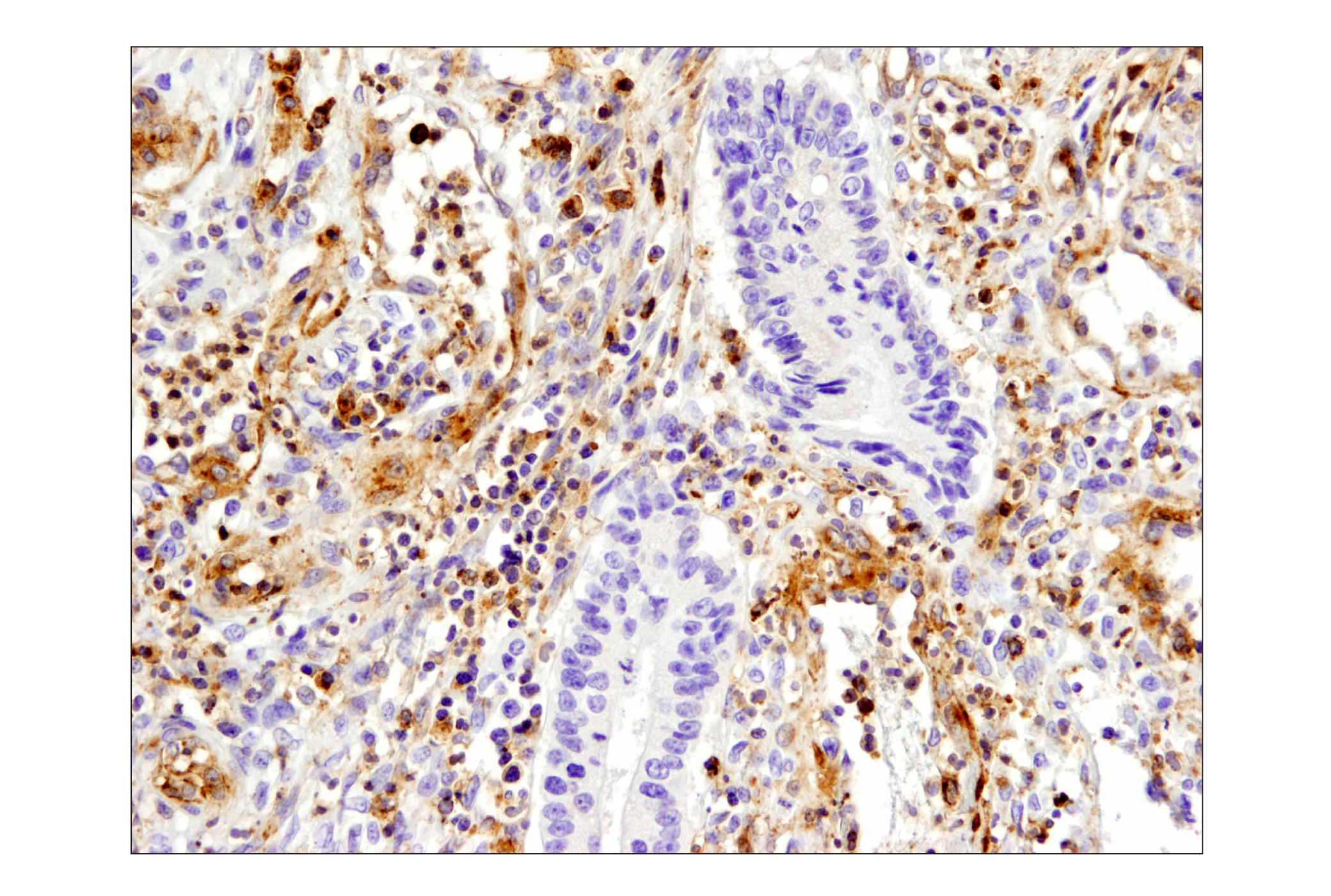
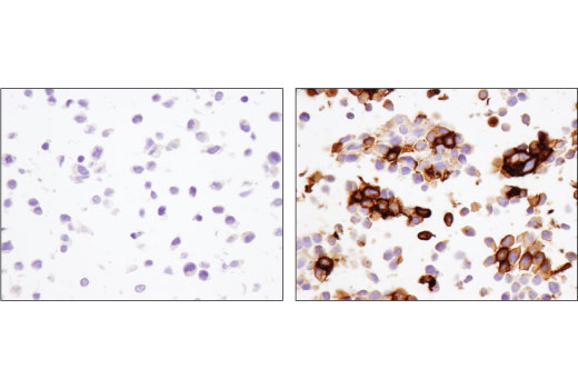
PD-1 (PDCD1, CD279), TIM-3 (HAVCR2), LAG3 (CD223), VISTA (PD-H1), and B7-H3 (CD276) are immune cell co-inhibitory receptors (also known as immune checkpoints) that negatively regulate T cell function, and dampen the immune response to pathogens and cancer. In addition to activated T cells, PD-1 is expressed by activated B-cells and monocytes. TIM-3 is expressed by exhausted T cells in the settings of chronic infection and cancer. Tumor-infiltrating macrophages and dendritic cells also express TIM-3. LAG3 is primarily expressed by activated CD4+ T cells, CD8+ T cells, FoxP3+ T regulatory cells (Tregs) and natural killer (NK) cells. Although primarily expressed by myeloid cells, VISTA is also expressed by CD4+, CD8+, and Treg cells. Research examining the biological function of B7-H3 suggested that B7-H3 can be both a positive and negative regulator of T cell response. B7-H3 is expressed by antigen presenting cells, activated T cells, and a few normal tissues, including placenta and prostate. Expression of B7-H3 is seen in several cancer types, including prostate, breast, colon, lung, and gastric cancers, and in endothelial cells from tumor associated vasculature. Therapeutic blockade of these immune checkpoint receptors is a promising strategy for neoplastic intervention by enabling anti-tumor immune responses (1-3).
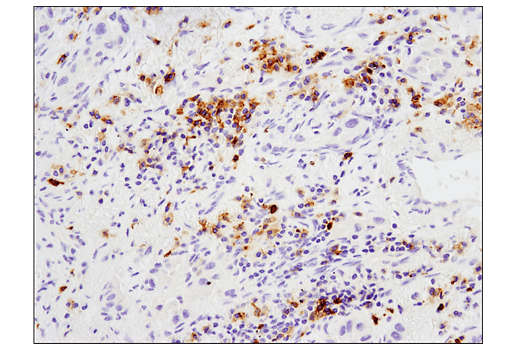
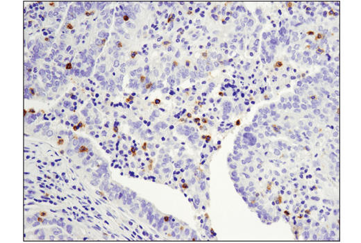
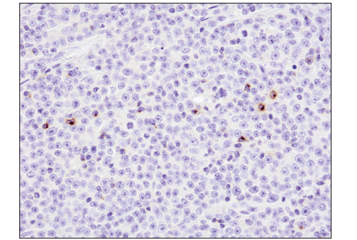
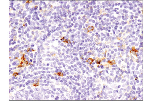
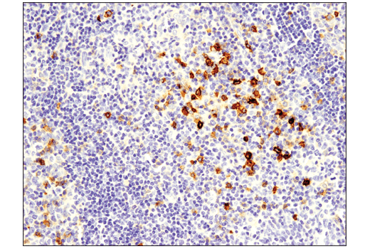
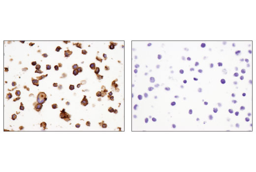
4-1BB (TNFRSF9, CD137), GITR (TNFRSF18), OX40 (TNFRSF4, CD134), and CD40 ligand (CD40L, CD154, TRAP, gp39) are immune cell co-stimulatory receptors that promote effector T cell survival and activation, and enable optimal immune responses to pathogens. 4-1BB is expressed in activated CD4+ and CD8+ T cells, natural killer cells and dendritic cells. GITR is expressed constitutively at high levels on Tregs, at low levels on naive and memory T cells, and is induced upon T cell activation. Studies show GITR can also be induced on NK cells, macrophages, and DCs. GITR ligation has been shown to induce CD8+ T cell activation, cytoxicity, and memory T cell survival, and conversely inhibit Treg suppressive function while promoting effector T cell resistance to Treg suppression. OX40 is primarily expressed on activated CD4+ and CD8+ T cells, while CD40L is primarily expressed on the surface of T cells, but has also been reported in blood platelets, mast cells, basophils, NK cells, and B cells. Research studies show that agonists of these co-stimulatory receptors augment anti-tumor immunity in several cancer types. Due to the combined effects on both Treg suppression and effector cell activation, GITR represents a unique opportunity for immunotherapeutic intervention in cancer. These pathways are an important area of interest in the study of cancer, vascular diseases, and inflammatory disorders (4-7).
- Schildberg, F.A. et al. (2016) Immunity 44, 955-72.
- Anderson, A.C. et al. (2016) Immunity 44, 989-1004.
- Callahan, M.K. et al. (2016) Immunity 44, 1069-78.
- Ward-Kavanagh, L.K. et al. (2016) Immunity 44, 1005-19.
- Ara, A. et al. (2018) Immunotargets Ther 7, 55-61.
- Knee, D.A. et al. (2016) Eur J Cancer 67, 1-10.
- Chester, C. et al. (2018) Blood 131, 49-57.
Background References
Trademarks and Patents
Limited Uses
Except as otherwise expressly agreed in a writing signed by a legally authorized representative of CST, the following terms apply to Products provided by CST, its affiliates or its distributors. Any Customer's terms and conditions that are in addition to, or different from, those contained herein, unless separately accepted in writing by a legally authorized representative of CST, are rejected and are of no force or effect.
Products are labeled with For Research Use Only or a similar labeling statement and have not been approved, cleared, or licensed by the FDA or other regulatory foreign or domestic entity, for any purpose. Customer shall not use any Product for any diagnostic or therapeutic purpose, or otherwise in any manner that conflicts with its labeling statement. Products sold or licensed by CST are provided for Customer as the end-user and solely for research and development uses. Any use of Product for diagnostic, prophylactic or therapeutic purposes, or any purchase of Product for resale (alone or as a component) or other commercial purpose, requires a separate license from CST. Customer shall (a) not sell, license, loan, donate or otherwise transfer or make available any Product to any third party, whether alone or in combination with other materials, or use the Products to manufacture any commercial products, (b) not copy, modify, reverse engineer, decompile, disassemble or otherwise attempt to discover the underlying structure or technology of the Products, or use the Products for the purpose of developing any products or services that would compete with CST products or services, (c) not alter or remove from the Products any trademarks, trade names, logos, patent or copyright notices or markings, (d) use the Products solely in accordance with CST Product Terms of Sale and any applicable documentation, and (e) comply with any license, terms of service or similar agreement with respect to any third party products or services used by Customer in connection with the Products.