| Product Includes | Product # | Quantity | Mol. Wt | Isotype/Source |
|---|---|---|---|---|
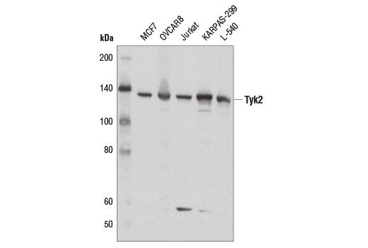
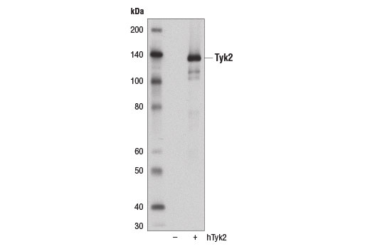
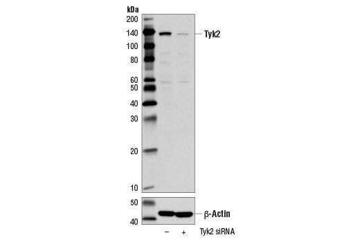
| Tyk2 (D4I5T) Rabbit mAb | 14193 | 20 µl | 134 kDa | Rabbit IgG |
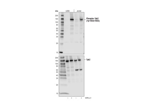
| Phospho-Tyk2 (Tyr1054/1055) (D7T8A) Rabbit mAb | 68790 | 20 µl | 134 kDa | Rabbit IgG |
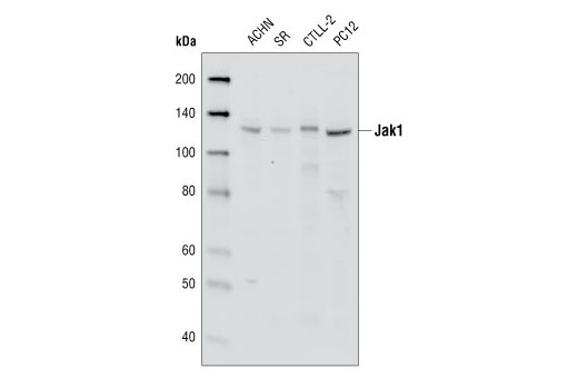
| Jak1 (6G4) Rabbit mAb | 3344 | 20 µl | 130 kDa | Rabbit IgG |
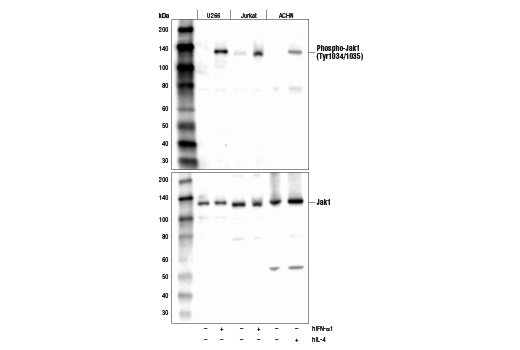
| Phospho-Jak1(Tyr1034/1035) (D7N4Z) Rabbit mAb | 74129 | 20 µl | 130 kDa | Rabbit IgG |
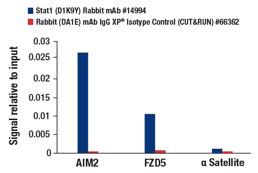
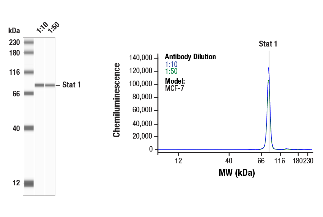
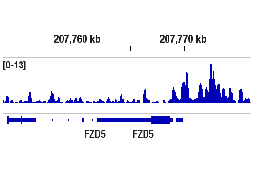
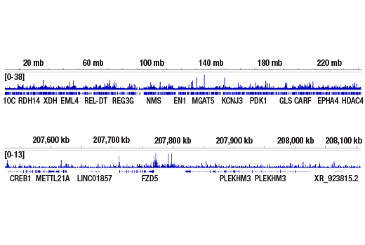
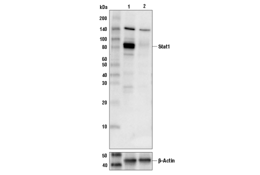
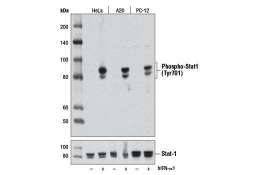
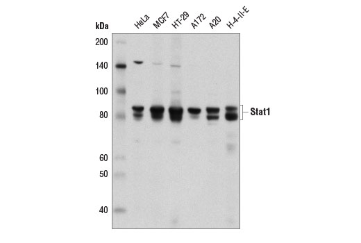
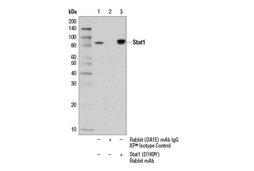
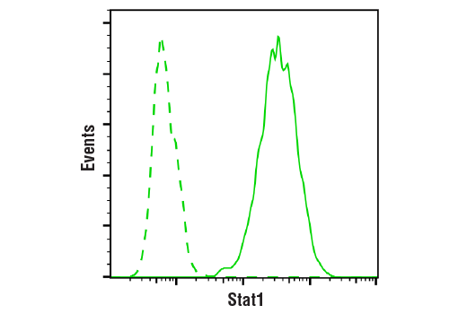
| Stat1 (D1K9Y) Rabbit mAb | 14994 | 20 µl | 84, 91 kDa | Rabbit IgG |
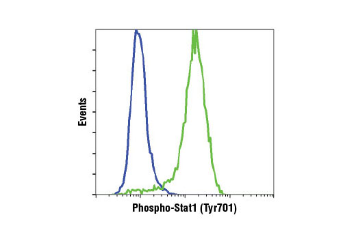
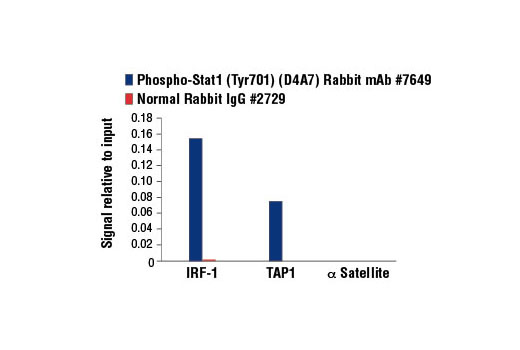
| Phospho-Stat1 (Tyr701) (D4A7) Rabbit mAb | 7649 | 20 µl | 84, 91 kDa | Rabbit IgG |
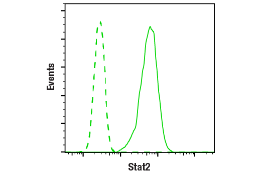
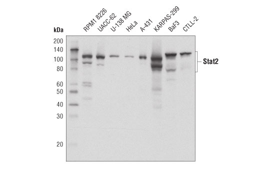
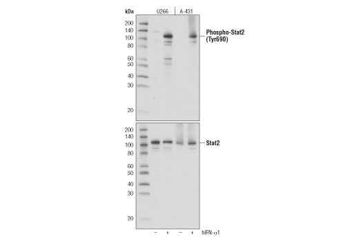
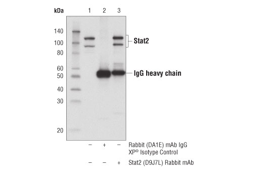
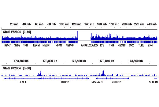
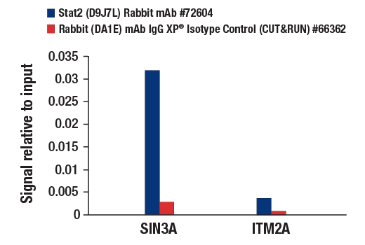
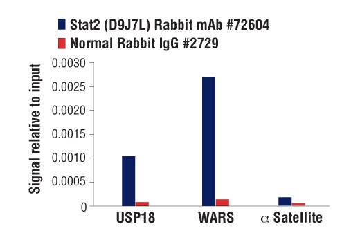
| Stat2 (D9J7L) Rabbit mAb | 72604 | 20 µl | 97, 113 kDa | Rabbit IgG |
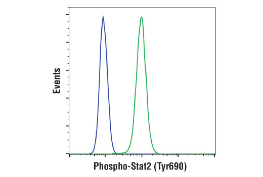
| Phospho-Stat2 (Tyr690) (D3P2P) Rabbit mAb | 88410 | 20 µl | 97, 113 kDa | Rabbit IgG |
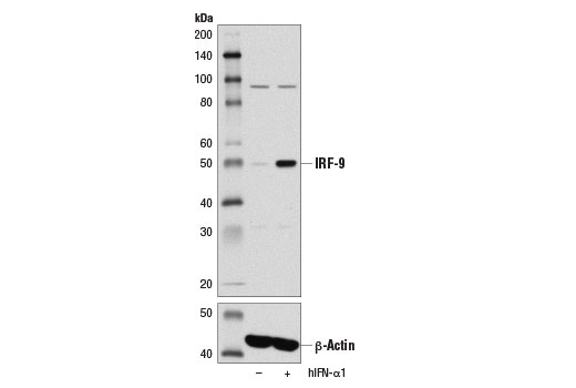
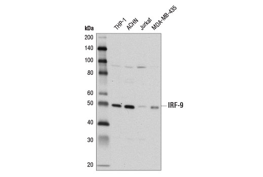
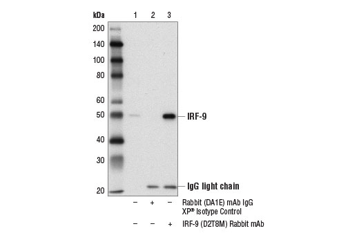
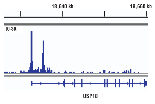
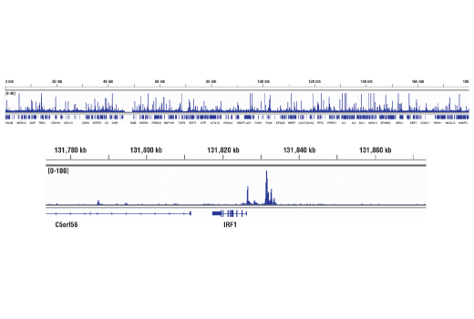
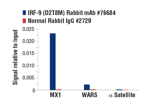
| IRF-9 (D2T8M) Rabbit mAb | 76684 | 20 µl | 48 kDa | Rabbit IgG |
| Anti-rabbit IgG, HRP-linked Antibody | 7074 | 100 µl | Goat |
Please visit cellsignal.com for individual component applications, species cross-reactivity, dilutions, protocols, and additional product information.
Description
The IFN (Type I/III) Signaling Pathway Antibody Sampler Kit provides an economical means of detecting the activation of the IFN (Type I/III) signaling pathway using phospho-specific and control antibodies. The kit includes enough antibodies to perform two western blot experiments with each primary antibody.
Storage
Background
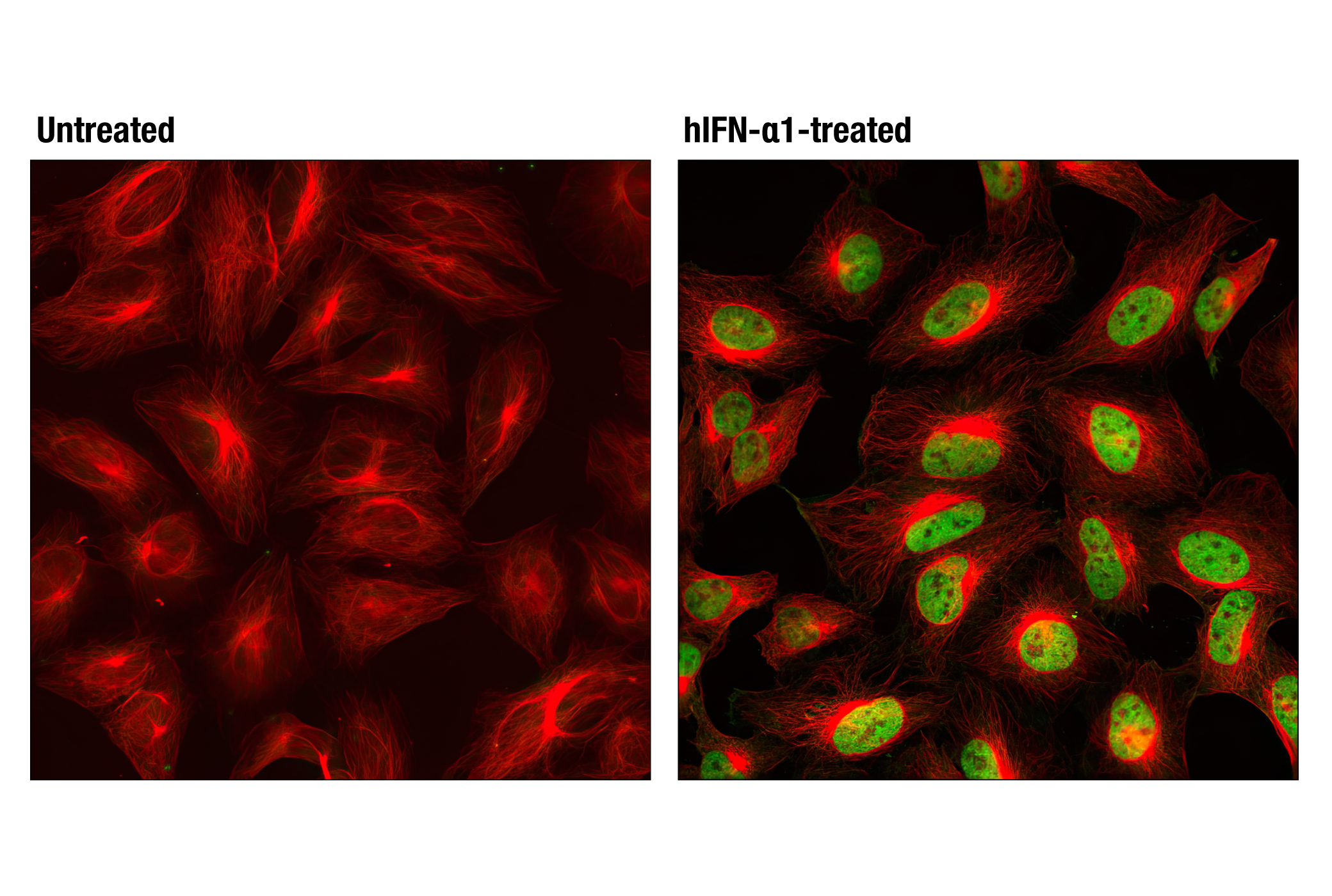
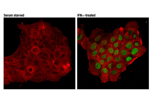
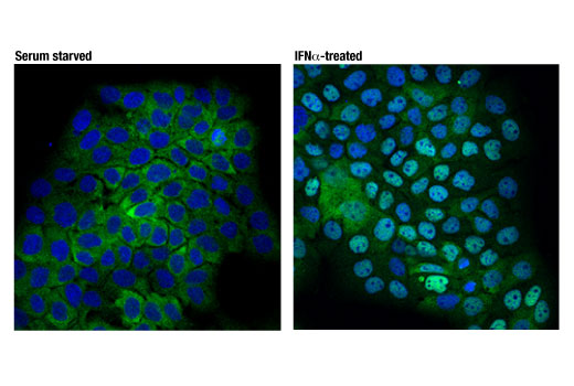
Originally discovered in the late 1950s for their antiviral activity, interferons (IFNs) have since been assigned diverse roles in many physiological and pathological processes. There are three families of IFNs: types I, II, and III. In humans, type I contains IFN-α (13 different subtypes), IFN-β (also known as IFN-β1), IFN-ε, IFN-κ, and IFN-ω. They bind to a receptor complex containing IFNAR1 and IFNAR2, which is broadly expressed on most cells. IFN-γ is the sole member of type II IFN. It signals through a receptor complex consisting of IFNγR1 and IFNγR2, which is also expressed on most cell types. Type III IFN, also known as interferon lambdas (IFN-λs), have four members in humans: IFN-λ1 (IL29), IFN-λ2 (IL28A), IFN-λ3 (IL28B), and IFN-λ4. IFN-λs signal through a heterodimeric receptor comprised of IFNλR1 and IL-10R2. While IL-10R2 is broadly expressed and shared by the IL-10 family cytokines, IFNλR1 expression is restricted to epithelial cells, neuronal cells, and subsets of myeloid cells (1-3). Engagement of all IFNs with their receptors initiates downstream signaling events, mainly, activation of the Jak–Stat signaling cascade. For type I and III IFNs, Jak1 and Tyk2 are phosphorylated and activated, leading to subsequent phosphorylation of Stat1 and Stat2. Phosphorylated Stat1 and Stat2 are released from the receptor complex and, together with IRF-9, they form so-called ISGF3 (interferon-stimulated gene factor 3) transcriptional complex. ISGF3 translocates to the nucleus, binds to the interferon-stimulated response element (ISRE) to initiate the transcription of a wide array of interferon-stimulated genes (ISGs) (4,5). On the other hand, IFN-γ induces phosphorylation and activation of Jak1 and Jak2, which subsequently phosphorylate Stat1. Phosphorylated Stat1 dimerizes, translocates to the nucleus, and binds to γ-interferon-activated site (GAS) to initiate the transcription of ISGs (6,7).
- Schneider, W.M. et al. (2014) Annu Rev Immunol 32, 513-45.
- Hemann, E.A. et al. (2017) Front Immunol 8, 1707.
- Walter, M.R. (2020) Front Immunol 11, 606489.
- Hervas-Stubbs, S. et al. (2011) Clin Cancer Res 17, 2619-27.
- Mesev, E.V. et al. (2019) Nat Microbiol 4, 914-924.
- Green, D.S. et al. (2017) J Biol Chem 292, 13925-13933.
- Ivashkiv, L.B. (2018) Nat Rev Immunol 18, 545-558.
Background References
Trademarks and Patents
Limited Uses
Except as otherwise expressly agreed in a writing signed by a legally authorized representative of CST, the following terms apply to Products provided by CST, its affiliates or its distributors. Any Customer's terms and conditions that are in addition to, or different from, those contained herein, unless separately accepted in writing by a legally authorized representative of CST, are rejected and are of no force or effect.
Products are labeled with For Research Use Only or a similar labeling statement and have not been approved, cleared, or licensed by the FDA or other regulatory foreign or domestic entity, for any purpose. Customer shall not use any Product for any diagnostic or therapeutic purpose, or otherwise in any manner that conflicts with its labeling statement. Products sold or licensed by CST are provided for Customer as the end-user and solely for research and development uses. Any use of Product for diagnostic, prophylactic or therapeutic purposes, or any purchase of Product for resale (alone or as a component) or other commercial purpose, requires a separate license from CST. Customer shall (a) not sell, license, loan, donate or otherwise transfer or make available any Product to any third party, whether alone or in combination with other materials, or use the Products to manufacture any commercial products, (b) not copy, modify, reverse engineer, decompile, disassemble or otherwise attempt to discover the underlying structure or technology of the Products, or use the Products for the purpose of developing any products or services that would compete with CST products or services, (c) not alter or remove from the Products any trademarks, trade names, logos, patent or copyright notices or markings, (d) use the Products solely in accordance with CST Product Terms of Sale and any applicable documentation, and (e) comply with any license, terms of service or similar agreement with respect to any third party products or services used by Customer in connection with the Products.