WB, IHC-Bond, IHC-P
H
Endogenous
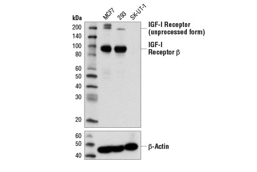
95, 200
Rabbit IgG
#P08069
3480
Product Information
Product Usage Information
| Application | Dilution |
|---|---|
| Western Blotting | 1:1000 |
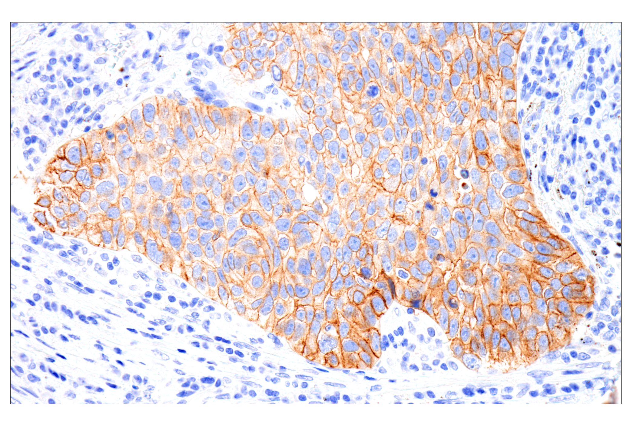
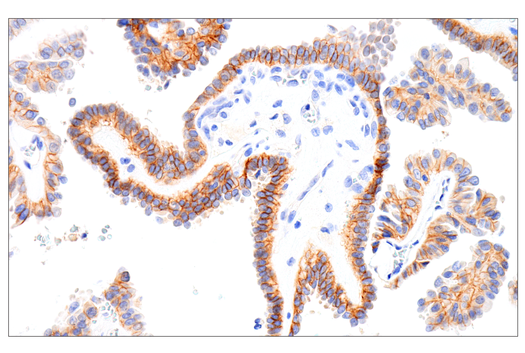
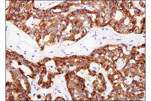
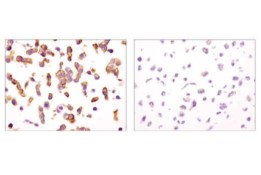
| IHC Leica Bond | 1:50 - 1:200 |
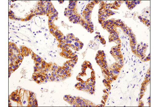
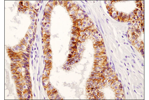
| Immunohistochemistry (Paraffin) | 1:400 |
Storage
Specificity / Sensitivity
Species Reactivity:
Human
Source / Purification
Monoclonal antibody is produced by immunizing animals with recombinant protein specific to human IGF-I receptor β protein.
Background
Type I insulin-like growth factor receptor (IGF-IR) is a transmembrane receptor tyrosine kinase that is widely expressed in many cell lines and cell types within fetal and postnatal tissues (1-3). Receptor autophosphorylation follows binding of the IGF-I and IGF-II ligands. Three tyrosine residues within the kinase domain (Tyr1131, Tyr1135, and Tyr1136) are the earliest major autophosphorylation sites (4). Phosphorylation of these three tyrosine residues is necessary for kinase activation (5,6). Insulin receptors (IRs) share significant structural and functional similarity with IGF-I receptors, including the presence of an equivalent tyrosine cluster (Tyr1146/1150/1151) within the kinase domain activation loop. Tyrosine autophosphorylation of IRs is one of the earliest cellular responses to insulin stimulation (7). Autophosphorylation begins with phosphorylation at Tyr1146 and either Tyr1150 or Tyr1151, while full kinase activation requires triple tyrosine phosphorylation (8).
- Adams, T.E. et al. (2000) Cell Mol Life Sci 57, 1050-93.
- Baserga, R. (2000) Oncogene 19, 5574-81.
- Scheidegger, K.J. et al. (2000) J Biol Chem 275, 38921-8.
- Hernández-Sánchez, C. et al. (1995) J Biol Chem 270, 29176-81.
- Lopaczynski, W. et al. (2000) Biochem Biophys Res Commun 279, 955-60.
- Baserga, R. (1999) Exp Cell Res 253, 1-6.
- White, M.F. et al. (1985) J Biol Chem 260, 9470-8.
- White, M.F. et al. (1988) J Biol Chem 263, 2969-80.
Species Reactivity
Species reactivity is determined by testing in at least one approved application (e.g., western blot).
Western Blot Buffer
IMPORTANT: For western blots, incubate membrane with diluted primary antibody in 5% w/v BSA, 1X TBS, 0.1% Tween® 20 at 4°C with gentle shaking, overnight.
Applications Key
WB: Western Blotting IHC-Bond: IHC Leica Bond IHC-P: Immunohistochemistry (Paraffin)
Cross-Reactivity Key
H: human M: mouse R: rat Hm: hamster Mk: monkey Vir: virus Mi: mink C: chicken Dm: D. melanogaster X: Xenopus Z: zebrafish B: bovine Dg: dog Pg: pig Sc: S. cerevisiae Ce: C. elegans Hr: horse GP: Guinea Pig Rab: rabbit All: all species expected
Trademarks and Patents
Limited Uses
Except as otherwise expressly agreed in a writing signed by a legally authorized representative of CST, the following terms apply to Products provided by CST, its affiliates or its distributors. Any Customer's terms and conditions that are in addition to, or different from, those contained herein, unless separately accepted in writing by a legally authorized representative of CST, are rejected and are of no force or effect.
Products are labeled with For Research Use Only or a similar labeling statement and have not been approved, cleared, or licensed by the FDA or other regulatory foreign or domestic entity, for any purpose. Customer shall not use any Product for any diagnostic or therapeutic purpose, or otherwise in any manner that conflicts with its labeling statement. Products sold or licensed by CST are provided for Customer as the end-user and solely for research and development uses. Any use of Product for diagnostic, prophylactic or therapeutic purposes, or any purchase of Product for resale (alone or as a component) or other commercial purpose, requires a separate license from CST. Customer shall (a) not sell, license, loan, donate or otherwise transfer or make available any Product to any third party, whether alone or in combination with other materials, or use the Products to manufacture any commercial products, (b) not copy, modify, reverse engineer, decompile, disassemble or otherwise attempt to discover the underlying structure or technology of the Products, or use the Products for the purpose of developing any products or services that would compete with CST products or services, (c) not alter or remove from the Products any trademarks, trade names, logos, patent or copyright notices or markings, (d) use the Products solely in accordance with CST Product Terms of Sale and any applicable documentation, and (e) comply with any license, terms of service or similar agreement with respect to any third party products or services used by Customer in connection with the Products.