WB, IHC-Bond, IHC-P
H
Endogenous
80
Rabbit IgG
#P01871
3507
Product Information
Product Usage Information
| Application | Dilution |
|---|---|
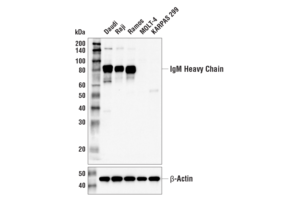
| Western Blotting | 1:1000 |
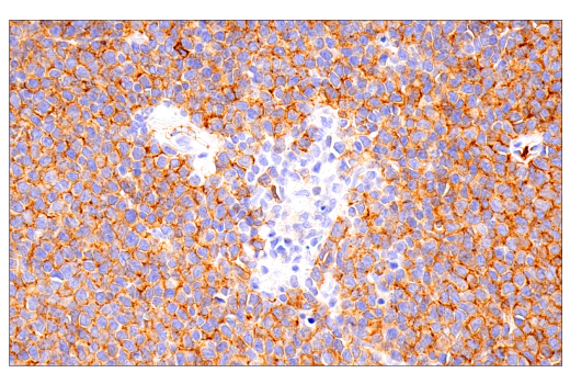
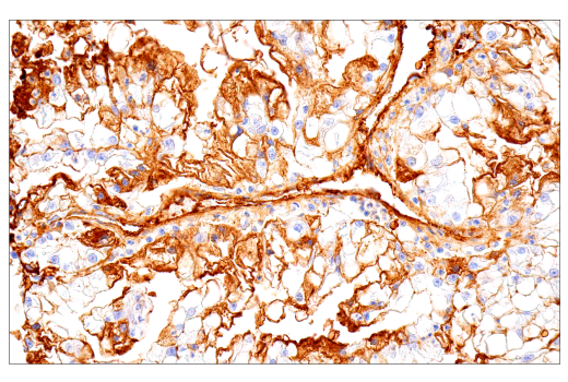
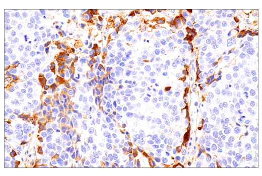
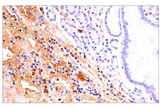
| IHC Leica Bond | 1:200 - 1:800 |
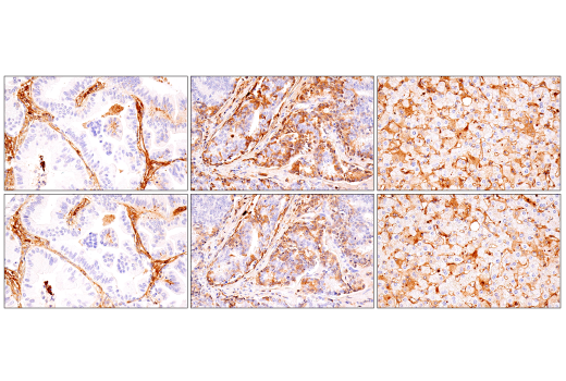
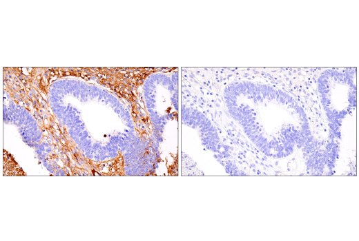
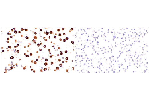
| Immunohistochemistry (Paraffin) | 1:200 - 1:800 |
Storage
For a carrier free (BSA and azide free) version of this product see product #94264.
Specificity / Sensitivity
Species Reactivity:
Human
Source / Purification
Monoclonal antibody is produced by immunizing animals with a synthetic peptide corresponding to residues surrounding Leu310 within the constant region of human IgM heavy chain protein.
Background
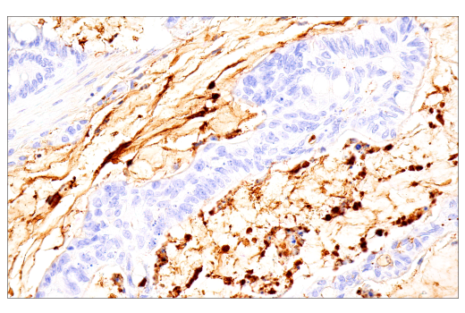
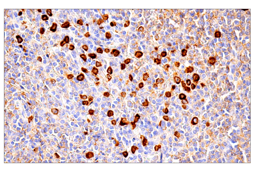
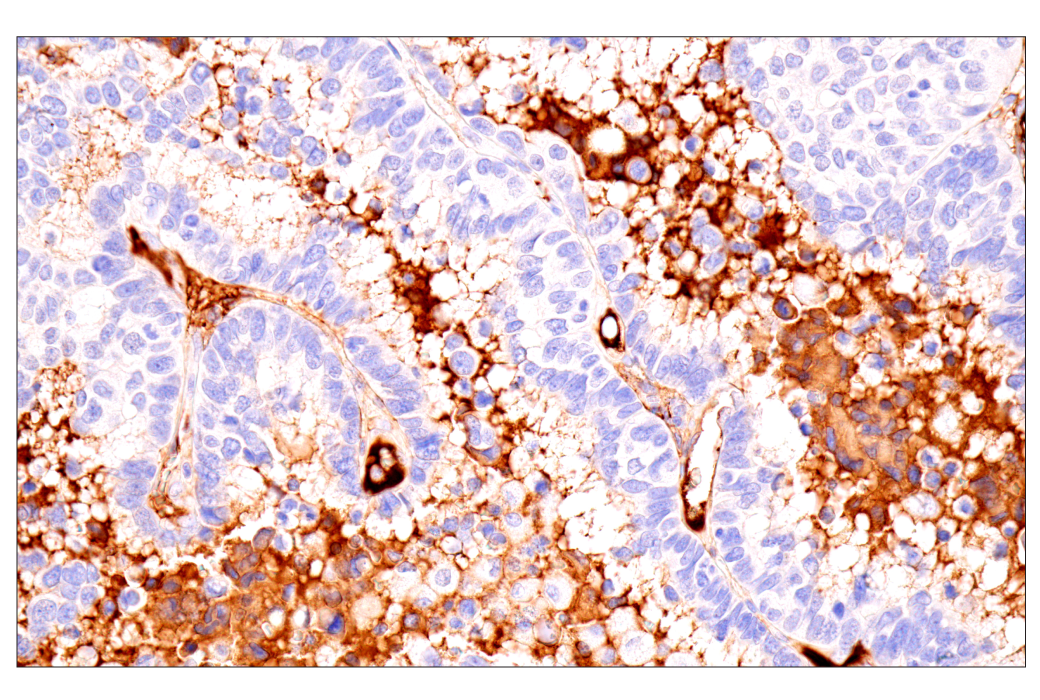
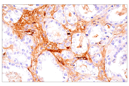
Immunoglobulin M (IgM) is one of the five major human immunoglobulin isotypes and is the first antibody to be secreted by plasmablasts in a humoral immune response after exposure to antigen. Structurally, IgM is the largest immunoglobulin and predominantly exists in pentameric form when secreted (1). Alternative splicing of the IgM heavy chain mRNA can generate an alternative form of the antibody, which facilitates its insertion into the plasma membrane of B cells to function in antigen recognition. IgM is the first Ig isotype to appear on the surface of developing B cells and is a major component of the B cell antigen receptor (BCR) signaling complex, which drives B cell survival and proliferation upon antigen-induced ligation (2,3). Research studies have shown that defects in the assembly of the BCR account for the low levels of surface IgM expression in B-chronic lymphocytic leukemia (4,5).
Species Reactivity
Species reactivity is determined by testing in at least one approved application (e.g., western blot).
Western Blot Buffer
IMPORTANT: For western blots, incubate membrane with diluted primary antibody in 5% w/v BSA, 1X TBS, 0.1% Tween® 20 at 4°C with gentle shaking, overnight.
Applications Key
WB: Western Blotting IHC-Bond: IHC Leica Bond IHC-P: Immunohistochemistry (Paraffin)
Cross-Reactivity Key
H: human M: mouse R: rat Hm: hamster Mk: monkey Vir: virus Mi: mink C: chicken Dm: D. melanogaster X: Xenopus Z: zebrafish B: bovine Dg: dog Pg: pig Sc: S. cerevisiae Ce: C. elegans Hr: horse GP: Guinea Pig Rab: rabbit All: all species expected
Trademarks and Patents
Limited Uses
Except as otherwise expressly agreed in a writing signed by a legally authorized representative of CST, the following terms apply to Products provided by CST, its affiliates or its distributors. Any Customer's terms and conditions that are in addition to, or different from, those contained herein, unless separately accepted in writing by a legally authorized representative of CST, are rejected and are of no force or effect.
Products are labeled with For Research Use Only or a similar labeling statement and have not been approved, cleared, or licensed by the FDA or other regulatory foreign or domestic entity, for any purpose. Customer shall not use any Product for any diagnostic or therapeutic purpose, or otherwise in any manner that conflicts with its labeling statement. Products sold or licensed by CST are provided for Customer as the end-user and solely for research and development uses. Any use of Product for diagnostic, prophylactic or therapeutic purposes, or any purchase of Product for resale (alone or as a component) or other commercial purpose, requires a separate license from CST. Customer shall (a) not sell, license, loan, donate or otherwise transfer or make available any Product to any third party, whether alone or in combination with other materials, or use the Products to manufacture any commercial products, (b) not copy, modify, reverse engineer, decompile, disassemble or otherwise attempt to discover the underlying structure or technology of the Products, or use the Products for the purpose of developing any products or services that would compete with CST products or services, (c) not alter or remove from the Products any trademarks, trade names, logos, patent or copyright notices or markings, (d) use the Products solely in accordance with CST Product Terms of Sale and any applicable documentation, and (e) comply with any license, terms of service or similar agreement with respect to any third party products or services used by Customer in connection with the Products.