WB, IF-IC, FC-FP
H
Endogenous
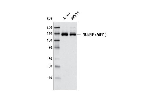
140
Rabbit
#Q9NQS7
3619
Product Information
Product Usage Information
| Application | Dilution |
|---|---|
| Western Blotting | 1:1000 |
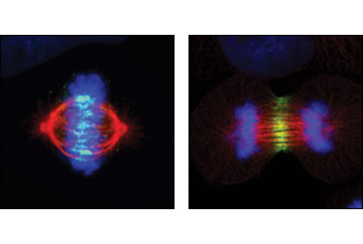
| Immunofluorescence (Immunocytochemistry) | 1:50 |
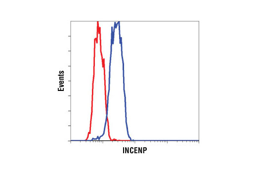
| Flow Cytometry (Fixed/Permeabilized) | 1:50 |
Storage
Specificity / Sensitivity
Species Reactivity:
Human
Source / Purification
Polyclonal antibodies are produced by immunizing animals with a synthetic peptide corresponding to amino acids surrounding Ala841 of human INCENP. Antibodies are purified by peptide affinity chromatography.
Background
INCENP (inner centromere protein antigens 135 kDa, 155 kDa) is a chromosomal passenger protein crucial for multiple events that mediate chromosome separation during mitosis (1). At prophase INCENP is associated with chromatin whereas during prometaphase and metaphase it translocates to the inner centromere (1). Depletion of INCENP results in aberrant chromosome alignment at the metaphase plate, incomplete chromosome separation, and disruption of proper spindle formation and cytokinesis (2). INCENP is part of the chromosomal passenger complex that also contains Aurora B, borealin and survivin (2). Aurora B and INCENP are mutually dependent on each other for proper localization (3), and in Drosophila cells and C.elegans embryos that lack INCENP or survivin, Aurora B cannot organize the kinetochores and the midbody (4,5). Phosphorylation on INCENP by CDK1 on Thr59 and Thr388 leads to the association of INCENP with Plk1, another important regulator of mitotic entry and exit (6). Interaction of INCENP with Plk1 is necessary for recruitment of Plk1 to the kinetochores, and the metaphase to anaphase transition (6). Interactions have also been reported between INCENP and heterochromatin protein 1α (HP1) (7) and β-tubulin (8).
- Carmena, M. and Earnshaw, W.C. (2006) Nat Cell Biol 8, 110-1.
- Carmena, M. and Earnshaw, W.C. (2003) Nat Rev Mol Cell Biol 4, 842-54.
- Kaitna, S. et al. (2000) Curr Biol 10, 1172-81.
- Speliotes, E.K. et al. (2000) Mol Cell 6, 211-23.
- Adams, R.R. et al. (2001) J Cell Biol 153, 865-80.
- Goto, H. et al. (2006) Nat Cell Biol 8, 180-7.
- Ainsztein, A.M. et al. (1998) J Cell Biol 143, 1763-74.
- Wheatley, S.P. et al. (2001) Exp Cell Res 262, 122-7.
Species Reactivity
Species reactivity is determined by testing in at least one approved application (e.g., western blot).
Western Blot Buffer
IMPORTANT: For western blots, incubate membrane with diluted primary antibody in 5% w/v BSA, 1X TBS, 0.1% Tween® 20 at 4°C with gentle shaking, overnight.
Applications Key
WB: Western Blotting IF-IC: Immunofluorescence (Immunocytochemistry) FC-FP: Flow Cytometry (Fixed/Permeabilized)
Cross-Reactivity Key
H: human M: mouse R: rat Hm: hamster Mk: monkey Vir: virus Mi: mink C: chicken Dm: D. melanogaster X: Xenopus Z: zebrafish B: bovine Dg: dog Pg: pig Sc: S. cerevisiae Ce: C. elegans Hr: horse GP: Guinea Pig Rab: rabbit All: all species expected
Trademarks and Patents
Limited Uses
Except as otherwise expressly agreed in a writing signed by a legally authorized representative of CST, the following terms apply to Products provided by CST, its affiliates or its distributors. Any Customer's terms and conditions that are in addition to, or different from, those contained herein, unless separately accepted in writing by a legally authorized representative of CST, are rejected and are of no force or effect.
Products are labeled with For Research Use Only or a similar labeling statement and have not been approved, cleared, or licensed by the FDA or other regulatory foreign or domestic entity, for any purpose. Customer shall not use any Product for any diagnostic or therapeutic purpose, or otherwise in any manner that conflicts with its labeling statement. Products sold or licensed by CST are provided for Customer as the end-user and solely for research and development uses. Any use of Product for diagnostic, prophylactic or therapeutic purposes, or any purchase of Product for resale (alone or as a component) or other commercial purpose, requires a separate license from CST. Customer shall (a) not sell, license, loan, donate or otherwise transfer or make available any Product to any third party, whether alone or in combination with other materials, or use the Products to manufacture any commercial products, (b) not copy, modify, reverse engineer, decompile, disassemble or otherwise attempt to discover the underlying structure or technology of the Products, or use the Products for the purpose of developing any products or services that would compete with CST products or services, (c) not alter or remove from the Products any trademarks, trade names, logos, patent or copyright notices or markings, (d) use the Products solely in accordance with CST Product Terms of Sale and any applicable documentation, and (e) comply with any license, terms of service or similar agreement with respect to any third party products or services used by Customer in connection with the Products.