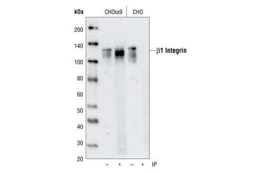
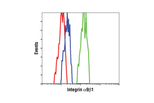
IP, FC-FP
H
Endogenous
150: alpha9, 130: beta1
Mouse IgG1
#P05556, #Q13797
3688, 3680
Product Information
Product Usage Information
| Application | Dilution |
|---|---|
| Immunoprecipitation | 1:50 |
| Flow Cytometry (Fixed/Permeabilized) | 1:400 |
Storage
Specificity / Sensitivity
Species Reactivity:
Human
Source / Purification
Monoclonal antibody is produced by immunizing animals with murine L cells transfected with human α9 integrin protein.
Background
Integrins are transmembrane glycoproteins that form heterodimers consisting of one α and one β subunit. The dimers act as receptors for extracellular matrix (ECM) proteins at sites of cell adhesion, and interact with focal adhesion (FA) proteins on the cytosolic side, forming the connection between the ECM and the actin cytoskeleton. Signaling to and from integrins regulates cell adhesion, motility, proliferation, apoptosis and gene expression, impacting cellular processes such as development, wound healing, immune response, invasion, metastasis and angiogenesis (reviewed in 1,2). α9β1 integrin is expressed in epithelial cells, smooth and skeletal muscle, neutrophils and hepatocytes (3). Its ligands include the ECM protein tenascin (4) and vascular cell adhesion molecule-1 (VCAM-1) (5). The cytoplasmic domain of α9 integrin binds the focal adhesion adaptor protein, paxillin, inhibiting cell spreading (6,7). Binding of the α9 cytoplasmic domain to spermidine/spermine N(1)-acetyltransferase (SSAT) mediates α9/β1 enhancement of cell migration (8). Physiological functions include development of the lymphatic system (9), possibly through binding to the lymphatic vascular endothelial growth factors VEGF-C and -D (10), neutrophil migration (5), and myogenic differentiation (11).
- Calderwood, D.A. et al. (2000) J Biol Chem 275, 22607-10.
- ffrench-Constant, C. and Colognato, H. (2004) Trends Cell. Biol. 14, 678-686.
- Palmer, E.L. et al. (1993) J. Cell Biol. 123, 1289-1297.
- Yokosaki, Y. et al. (1994) J. Biol. Chem. 269, 26691-26696.
- Taooka, Y. et al. (1999) J. Cell Biol. 145, 413-420.
- Young, B.A. et al. (2001) Mol. Biol. Cell 12, 3214-3225.
- Liu, S. et al. (2001) J. Biol. Chem. 276, 37086-37092.
- Chen, C. et al. (2004) J. Cell Biol. 167, 161-170.
- Huang, X.Z. et al. (2000) Mol. Cell Biol. 20, 5208-5215.
- Vlahakis, N.E. et al. (2005) J. Biol. Chem. 280, 4544-4552.
- Lafuste, P. et al. (2005) Mol. Biol. Cell 16, 861-870.
Species Reactivity
Species reactivity is determined by testing in at least one approved application (e.g., western blot).
Applications Key
IP: Immunoprecipitation FC-FP: Flow Cytometry (Fixed/Permeabilized)
Cross-Reactivity Key
H: human M: mouse R: rat Hm: hamster Mk: monkey Vir: virus Mi: mink C: chicken Dm: D. melanogaster X: Xenopus Z: zebrafish B: bovine Dg: dog Pg: pig Sc: S. cerevisiae Ce: C. elegans Hr: horse GP: Guinea Pig Rab: rabbit All: all species expected
Trademarks and Patents
Limited Uses
Except as otherwise expressly agreed in a writing signed by a legally authorized representative of CST, the following terms apply to Products provided by CST, its affiliates or its distributors. Any Customer's terms and conditions that are in addition to, or different from, those contained herein, unless separately accepted in writing by a legally authorized representative of CST, are rejected and are of no force or effect.
Products are labeled with For Research Use Only or a similar labeling statement and have not been approved, cleared, or licensed by the FDA or other regulatory foreign or domestic entity, for any purpose. Customer shall not use any Product for any diagnostic or therapeutic purpose, or otherwise in any manner that conflicts with its labeling statement. Products sold or licensed by CST are provided for Customer as the end-user and solely for research and development uses. Any use of Product for diagnostic, prophylactic or therapeutic purposes, or any purchase of Product for resale (alone or as a component) or other commercial purpose, requires a separate license from CST. Customer shall (a) not sell, license, loan, donate or otherwise transfer or make available any Product to any third party, whether alone or in combination with other materials, or use the Products to manufacture any commercial products, (b) not copy, modify, reverse engineer, decompile, disassemble or otherwise attempt to discover the underlying structure or technology of the Products, or use the Products for the purpose of developing any products or services that would compete with CST products or services, (c) not alter or remove from the Products any trademarks, trade names, logos, patent or copyright notices or markings, (d) use the Products solely in accordance with CST Product Terms of Sale and any applicable documentation, and (e) comply with any license, terms of service or similar agreement with respect to any third party products or services used by Customer in connection with the Products.