WB, IHC-P, FC-FP
H
Endogenous
210
Rabbit IgG
#P16144
3691
Product Information
Product Usage Information
| Application | Dilution |
|---|---|
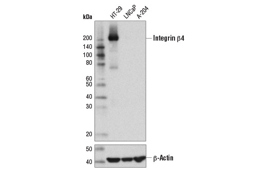
| Western Blotting | 1:1000 |
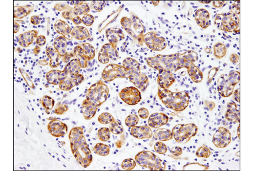
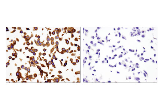
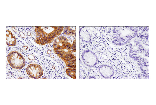
| Immunohistochemistry (Paraffin) | 1:200 |
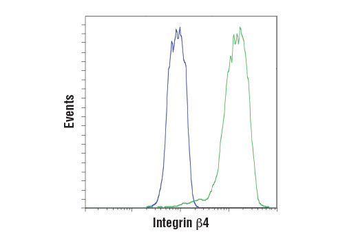
| Flow Cytometry (Fixed/Permeabilized) | 1:100 |
Storage
Specificity / Sensitivity
Species Reactivity:
Human
Source / Purification
Monoclonal antibody is produced by immunizing animals with a synthetic peptide corresponding to residues near the carboxy terminus of human integrin β4 protein.
Background
Integrins are α/β heterodimeric cell surface receptors that play a pivotal role in cell adhesion and migration, as well as in growth and survival (1,2). The integrin family contains at least 18 α and 8 β subunits that form 24 known integrins with distinct tissue distribution and overlapping ligand specificities (3). Integrins not only transmit signals to cells in response to the extracellular environment (outside-in signaling), but also sense intracellular cues to alter their interaction with extracellular environment (inside-out signaling) (1,2).
Integrin β4 pairs with integrin α6 on the cell surface membrane to form the integrin α6β4 heterodimer, an important laminin receptor that is essential for hemidesmosome formation and the support of stable adhesions between basal epithelial cells and the basement membrane (4,5). Integrin β4 is an important component in several growth factor induced signaling pathways that are involved in tumorigenesis and invasive cell growth (6,7).
- Liu, S. et al. (2000) J Cell Sci 113 ( Pt 20), 3563-71.
- Hood, J.D. and Cheresh, D.A. (2002) Nat Rev Cancer 2, 91-100.
- van der Flier, A. and Sonnenberg, A. (2001) Cell Tissue Res 305, 285-98.
- Schaapveld, R.Q. et al. (1998) J Cell Biol 142, 271-84.
- Litjens, S.H. et al. (2006) Trends Cell Biol 16, 376-83.
- Guo, W. et al. (2006) Cell 126, 489-502.
- Bertotti, A. et al. (2006) J Cell Biol 175, 993-1003.
Species Reactivity
Species reactivity is determined by testing in at least one approved application (e.g., western blot).
Western Blot Buffer
IMPORTANT: For western blots, incubate membrane with diluted primary antibody in 5% w/v nonfat dry milk, 1X TBS, 0.1% Tween® 20 at 4°C with gentle shaking, overnight.
Applications Key
WB: Western Blotting IHC-P: Immunohistochemistry (Paraffin) FC-FP: Flow Cytometry (Fixed/Permeabilized)
Cross-Reactivity Key
H: human M: mouse R: rat Hm: hamster Mk: monkey Vir: virus Mi: mink C: chicken Dm: D. melanogaster X: Xenopus Z: zebrafish B: bovine Dg: dog Pg: pig Sc: S. cerevisiae Ce: C. elegans Hr: horse GP: Guinea Pig Rab: rabbit All: all species expected
Trademarks and Patents
Limited Uses
Except as otherwise expressly agreed in a writing signed by a legally authorized representative of CST, the following terms apply to Products provided by CST, its affiliates or its distributors. Any Customer's terms and conditions that are in addition to, or different from, those contained herein, unless separately accepted in writing by a legally authorized representative of CST, are rejected and are of no force or effect.
Products are labeled with For Research Use Only or a similar labeling statement and have not been approved, cleared, or licensed by the FDA or other regulatory foreign or domestic entity, for any purpose. Customer shall not use any Product for any diagnostic or therapeutic purpose, or otherwise in any manner that conflicts with its labeling statement. Products sold or licensed by CST are provided for Customer as the end-user and solely for research and development uses. Any use of Product for diagnostic, prophylactic or therapeutic purposes, or any purchase of Product for resale (alone or as a component) or other commercial purpose, requires a separate license from CST. Customer shall (a) not sell, license, loan, donate or otherwise transfer or make available any Product to any third party, whether alone or in combination with other materials, or use the Products to manufacture any commercial products, (b) not copy, modify, reverse engineer, decompile, disassemble or otherwise attempt to discover the underlying structure or technology of the Products, or use the Products for the purpose of developing any products or services that would compete with CST products or services, (c) not alter or remove from the Products any trademarks, trade names, logos, patent or copyright notices or markings, (d) use the Products solely in accordance with CST Product Terms of Sale and any applicable documentation, and (e) comply with any license, terms of service or similar agreement with respect to any third party products or services used by Customer in connection with the Products.