WB, IF-IC
H Mk
Endogenous
78-105
Rabbit
#P51617
3654
Product Information
Product Usage Information
| Application | Dilution |
|---|---|
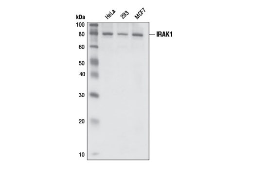
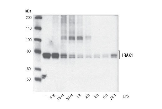
| Western Blotting | 1:1000 |
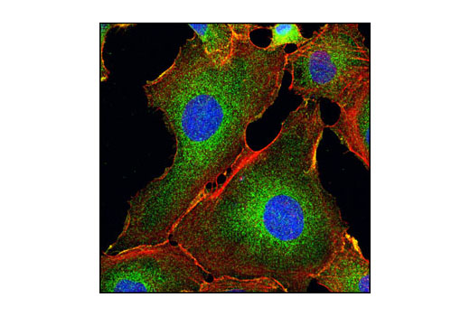
| Immunofluorescence (Immunocytochemistry) | 1:50 |
Storage
Specificity / Sensitivity
Species Reactivity:
Human, Monkey
Source / Purification
Polyclonal antibodies are produced by immunizing animals with a synthetic peptide corresponding to residues surrounding glycine 696 of human IRAK1. Antibodies are purified by protein A and peptide affinity chromatography.
Background
Interleukin-1 (IL-1) receptor-associated kinase (IRAK) is a serine/threonine-specific kinase that can be coprecipitated in an IL-1-inducible manner with the IL-1 receptor (1). The mammalian family of IRAK molecules contains four members (IRAK1, IRAK2, IRAK3/IRAK-M, and IRAK4). The binding of IL-1 to IL-1 receptor type I (IL-1RI) initiates the formation of a complex that includes IL-1RI, AcP, MyD88, and IRAKs (2). IRAK undergoes autophosphorylation shortly after IL-1 stimulation. The subsequent events involve IRAK dissociation from the IL-1RI complex, its ubiquitination, and its association with two membrane-bound proteins: TAB2 and TRAF6. The resulting IRAK-TRAF6-TAB2 complex is then released into the cytoplasm where it activates protein kinase cascades, including TAK1, IKKs, and the stress-activated kinases (3).
Species Reactivity
Species reactivity is determined by testing in at least one approved application (e.g., western blot).
Western Blot Buffer
IMPORTANT: For western blots, incubate membrane with diluted primary antibody in 5% w/v BSA, 1X TBS, 0.1% Tween® 20 at 4°C with gentle shaking, overnight.
Applications Key
WB: Western Blotting IF-IC: Immunofluorescence (Immunocytochemistry)
Cross-Reactivity Key
H: human M: mouse R: rat Hm: hamster Mk: monkey Vir: virus Mi: mink C: chicken Dm: D. melanogaster X: Xenopus Z: zebrafish B: bovine Dg: dog Pg: pig Sc: S. cerevisiae Ce: C. elegans Hr: horse GP: Guinea Pig Rab: rabbit All: all species expected
Trademarks and Patents
Limited Uses
Except as otherwise expressly agreed in a writing signed by a legally authorized representative of CST, the following terms apply to Products provided by CST, its affiliates or its distributors. Any Customer's terms and conditions that are in addition to, or different from, those contained herein, unless separately accepted in writing by a legally authorized representative of CST, are rejected and are of no force or effect.
Products are labeled with For Research Use Only or a similar labeling statement and have not been approved, cleared, or licensed by the FDA or other regulatory foreign or domestic entity, for any purpose. Customer shall not use any Product for any diagnostic or therapeutic purpose, or otherwise in any manner that conflicts with its labeling statement. Products sold or licensed by CST are provided for Customer as the end-user and solely for research and development uses. Any use of Product for diagnostic, prophylactic or therapeutic purposes, or any purchase of Product for resale (alone or as a component) or other commercial purpose, requires a separate license from CST. Customer shall (a) not sell, license, loan, donate or otherwise transfer or make available any Product to any third party, whether alone or in combination with other materials, or use the Products to manufacture any commercial products, (b) not copy, modify, reverse engineer, decompile, disassemble or otherwise attempt to discover the underlying structure or technology of the Products, or use the Products for the purpose of developing any products or services that would compete with CST products or services, (c) not alter or remove from the Products any trademarks, trade names, logos, patent or copyright notices or markings, (d) use the Products solely in accordance with CST Product Terms of Sale and any applicable documentation, and (e) comply with any license, terms of service or similar agreement with respect to any third party products or services used by Customer in connection with the Products.