WB, IP
H M R
Endogenous
46
Rabbit
#Q13572
3705
Product Information
Product Usage Information
| Application | Dilution |
|---|---|
| Western Blotting | 1:1000 |
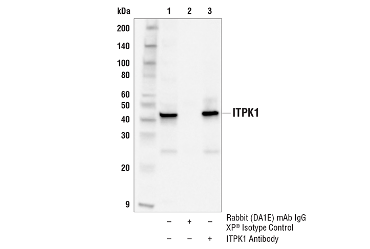
| Immunoprecipitation | 1:50 |
Storage
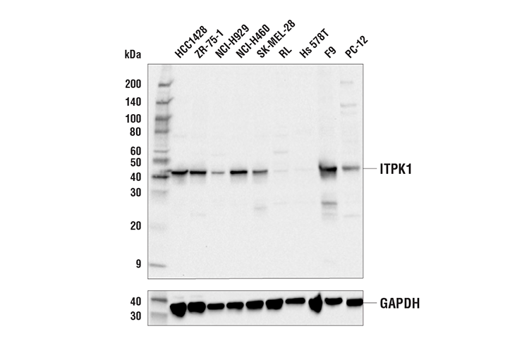
Specificity / Sensitivity
Species Reactivity:
Human, Mouse, Rat
Source / Purification
Polyclonal antibodies are produced by immunizing animals with a synthetic peptide corresponding to residues surrounding Ala382 of human ITPK1 protein. Antibodies are purified by peptide affinity chromatography.
Background
Inositol phosphates (IPs) are key second messengers produced from receptor activated phospholipase c (PLC) isoforms as well as a family of inositol kinases including ITPK1, IPMK, IPK1, IP3K, IP6K, and VIP1 (1,2). Lipid-derived inositol 1,3,4-trisphosphate (Ins(1,3,4)P3) produced from PLC is a well known regulator of calcium signaling. Cytosolic Ins(1,3,4)P3 is further modified to higher order IPs and pyrophosphates with roles in numerous physiological processes, including energy metabolism, ion channel regulation, transcriptional regulation, and cell death.ITPK1 (inositol 1,3,4-trisphosphate 5/6-kinase) is a reversible, poly-specific IP kinase that phosphorylates Ins(1,3,4)P3 with potential physiological implications including acting as a modifier of cystic fibrosis (3,4). ITPK1 can add either a 5- or 6-phosphate to Ins(1,3,4)P3, add a 1-phosphate to Ins(3,4,5,6)P4, or dephosphorylate Ins(1,3,4,5,6)P5 to Ins(3,4,5,6)P4 (5,6). ITPK1 inhibits TNF-alpha induced cell death, though the mechanism is unclear (7). ITPK1 as well as IPMK were associated with essential roles in MLKL-dependent necroptosis, a programmed process for necrotic cell death (8). MLKL directly interacts with higher order IPs like IP6, and is important for oligomerization of phosphorylated MLKL and subsequent plasma membrane localization. Mice generated with reduced expression of ITPK1 have neural tube defects (9). Furthermore, polymorphisms in the maternal ITPK1 gene have been associated as risk factors for neural tube defects resulting in spina bifida (10).
- Hatch, A.J. and York, J.D. (2010) Cell 143, 1030-1030.e1.
- Otto, J.C. et al. (2007) Proc Natl Acad Sci U S A 104, 15653-8.
- Desfougères, Y. et al. (2019) Proc Natl Acad Sci U S A 116, 24551-61.
- Chamberlain, P.P. et al. (2007) J Biol Chem 282, 28117-25.
- Yang, L. et al. (2006) J Cell Sci 119, 1320-8.
- Ho, M.W. et al. (2002) Curr Biol 12, 477-82.
- Sun, Y. et al. (2003) J Biol Chem 278, 43645-53.
- Dovey, C.M. et al. (2018) Mol Cell 70, 936-948.e7.
- Wilson, M.P. et al. (2009) Proc Natl Acad Sci U S A 106, 9831-5.
- Guan, Z. et al. (2014) PLoS One 9, e86145.
Species Reactivity
Species reactivity is determined by testing in at least one approved application (e.g., western blot).
Western Blot Buffer
IMPORTANT: For western blots, incubate membrane with diluted primary antibody in 5% w/v BSA, 1X TBS, 0.1% Tween® 20 at 4°C with gentle shaking, overnight.
Applications Key
WB: Western Blotting IP: Immunoprecipitation
Cross-Reactivity Key
H: human M: mouse R: rat Hm: hamster Mk: monkey Vir: virus Mi: mink C: chicken Dm: D. melanogaster X: Xenopus Z: zebrafish B: bovine Dg: dog Pg: pig Sc: S. cerevisiae Ce: C. elegans Hr: horse GP: Guinea Pig Rab: rabbit All: all species expected
Trademarks and Patents
Limited Uses
Except as otherwise expressly agreed in a writing signed by a legally authorized representative of CST, the following terms apply to Products provided by CST, its affiliates or its distributors. Any Customer's terms and conditions that are in addition to, or different from, those contained herein, unless separately accepted in writing by a legally authorized representative of CST, are rejected and are of no force or effect.
Products are labeled with For Research Use Only or a similar labeling statement and have not been approved, cleared, or licensed by the FDA or other regulatory foreign or domestic entity, for any purpose. Customer shall not use any Product for any diagnostic or therapeutic purpose, or otherwise in any manner that conflicts with its labeling statement. Products sold or licensed by CST are provided for Customer as the end-user and solely for research and development uses. Any use of Product for diagnostic, prophylactic or therapeutic purposes, or any purchase of Product for resale (alone or as a component) or other commercial purpose, requires a separate license from CST. Customer shall (a) not sell, license, loan, donate or otherwise transfer or make available any Product to any third party, whether alone or in combination with other materials, or use the Products to manufacture any commercial products, (b) not copy, modify, reverse engineer, decompile, disassemble or otherwise attempt to discover the underlying structure or technology of the Products, or use the Products for the purpose of developing any products or services that would compete with CST products or services, (c) not alter or remove from the Products any trademarks, trade names, logos, patent or copyright notices or markings, (d) use the Products solely in accordance with CST Product Terms of Sale and any applicable documentation, and (e) comply with any license, terms of service or similar agreement with respect to any third party products or services used by Customer in connection with the Products.